Abstract
We have genetically engineered an attenuated yellow fever (YF) virus to carry and express foreign antigenic sequences and evaluated the potential of this type of recombinant virus to serve as a safe and effective tumor vaccine. Live-attenuated YF vaccine is one of the most effective viral vaccines available today. Important advantages include its ability to induce long-lasting immunity, its safety, its affordability, and its documented efficacy. In this study, recombinant live-attenuated (strain 17D) YF viruses were constructed to express a cytotoxic T-lymphocyte epitope derived from chicken ovalbumin (SIINFEKL). These recombinant viruses replicated comparably to the 17D vaccine strain in cell culture and stably expressed the ovalbumin antigen, and infected cells presented the antigen in the context of major histocompatibility complex class I. Inoculation of mice with recombinant YF virus elicited SIINFEKL-specific CD8+ lymphocytes and induced protective immunity against challenge with lethal doses of malignant melanoma cells expressing ovalbumin. Furthermore, active immunotherapy with recombinant YF viruses induced regression of established solid tumors and pulmonary metastases. Thus, recombinant YF viruses are attractive viral vaccine vector candidates for the development of therapeutic anticancer vaccines.
Yellow fever (YF) virus 17D is an extremely safe and effective live viral vaccine, prepared from infected chicken embryos under standards developed by the World Health Organization. After vaccination, immunity is elicited within 10 days in over 95% of vaccinees (42) and neutralizing antibodies directed against the virus can be detected for more than 35 years (40). The vaccine safety record is outstanding: serious adverse reactions to YF virus 17D vaccine are extremely uncommon, and reversion to wild type is virtually nonexistent (4, 52).
YF virus is an enveloped, positive-stranded RNA virus and a member of the Flavivirus genus within the family Flaviviridae. The genome is approximately 11 kb in length and encodes a single polypeptide (51). This polypeptide precursor is proteolytically processed during and after translation, generating the functional proteins necessary for viral replication. Processing is mediated by cellular and viral proteases that recognize short specific amino acid sequences present at the junctions of the viral proteins. The viral protease NS2B-NS3 mediates most of the cleavages of the nonstructural proteins in the cytosol of an infected cell (3, 13, 15).
Given the favorable properties of YF virus 17D vaccine, a few laboratories are exploring the possibility of using chimeric YF viruses in which sequences encoding structural proteins are replaced by those derived from either Japanese encephalitis virus or Dengue virus. Promising results from these studies have been obtained with nonhuman primate models (14, 23, 24, 35, 37). Here we report a novel strategy to exploit the outstanding properties of the YF virus vaccine and demonstrate that inoculation of recombinant YF virus has therapeutic effect in a murine cancer model.
Tumor-specific cytotoxic T lymphocytes (CTLs) can prevent or eradicate tumors in a number of experimental systems and in patients with cancer (22, 26, 27). Clinical trials have demonstrated that 35% of patients with melanoma treated with specific, tumor-reactive lymphocytes can achieve either partial or complete tumor regression (46). The antigens recognized by the T cells have, in some cases, been identified (9, 10). Although cancer cells express a number of tumor-associated antigens (TAAs), CTLs directed against TAAs are not always elicited by the growing tumor, and as a consequence, the immune system fails to control tumor growth.
In contrast to tumor cells, viruses are efficient inducers of cellular immune responses. Thus, activation of the tumor-directed CTL response by vaccination with recombinant viruses expressing TAAs has been proposed for the prevention and treatment of malignancies. Viral vaccine vectors that have been successfully used in experimental cancer models include poxviruses, adenoviruses, picornaviruses, and influenza viruses (12, 16, 32, 43, 43a). However, each vaccine vector presents its own set of beneficial and adverse properties, and therefore, the search for new vectors continues to be an active area of research. In fact, clinical use of some vectors currently under study may be limited by their record of safety, efficacy, potential oncogenicity, or induction of immunosuppression. In addition, preexisting immunity against the vector may hinder the potency of treatment (16, 45), and therefore alternative viral vectors are needed. Finally, the combined use of multiple vaccine vectors expressing several TAAs may enhance the therapeutic effect of vaccination.
In this report, we describe the construction of a novel type of recombinant viruses based on YF virus 17D. These viruses are able to replicate without the need of a helper virus and stably carry and express a short foreign antigenic sequence (SIINFEKL, a CTL epitope from chicken ovalbumin). Cells infected with the recombinant virus presented the foreign peptide in a major histocompatibility complex (MHC) class I-restricted manner. Inoculation of the virus in mice elicited a CD8+ T-cell population that was specific for the inserted antigen. Importantly, immunization of mice with YF virus recombinants generated preventive and therapeutic immune responses that protected mice against lethal challenge with malignant melanoma cells expressing ovalbumin.
MATERIALS AND METHODS
Plasmids and PCR fragments.
Plasmids pYF5′3′ and pYFM5.2, which bear the complete YF virus 17D sequence, were kindly provided by Charles Rice. A full-length viral RNA can be generated from these plasmids by an in vitro ligation procedure (44). Briefly, we inserted into the viral cDNA a PCR-generated DNA fragment encoding a 13-amino-acid peptide from chicken ovalbumin flanked by BssHII and BstEII or ClaI and NdeI restriction enzyme sites and then by the viral peptidase (NS2B-NS3 complex) recognition site. We inserted the fragment at the following sites of the viral cDNA: the N terminus of the viral polypeptide or between proteins C and prM, NS2A and NS2B, NS2B and NS3, NS3 and NS4A, and NS4A and NS4B. To insert foreign sequences within the structural region of the genome, PCR fragments containing the foreign sequences were cloned into plasmid pYF5′3′ at each different location. To generate recombinants in the nonstructural part of the genome, 8-kb PCR fragments corresponding to the YF virus sequences in pYFM5.2 and containing the inserts of interest were produced. These PCR fragments were used as substitutes for plasmid pYFM5.2 in the in vitro ligation reaction described below.
Generation of viruses from plasmids.
Production of a molecular clone of YF virus 17D was carried out in a manner similar to that of a procedure originally published by Rice and coworkers (44). Briefly, 5 μg of each plasmid (or the corresponding sequences generated by PCR) was digested with the restriction enzymes AatII and ApaI. After digestion, the plasmids containing the 5′ and 3′ ends of the viral genome and the fragment from YFM5.2 corresponding to the middle region were each purified using low-melting-point agarose gel electrophoresis and ligated in equimolar concentrations for 4 h at 16°C. Ligase was inactivated by incubation for 20 min at 60°C. The ligated DNA was then digested with XhoI and used as the template for in vitro transcription by SP6 RNA polymerase (Promega, Madison, Wis.) in the presence of m7GpppAmp (New England Biolabs, Beverly, Mass.). Without further purification, synthetic RNA was transfected into BHK-21 cells by electroporation (electro cell manipulator 600; BTX, San Diego, Calif.).
Viral stocks.
Cytopathic effect (CPE) was observed 3 to 5 days following transfection. Viruses were cloned from individual plaques produced in BHK-21 cells. To generate viral stocks, cloned viruses were propagated in SW13 cells; supernatants of infected cells were cleared, aliquoted, titrated, and stored at −70°C.
Single-step growth curves.
Subconfluent SW13 cell monolayers were washed once with phosphate-buffered saline (PBS) and infected at a multiplicity of infection (MOI) of 5 PFU/cell. After a 2-h incubation period at 37°C, the cells were washed twice with PBS and then covered with L-15 medium supplemented with 10% fetal calf serum. Infected cell cultures were incubated at 37°C for several days, and 100-μl aliquots were recovered every 6 h for a period of 6 days or until total CPE occurred. Titers were determined by plaque assay.
Analysis of viral RNA by RT-PCR.
After subsequent passages of recombinant viruses on SW13 cells, total cytoplasmic RNA was obtained from infected cells by following the method employed by Chomczynski and Sacchi (17). Reverse transcription (RT) was carried out with Superscript (Gibco-BRL) by using random hexamers and a specific primer (ATCGCGGACCGAGTGGTTTTGTGTTTGTCATCCAAAGGTCTGCTTATTCTTGAGC) and following the manufacturer's recommended protocol. After 1 h of incubation at 42°C, 2 μl of each reaction product was used as the template in a PCR with RTth (Perkin-Elmer) and specific primers flanking the sequence to be studied (CAATGAGGCACTCGCAGCAGCTGG and TGCCCTAGCTCTGTGCGCTGCCC in YF virus pOva-8). The amplified PCR product was analyzed by restriction enzyme digestion and/or DNA sequencing.
Cell lines.
In addition to the BHK-21 and SW13 (human adenocarcinoma, adrenal cortex, ATCC CCL-105, passage 18) cells already described, the following cell lines were used in the antigen presentation assay or in the tumor rejection challenge: B3Z (T-cell hybridoma), EL-4 (thymoma), and EL-4 SL8 cells. B3Z cells and EL-4 SL8 cells were kindly provided by Nilabh Shastri, University of California, Berkeley. EL-4 SL8 cells stably express and present the ovalbumin (Ova) CTL epitope (SIINFEKL). B3Z is a murine T-cell hybridoma specific for Kb+SIINFEKL, which is transfected with the lacZ reporter gene under the transcriptional control of the interleukin 2 enhancer element. Thus, B3Z cell activation can easily be detected by the expression of β-galactosidase (25). The C57BL/6-derived melanoma cell lines B16F0 and B16-Ova were a kind gift from Kenneth Rock, University of Massachusetts. B16-Ova (Mo5.20.10) is a cell line that stably expresses ovalbumin and that is constructed by transfection of B16F0 cells with plasmid pAc-neo-Ova (22). HeLa Kb cells (a kind gift from Nilabh Shastri) were grown in RPMI 1640 supplemented with 10% fetal calf serum, 2 mM l-glutamine, and 1% penicillin–streptomycin and were constantly selected with 0.5 mg of G418 per ml. Cos Kb cells, transfected with sequences encoding the murine H-2 Kb molecule (Ken L. Rock, unpublished data), were grown in the same medium but permanently selected with G418 at 1 mg/ml.
Antigen presentation assay.
HeLa Kb and Cos Kb cells were mock infected or infected with YF virus 17D and recombinant viruses at an MOI of 10 PFU/cell. After a 48-h incubation period at 37°C, the infected cells or the same number of uninfected control cells was cocultured with 5 × 10⁴ B3Z cells for 16 h at 37°C. To determine the expression of β-galactosidase, cultures were washed with PBS and then fixed with 1% formaldehyde–0.2% glutaraldehyde for 5 min at 4°C. Cells were washed again and incubated with a solution consisting of 1 mg of X-Gal (5-bromo-4-chloro-3-indolyl-β-d-galactopyranoside), 5 mM potassium ferrocyanide, 5 mM potassium ferricyanide, and 2 mM MgCl2 in PBS. They were then incubated overnight at 37°C and examined microscopically for the presence of β-galactosidase activity (blue cells). As controls, we used EL-4 and EL-4 SL8, a cell line that constitutively expresses the SIINFEKL peptide. Cocultivation with EL-4 SL8 but not with EL-4 induced β-galactosidase production.
CD8+ T-cell responses. (i) Immunizations.
Groups of three C57BL/6 mice were inoculated intravenously (i.v.) once with PBS or with 107 PFU of either YF virus 17D or recombinant YF virus pOva-8 per mouse. Seven days later, all mice were sacrificed and their spleens were removed and dispersed to single-cell suspensions.
(ii) Restimulations and tetramer binding assays.
Splenocytes (3 × 10⁶) were restimulated by coculturing them for 5 days with 105 SIINFEKL-expressing EL-4 cells (EL-4 SL8) irradiated at 6,000 rads. SIINFEKL-MHC class I tetramers were the kind gift of John Altman (Emory University, Atlanta, Ga.). At day 5, cells were stained as described by Altman et al. (2) and analyzed by flow cytometry.
Immunizations and tumor challenge.
C57BL/6 mice (H-2Kb) were purchased from the Jackson Laboratory and used between 6 and 8 weeks of age. Groups of 5 or 10 mice were immunized intraperitoneally (i.p.), subcutanously (s.c.), intramuscularly (i.m.), or i.v. with 3 × 10⁵ PFU of YF virus 17D or YF virus pOva-8 (Ova-expressing recombinant 17D virus) per mouse. All groups were boosted with the same dose 2 weeks later. Nonimmunized mice were used as naïve controls. Melanoma cells were harvested by incubation in Ca2+- and Mg2+-free PBS for 5 min, viable cells were counted by trypan blue exclusion, and 30 days postinfection all mice were challenged with an s.c. injection of 5 × 10⁴ B16-Ova or B16F0 melanoma cells. The sizes of tumors were determined twice a week and expressed as tumor area corresponding to the largest perpendicular diameter in square centimeters. Animals that developed tumors greater than 2.0 cm2 were sacrificed.
Immunotherapy of solid tumors.
Mice were injected s.c. with 5 × 10⁴ B16-Ova cells. Treatment was started at day 0, 5, or 10 postimplantation of tumor cells and consisted of three s.c. injections of 4 × 10⁵ PFU of YF virus pOva-8 or 17D given in 5-day intervals. A control group was left untreated. Mice were observed for tumor development every three days, and tumors larger than 0.3 cm2 were scored as positive.
Immunotherapy of experimental pulmonary metastasis.
Mice were injected i.v. with either 5 × 10⁴ or 1 × 10⁵ B16-Ova cells. Immunotherapy was performed as described above for solid tumors. On day 30, 10 mice of each group were sacrificed and then their lungs were removed, placed for 5 min in 3% H2O2 in H2O, and fixed in Bouin's solution (Sigma Diagnostics, St. Louis, Mo.). The H2O2 treatment facilitates the analysis of metastasis under a dissection microscope by inflating the lungs and bleaching hemorrhages which otherwise might be mistaken for metastases.
RESULTS
Generation of YF virus 17D recombinants expressing a chicken ovalbumin T-cell epitope.
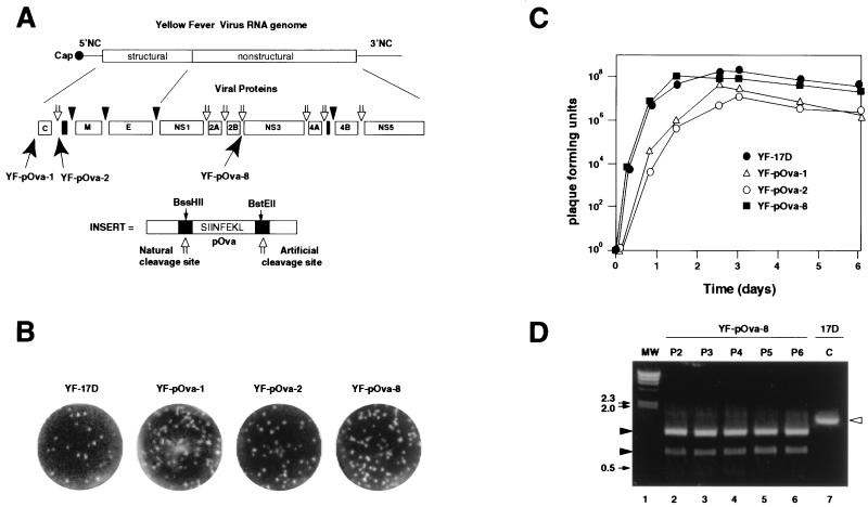
Our strategy to engineer YF virus recombinants was previously employed to engineer poliovirus recombinants. It uses basic aspects of the viral life cycle and permits the generation of replication-competent recombinant viruses that are able to replicate without the need of a helper virus (5). Foreign sequences (flanked by protease recognition sites) are inserted in frame at different positions within the YF virus polyprotein precursor. In this way, the viral protease recognizes and cleaves the flanking proteolytic sites, freeing the exogenous antigenic sequences from the rest of the YF virus polyprotein, and all of the YF virus proteins are produced correctly and viral replication proceeds normally (Fig. 1A). We introduced at several positions of the viral genome sequences encoding a chicken ovalbumin CTL epitope followed by an 8-amino-acid cleavage site for the viral protease NS2B-NS3 (Fig. 1A). The entire inserted sequence was 84 nucleotides in length and encoded the amino acid sequence SIINFEKL, an epitope restricted to the murine MHC class I molecule H-2 Kb (47).
FIG. 1.
Recombinant YF virus vectors expressing an MHC class I epitope derived from chicken ovalbumin. (A) Schematic diagram of a YF virus vector, YF virus pOva (YF-pOva), and strategy for expression of chicken ovalbumin. The top bar represents YF virus vector genomic RNA. The boxes below represent mature viral proteins. Open arrows indicate NS2B-NS3 cleavage sites, and black triangles indicate cellular signal peptidase cleavage sites. Nucleotide sequences encoding the ovalbumin Kb epitope SIINFEKL and flanking viral protease cleavage sites (see Materials and Methods) were inserted at the N terminus or at the junctions between C and prM and NS2B and NS3 (indicated by black arrows). Following translation, the viral polyprotein was proteolytically processed, resulting in the release of the foreign peptide and the generation of mature and functional viral proteins. NC, noncoding. (B) Plaque assay of parental strain 17D and vectors using BHK cells. (C) One-step growth curves of parental YF virus strain 17D (YF-17D) and three YF virus vectors: pOva-1, pOva-2, and pOva-8. SW13 cell monolayers were infected (MOI = 5) with 17D, pOva-1, pOva-2, or pOva-8. Virus production (PFU per milliliter) was determined at each time point by plaque assay. (D) Analysis of the stability of the pOva-8 vector by RT-PCR. SW13 cells were infected at low level (MOI < 1) with pOva-8 obtained after two, three, four, five, and six successive passages (P2, P3, P4, P5, and P6, respectively) in SW13 cells. The presence of the Ova insert was analyzed by RT-PCR using total cytoplasmic RNA of infected cells as a template for RT. The PCR product was digested with BstEII. The presence of the restriction site confirmed the presence of the foreign sequence in the viral genome. Molecular weight (MW) markers indicate relative mobilities. Bands with electrophoresis mobilities corresponding to those of 17D sequences are indicated by an open arrowhead, and mobilities corresponding to those of recombinant virus are indicated by filled arrowheads. C, YF virus 17D control.
Recombinant live YF viruses were recovered by transfection of BHK cells with in vitro-synthesized RNA. Insertion of exogenous sequences at three sites in the genome, the amino terminus and the C-prM and NS2B-NS3 junctions, yielded viable recombinant viruses (the resulting viruses were named YF-pOva-1, YF-pOva-2, and YF-pOva-8, respectively). In contrast, insertion at the NS2A-NS2B, NS3-NS4A, and NS4A-NS4B junctions abolished viral replication (as determined by plaque assay [data not shown]). The viruses were isolated from individual plaques, and viral stocks were generated by two sequential passages in SW13 cells.
Plaque assays and one-step growth curves showed that the recombinants YF-pOva-1, YF-pOva-2, and YF-pOva-8 replicate at rates remarkably similar to that of the parental 17D strain (Fig. 1B and C). Recombinant YF-pOva-8 replicated with kinetics identical to those of 17D and achieved nearly equivalent titers. Recombinant YF-pOva-1 and YF-pOva-2 replicated more slowly than the parental strain 17D, exhibiting a lag in replication, and by 3 days postinfection achieved only 10 to 20% of the titer of YF virus 17D (Fig. 1C). In view of the better replication properties of YF-pOva-8, this recombinant was used in all subsequent experiments aimed at characterizing the immunogenicity of YF virus vectors.
The ability of viral vectors to retain the inserted sequence is restricted in certain positive-strand RNA viruses by the high frequency of RNA recombination (38, 49). Therefore, we examined the presence of the inserted sequence by RT-PCR and diagnostic restriction enzyme digestion. The inserted sequences of pOva-8 were retained after six passages in BHK-21 cells (Fig. 1D). Thus, insertion of a foreign sequence between the NS2B and NS3 coding sequences of the YF virus genome does not seriously compromise viral replication.
Cells infected with YF-pOva-8 present the Ova peptide SIINFEKL in an MHC class I-restricted manner.
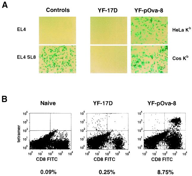
Next, we determined whether cells infected with recombinant YF viruses are able to express and present the foreign antigen in an MHC class I-restricted manner. We infected HeLa Kb and Cos Kb cells, which are transfected with sequences encoding the murine H-2 Kb molecule, with YF-pOva-8 and cocultivated the infected cells with a T-cell hybridoma that recognizes SIINFEKL presented in the context of Kb MHC class I molecules. The antigen is initially expressed in the cytoplasm of the infected cell, but it should be transported and presented on the surface of the cell through the MHC class I pathway. We used the T-cell hybridoma cell line B3Z, which carries a lacZ reporter gene under the transcriptional control of the interleukin-2 enhancer element NF-AT (25). Thus, T-cell receptor-specific stimulation of B3Z cells can be measured by the production of β-galactosidase activity.
Both HeLa Kb and Cos Kb cells infected with YF-pOva-8 activated the hybridoma T cells (Fig. 2A, right images). In contrast, cocultivation of B3Z cells with HeLa Kb and Cos Kb cells infected with parental YF virus 17D did not produce β-galactosidase activity (Fig. 2A, middle images). As controls, we cocultivated B3Z cells with EL-4 (negative control) or EL-4 SL8 cells, a cell line that stably expresses SIINFEKL (positive control) (Fig. 2A, left images). These results demonstrate that the foreign antigen encoded by the recombinant YF virus is expressed, processed, and presented in an MHC class I-restricted manner. In addition, Western blotting using polyclonal serum against YF virus indicated that the YF virus proteins are correctly produced and processed in YF-pOva-8-infected cells. This result suggests that the foreign antigen was appropriately cleaved away from the viral polyprotein (data not shown).
FIG. 2.
YF-pOva-8 infection activates CD8+ T-cell hybridomas in vitro and elicits specific CD8+ in vivo. (A) The CD8+ T-cell hybridoma cell line B3Z, which expresses β-galactosidase upon activation, was incubated with either a negative control (uninfected EL-4 cells) or a positive control (El-4 SL8 cells, which are stably transfected with SIINFEKL) (left images). B3Z cells were also cocultivated with HeLa Kb and Cos Kb cells infected with YF virus 17D and YF-pOva-8 (middle and right images, respectively). After 16 h at 37°C, cells were fixed and stained with X-Gal. Activated lacZ-positive T cells are stained blue, and unstained lacZ-negative cells are antigen-presenting cells and nonactivated T-cell hybridomas. (B) Tetrameric SIINFEKL-murine MHC class I molecule H-2 Kb complex bound to CD8+ splenocytes. Flow cytometry histograms illustrating tetramer binding to gated CD8+ T lymphocytes of naïve mice or mice infected with parental YF virus 17D or recombinant YF virus pOva-8 are shown. In this case, splenocytes were restimulated by cocultivation for 5 days with EL-4 cells expressing SIINFEKL (EL-4 SL8). The values indicate percentages of CD8+ T lymphocytes that bound the tetramer. FITC, fluorescein isothiocyanate.
Induction of Ova-specific CD8+ T cells by recombinant YF- pOva-8.
To determine whether recombinant YF viruses are able to induce specific CD8+ T lymphocytes, mice were inoculated one time with either YF-pOva-8, parental YF virus 17D, or PBS (naïve). Splenocytes obtained 7 days after immunization were restimulated by cocultivation with a cell line that expresses SIINFEKL (EL-4 SL8) and monitored for the development of a CD8+ T-lymphocyte population that bound MHC class I SIINFEKL tetramers. Splenocytes from naïve mice, or mice immunized with YF virus 17D, failed to produce SIINFEKL-specific T cells under these conditions (Fig. 2B). In contrast, a significant percentage of CD8+ T cells (8.75%) obtained from mice immunized with YF-pOva-8 were specific for SIINFEKL (Fig. 2B).
Protective immunity in vivo.
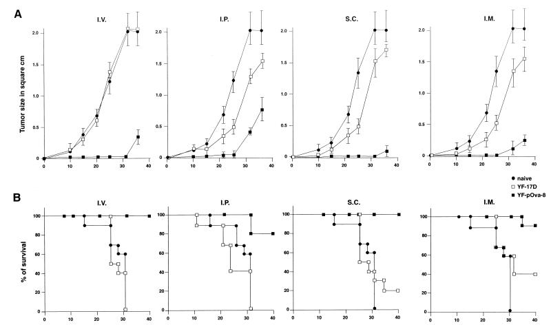
To evaluate whether the vector induces protective CTL immunity, we used an established tumor model in which CTLs play an essential role in protecting the host from challenge with a lethal dose of malignant melanoma cells (22). Mice were immunized twice with YF-pOva-8 or YF virus 17D and then challenged 30 days later with B16-Ova, a tumor cell line derived from B16 F0, which stably expresses chicken ovalbumin. Inoculation of B16-Ova cells produced tumors that grow rapidly and killed naïve mice and mice inoculated with parental 17D virus in a few weeks (Fig. 3). In contrast, immunization with YF-pOva-8 protected animals against lethal challenge with B16-Ova. Immunization protected mice from local tumor growth (Fig. 3A) and also from death (Fig. 3B).
FIG. 3.
Immunization with a YF virus vector induces protective and antigen-specific immunity to melanoma B16 cells expressing Ova. C57BL/6 mice were immunized either twice every two weeks i.p., i.v., i.m., or s.c. with YF-pOva-8 (3 × 10⁵ PFU/mouse). As a control, mice were inoculated with either the parental YF virus 17D or saline (naïve). Thirty days after the first immunization (day 0) animals were challenged with 5 × 10⁴ B16-Ova cells. (A) Local tumor growth. The size of the tumor was determined every 5 days and is plotted as the average tumor area ± the standard deviation in square centimeters versus time postchallenge (days). (B) Survival is plotted as the percentage of surviving animals versus time. All experiments included 10 mice per group and were repeated three times.
We administered the recombinant YF-pOva-8 by four different routes—s.c., i.m., i.p., and i.v.—to compare the efficiencies of the protective immune responses. s.c. and i.v. inoculation elicited the most potent protective responses. All of the animals vaccinated with YF-pOva-8 were protected at the time when 100% of the control mice had died. i.p. and i.m. inoculations were slightly less efficient; in these groups, 10 to 20% of the mice developed tumors and died. The vaccine effect was specific for SIINFEKL because mice vaccinated with YF-pOva-8 were not protected against challenge with parental B16 melanoma cells, which do not express Ova (data not shown). However, we observed a slight delay in tumor growth in mice inoculated with YF virus 17D when the virus was administered i.n., s.c., or i.m. (Fig. 3A). This effect may be due to increased cytokine production or other immunological responses induced by YF virus replication. Indeed, it has been shown previously that gamma and alpha interferon have antitumor and anticellular activities on B16 melanoma cells (6, 7, 30).
Active immunotherapy of established tumors.
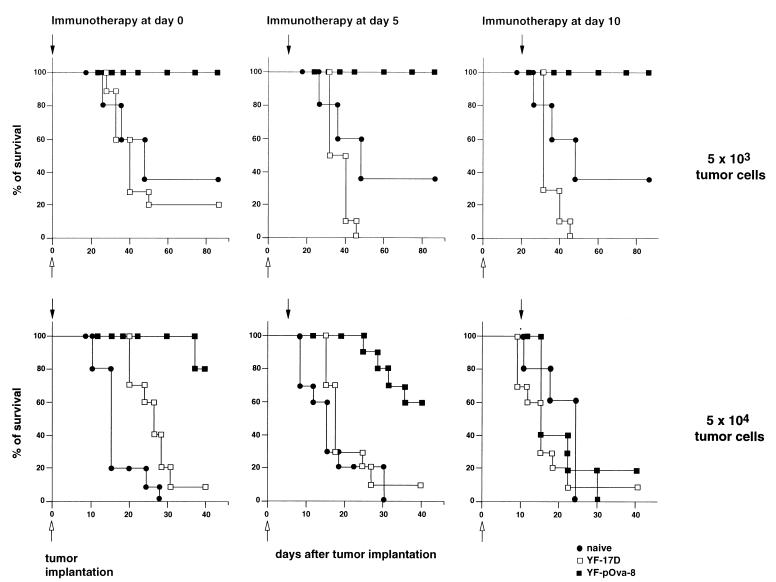
Next, we determined whether immunization with a recombinant YF virus is able to induce regression of established tumors. Mice were inoculated s.c. with either 5 × 10³ or 5 × 10⁴ B16-Ova tumor cells and subsequently infected with YF-pOva-8 at the day of tumor cell implantation (day 0), 5 days postimplantation of tumor cells (day 5), or 10 days postimplantation of tumor cells (day 10). Inoculation of 5 × 10³ B16-Ova tumor cells produced tumors in about 60% of naïve mice. In contrast, mice were completely protected by immunization with YF-pOva-8, even if treatment was started 10 days after tumor implantation (Fig. 4). For animals inoculated with the higher doses of tumor cells (5 × 10⁴), 80% of the animals immunized with YF-pOva-8 at day 0 remained tumor free 45 days after tumor injection while those injected with parental YF virus 17D or saline developed tumors and died within 3 to 4 weeks (Fig. 4). Immunization with 17D slightly delayed tumor growth relative to that with saline, but the effect was minimal and 90% of the animals developed tumors 30 days after tumor cell implantation. Immunization with recombinant YF-pOva-8 5 days postimplantation resulted in partial protection (60% of the mice remained tumor free). Immunization at day 10 had little or no effect on tumor growth. These results demonstrate that treatment of established tumors can be achieved by immunization with YF virus recombinants but that successful treatment depends on the tumor burden at the time at which immunotherapy is started.
FIG. 4.
Inoculation of the recombinant YF-pOva-8 eliminates established B16 solid tumors. C57BL/6 mice were injected s.c. with melanoma B16-Ova cells (5 × 10³ or 5 × 10⁴ cells/mouse) at day 0 (tumor implantation). Animals received three s.c. inoculations every 3 days of either PBS (naïve), the parental virus 17D (YF-17D), or YF-pOva-8 (4 × 10⁵ PFU/mouse). Viruses were administered at the day of tumor cell implantation (day 0) or 5 (day 5) or 10 (day 10) days after tumor cell inoculation. Mice were monitored for evidence of tumor growth by palpation and inspection twice a week.
Active immunotherapy of pulmonary metastasis.
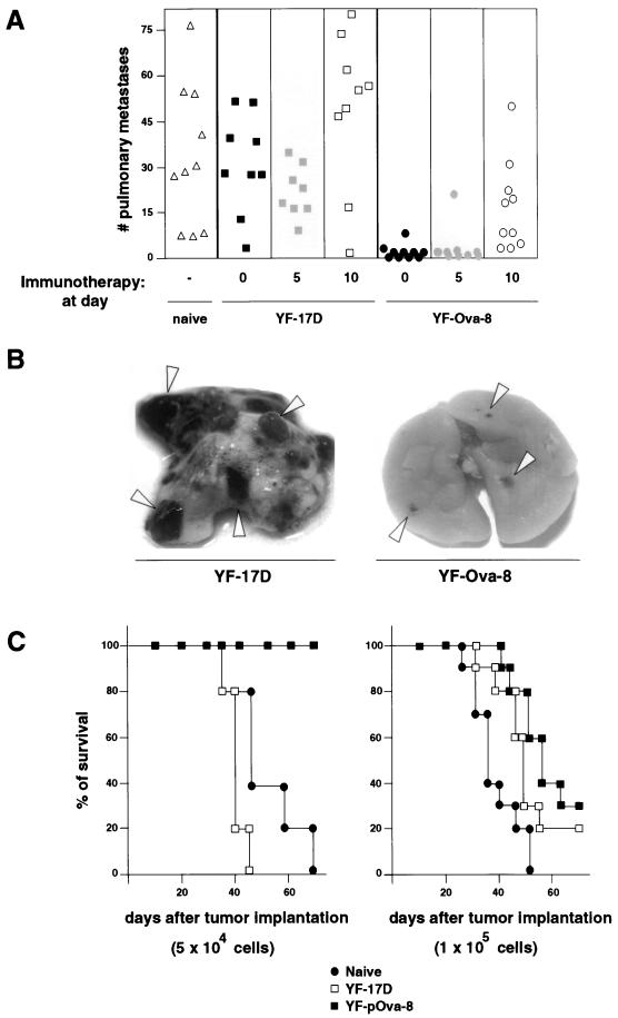
B16 melanoma cells, when injected into the tail vein of syngenic mice, reproducibly metastasize to the lungs (41). This provides a model to evaluate whether YF virus recombinants are able to elicit effective antimetastasis responses. Mice were inoculated i.v. with either 5 × 10⁴ or 1 × 10⁵ B16-Ova cells, and at day 0, 5, or 10 postinoculation, animals were immunized s.c. with YF-pOva-8 or the control virus. Immunization with YF-pOva-8 substantially reduced both the size and number of lung metastases (Fig. 5A and B) and prevented death (Fig. 5C). Ten weeks after implantation of 5 × 10⁴ tumor cells, 100% of the animals immunized at day 0 were healthy (Fig. 5C). Protection dropped to 80% when immunization was started at day 5 and dropped further to 20% when animals were immunized starting at day 10 (data not shown). Inoculation with YF virus 17D had no protective effect, and metastases developed with the same kinetics as in animals inoculated with saline (Fig. 5A and B). When mice were inoculated with a higher dose of tumor cells (105 cells), only 40% of mice treated at day 0 were protected. These results underline the importance of starting immunotherapy when the tumor burden is low. Nonetheless, these results demonstrate that recombinant YF viruses expressing a single CTL epitope are able to elicit a therapeutic antitumor response in mice.
FIG. 5.
Infection with YF-pOva-8 is an effective immunotherapeutic treatment for pulmonary metastases. Mice were inoculated i.v. with 5 × 10⁴ or 1 × 10⁵ B16-Ova tumor cells and then inoculated s.c. with YF-pOva-8 (3 × 10⁵ PFU/mouse) or controls (naïve or YF virus 17D) at 0, 5, or 10 days postinjection of tumor cells. (A) The lungs of treated mice were evaluated in a coded, blind manner for pulmonary metastases 30 days after the tumor cell inoculation. The number of pulmonary metastases is shown for individual mice. (B) Pictures of lungs treated with YF-pOva-8 or 17D at day 30 after tumor cell inoculation. (C) Survival is plotted as the percentage of surviving animals versus time. All experiments included 10 mice per group.
DISCUSSION
We have engineered the genome of YF virus to generate a replication-competent vaccine vector that carries and expresses antigenic sequences derived from other pathogens or tumors. Insertion of antigenic sequences can be tolerated at several positions within the virus genome without abrogation of viral replication. Viruses that carried insertions at the junction between NS2B and NS3 replicated with wild-type kinetics. Insertion of exogenous sequences at other sites in the genome, such as at the amino terminus and C-prM junction, also yielded viable chimeric viruses (Fig. 1B and C); however, insertions at the NS2A-NS2B, NS3-NS4A, and NS4A-NS4B junctions abolished viral replication. We have not yet determined the upper size limit of tolerated sequences at the NS2B-NS3 position; however, our previous work with poliovirus, in which we inserted as many as 1,089 nucleotides, suggests that small RNA viruses can tolerate relatively large insertions. Indeed, we have already successfully constructed chimeric YF viruses that stably carry 2,000 nucleotides of foreign sequences (these results will be reported elsewhere).
Because many RNA viruses have a high frequency of RNA recombination, one concern of using YF virus as a vaccine vector is that inserted sequences may be genetically unstable. Indeed, previous attempts to engineer picornaviruses have been limited by a high frequency of deletion after only a few rounds of replication in tissue culture (1, 11, 31, 38, 49). However, the results presented here suggest that YF virus recombinants carrying small insertions retain the foreign sequence for at least six passages in tissue culture (Fig. 1D).
As mentioned previously, important advantages of the live YF virus vaccine include its documented efficacy, ease of administration, economy of delivery, and ability to induce long-lasting immunity (33, 34, 36). The vaccine has a very good safety record, as serious adverse reactions are extremely uncommon. Allergic reactions occur at a very low rate (approximately 1 in 1 million). Vaccination usually results in a low-level viremia lasting 1 to 2 days and beginning 3 to 4 days after inoculation. The low magnitude of viremia and the fact that Aedes aegypti, the mosquito responsible for its natural transmission, is refractory to oral infection with 17D virus, preclude the possibility of natural transmission (and possible reversion) of the vaccine virus.
Thus, the potential adaptation of YF virus as a vaccine vector to express antigens from other pathogens deserves special attention. Recently, a system for the expression of heterologous genes based on self-replicating RNAs derived from a related flavivirus (Kunjin virus) has been described. Noncytopathic Kunjin virus replicon vectors were developed to express a number of foreign proteins, and these replicons can be packaged into virus-like particles (29, 50). The immunogenic capacity of this type of vector, however, has not been assessed yet. In a second approach, a YF virus-based candidate vaccine was constructed by replacing the envelope gene of YF virus by those of Japanese encephalitis virus and dengue virus. Both mice and monkeys inoculated with this chimeric virus developed neutralizing antibodies and were protected against challenge with a virulent Japanese encephalitis virus and dengue virus (14, 24, 37). These results demonstrate that YF virus can be used to generate protective immunity against a heterologous virus.
In this study we have generated novel replication-competent YF viruses that carry and express the well-characterized T-cell epitope SIINFEKL. Immunization of mice with YF virus expressing this model antigen induces a specific CD8+ cell population and protects animals from lethal challenge with an aggressive lethal melanoma cell line. Furthermore, active immunotherapy with YF-pOva-8 significantly reduced the numbers of established s.c. tumors and experimental pulmonary metastases. It has been shown that specific CTLs directed against SIINFEKL are essential for the eradication of B16-Ova-induced tumors (22). Since YF-pOva-8 expresses only the SIINFEKL epitope and immunization with YF virus 17D showed minimal protection from tumor challenge, the protective effect of pOva-8 inoculation must be mediated by specific CTLs directed against the inserted sequence.
The B16 tumor model has been previously used to study other forms of immunotherapy, like adoptive transfer of in vitro-expanded tumor-specific T cells (8) and immunization with genetically engineered tumor cells (19–21, 48), DNA-pulsed fibroblasts (18), or tumor extract-pulsed dendritic cells (53). Although all these experimental cancer therapies have shown significant therapeutic effects in mice, their use in humans may prove impractical. In particular, the manipulation and expansion of the appropriate cells may require complicated tissue culture manipulation and can be technically difficult. In contrast, the use of the YF virus or other vaccine viral vectors would be much simpler and more cost-effective.
Finding therapeutically valuable tumor-associated antigens has proven difficult. However, in melanoma patients, CD8+ and CD4+ T cells specific for antigens expressed by the tumor can frequently be found, but they are not normally effective at protecting from disease (26–28). Furthermore, most of the identified human TAAs are derived from melanoma cells and are also expressed in normal melanocytes. Therefore, the antigen expressed by the recombinant YF virus vector should be able to break tolerance, which could result in autoimmunity. Indeed, with a different viral vector, it has recently been shown that immunization of mice with a recombinant vaccinia virus expressing a melanoma-associated antigen, TRP-1, induces vitiligo disease (autoimmune depigmentation of patches of skin and hair) and effective antitumor immunity in mice (39). Because of the need for effective anticancer immunotherapies, the induction of immunity against tissue-specific antigens may be an approach in which the autoimmune side effects may represent acceptable collateral damage.
In summary, we have developed a novel viral vaccine vector that potentially can express antigens derived from other pathogens or tumors. Recombinant YF virus expressing a single antigenic determinant can elicit powerful cellular immune responses and have therapeutic efficacy against established tumors. We hope that these viruses can be employed by themselves or in prime-boost combinations with other recombinant viruses to develop a safe and effective protocol for the prevention and treatment of other infectious diseases or human cancers.
ACKNOWLEDGMENTS
We thank C. Rice for the gift of YF virus cDNA molecular clones, N. Shastry for the B3Z hybridoma cell line, and J. D. Altman for the gift of specific Ova tetramers. We are grateful to Rob Sadler, who never hesitated to help with hundreds of injections, and to Shane Crotty and Jody Baron for useful comments on the manuscript.
This work was supported by Public Health Service grant AI44343 to R.A.
Footnotes
Dedicated to the memory of Rob Sadler (1962–1999).
REFERENCES
- 1.Alexander L, Lu H H, Gromeier M, Wimmer E. Dicistronic polioviruses as expression vectors for foreign genes. AIDS Res Hum Retroviruses. 1994;10(Suppl. 2):S57–S60. [PubMed] [Google Scholar]
- 2.Altman J D, Moss P A H, Goulder P J R, Barouch D H, McHeyzer-Williams M G, Bell J I, McMichael A J, Davis M M. Phenotypic analysis of antigen-specific T lymphocytes. Science. 1996;274:94–96. . (Erratum, 280:1821, 1998.) [PubMed] [Google Scholar]
- 3.Amberger V R, Paganetti P A, Seulberger H, Eldering J A, Schwab M E. Characterization of a membrane-bound metalloendoprotease of rat C6 glioblastoma cells. Cancer Res. 1994;54:4017–4025. [PubMed] [Google Scholar]
- 4.American Medical Association. Fatal viral encephalitis following 17D yellow fever vaccine inoculation. Report of a case in a 3-year-old child. JAMA. 1966;198:671–672. doi: 10.1001/jama.1966.03110190153047. [DOI] [PubMed] [Google Scholar]
- 5.Andino R, Silvera D, Suggett S D, Achacoso P L, Miller C J, Baltimore D, Feinberg M B. Engineering poliovirus as a vaccine vector for the expression of diverse antigens. Science. 1994;265:1448–1451. doi: 10.1126/science.8073288. [DOI] [PubMed] [Google Scholar]
- 6.Arany I, Fleischmann C M, Tyring S K, Fleischmann W R. Interferon regulates expression of mda-6/WAF1/CIP1 and cyclin-dependent kinases independently from p53 in B16 murine melanoma cells. Biochem Biophys Res Commun. 1997;233:678–680. doi: 10.1006/bbrc.1997.6516. [DOI] [PubMed] [Google Scholar]
- 7.Arany I, Fleischmann C M, Tyring S K, Fleischmann W R., Jr Lack of mda-6/WAF1/CIP1-mediated inhibition of cyclin-dependent kinases in interferon-alpha resistant murine B16 melanoma cells. Cancer Lett. 1997;119:237–240. doi: 10.1016/s0304-3835(97)00288-7. [DOI] [PubMed] [Google Scholar]
- 8.Bohm W, Thoma S, Leithauser F, Moller P, Schirmbeck R, Reimann J. T cell-mediated, IFN-gamma-facilitated rejection of murine B16 melanomas. J Immunol. 1998;161:897–908. [PubMed] [Google Scholar]
- 9.Boon T. Tumor antigens recognized by cytolytic T lymphocytes: present perspectives for specific immunotherapy. Int J Cancer. 1993;54:177–180. doi: 10.1002/ijc.2910540202. [DOI] [PubMed] [Google Scholar]
- 10.Boon T, De Plaen E, Lurquin C, Van den Eynde B, van der Bruggen P, Traversari C, Amar-Costesec A, Van Pel A. Identification of tumour rejection antigens recognized by T lymphocytes. Cancer Surv. 1992;13:23–37. [PubMed] [Google Scholar]
- 11.Burke K L, Almond J W, Evans D J. Antigen chimeras of poliovirus. Prog Med Virol. 1991;38:56–68. [PubMed] [Google Scholar]
- 12.Carroll M W, Overwijk W W, Chamberlain R S, Rosenberg S A, Moss B, Restifo N P. Highly attenuated modified vaccinia virus Ankara (MVA) as an effective recombinant vector: a murine tumor model. Vaccine. 1997;15:387–394. doi: 10.1016/s0264-410x(96)00195-8. [DOI] [PMC free article] [PubMed] [Google Scholar]
- 13.Chambers T J, Grakoui A, Rice C M. Processing of the yellow fever virus nonstructural polyprotein: a catalytically active NS3 proteinase domain and NS2B are required for cleavages at dibasic sites. J Virol. 1991;65:6042–6050. doi: 10.1128/jvi.65.11.6042-6050.1991. [DOI] [PMC free article] [PubMed] [Google Scholar]
- 14.Chambers T J, Nestorowicz A, Mason P W, Rice C M. Yellow fever/Japanese encephalitis chimeric viruses: construction and biological properties. J Virol. 1999;73:3095–3101. doi: 10.1128/jvi.73.4.3095-3101.1999. [DOI] [PMC free article] [PubMed] [Google Scholar]
- 15.Chambers T J, Weir R C, Grakoui A, McCourt D W, Bazan J F, Fletterick R J, Rice C M. Evidence that the N-terminal domain of nonstructural protein NS3 from yellow fever virus is a serine protease responsible for site-specific cleavages in the viral polyprotein. Proc Natl Acad Sci USA. 1990;87:8898–8902. doi: 10.1073/pnas.87.22.8898. [DOI] [PMC free article] [PubMed] [Google Scholar]
- 16.Chen P W, Wang M, Bronte V, Zhai Y, Rosenberg S A, Restifo N P. Therapeutic antitumor response after immunization with a recombinant adenovirus encoding a model tumor-associated antigen. J Immunol. 1996;156:224–231. [PMC free article] [PubMed] [Google Scholar]
- 17.Chomczynski P, Sacchi N. Single-step method of RNA isolation by acid guanidinium thiocyanate–phenol-chloroform extraction. Anal Biochem. 1987;162:156–159. doi: 10.1006/abio.1987.9999. [DOI] [PubMed] [Google Scholar]
- 18.de Zoeten E F, Carr-Brendel V, Cohen E P. Resistance to melanoma in mice immunized with semiallogeneic fibroblasts transfected with DNA from mouse melanoma cells. J Immunol. 1998;160:2915–2922. [PubMed] [Google Scholar]
- 19.Dranoff G, Jaffee E, Lazenby A, Golumbek P, Levitsky H, Brose K, Jackson V, Hamada H, Pardoll D, Mulligan R C. Vaccination with irradiated tumor cells engineered to secrete murine granulocyte-macrophage colony-stimulating factor stimulates potent, specific, and long-lasting anti-tumor immunity. Proc Natl Acad Sci USA. 1993;90:3539–3543. doi: 10.1073/pnas.90.8.3539. [DOI] [PMC free article] [PubMed] [Google Scholar]
- 20.Dranoff G, Soiffer R, Lynch T, Mihm M, Jung K, Kolesar K, Liebster L, Lam P, Duda R, Mentzer S, Singer S, Tanabe K, Johnson R, Sober A, Bhan A, Clift S, Cohen L, Parry G, Rokovich J, Richards L, Drayer J, Berns A, Mulligan R C. A phase I study of vaccination with autologous, irradiated melanoma cells engineered to secrete human granulocyte-macrophage colony stimulating factor. Hum Gene Ther. 1997;8:111–123. doi: 10.1089/hum.1997.8.1-111. [DOI] [PubMed] [Google Scholar]
- 21.Ellem K A, O'Rourke M G, Johnson G R, Parry G, Misko I S, Schmidt C W, Parsons P G, Burrows S R, Cross S, Fell A, Li C L, Bell J R, Dubois P J, Moss D J, Good M F, Kelso A, Cohen L K, Dranoff G, Mulligan R C. A case report: immune responses and clinical course of the first human use of granulocyte/macrophage-colony-stimulating-factor-transduced autologous melanoma cells for immunotherapy. Cancer Immunol Immunother. 1997;44:10–20. doi: 10.1007/s002620050349. [DOI] [PMC free article] [PubMed] [Google Scholar]
- 22.Falo L D, Jr, Kovacsovics-Bankowski M, Thompson K, Rock K L. Targeting antigen into the phagocytic pathway in vivo induces protective tumour immunity. Nat Med. 1995;1:649–653. doi: 10.1038/nm0795-649. [DOI] [PubMed] [Google Scholar]
- 23.Guirakhoo F, Weltzin R, Chambers T J, Zhang Z X, Soike K, Ratterree M, Arroyo J, Georgakopoulos K, Catalan J, Monath T P. Recombinant chimeric yellow fever-dengue type 2 virus is immunogenic and protective in nonhuman primates. J Virol. 2000;74:5477–5485. doi: 10.1128/jvi.74.12.5477-5485.2000. [DOI] [PMC free article] [PubMed] [Google Scholar]
- 24.Guirakhoo F, Zhang Z X, Chambers T J, Delagrave S, Arroyo J, Barrett A D, Monath T P. Immunogenicity, genetic stability, and protective efficacy of a recombinant, chimeric yellow fever-Japanese encephalitis virus (ChimeriVax-JE) as a live, attenuated vaccine candidate against Japanese encephalitis. Virology. 1999;257:363–372. doi: 10.1006/viro.1999.9695. [DOI] [PubMed] [Google Scholar]
- 25.Karttunen J, Sanderson S, Shastri N. Detection of rare antigen-presenting cells by the lacZ T-cell activation assay suggests an expression cloning strategy for T-cell antigens. Proc Natl Acad Sci USA. 1992;89:6020–6024. doi: 10.1073/pnas.89.13.6020. [DOI] [PMC free article] [PubMed] [Google Scholar]
- 26.Kawakami Y, Eliyahu S, Delgado C H, Robbins P F, Rivoltini L, Topalian S L, Miki T, Rosenberg S A. Cloning of the gene coding for a shared human melanoma antigen recognized by autologous T cells infiltrating into tumor. Proc Natl Acad Sci USA. 1994;91:3515–3519. doi: 10.1073/pnas.91.9.3515. [DOI] [PMC free article] [PubMed] [Google Scholar]
- 27.Kawakami Y, Eliyahu S, Delgado C H, Robbins P F, Sakaguchi K, Appella E, Yannelli J R, Adema G J, Miki T, Rosenberg S A. Identification of a human melanoma antigen recognized by tumor-infiltrating lymphocytes associated with in vivo tumor rejection. Proc Natl Acad Sci USA. 1994;91:6458–6462. doi: 10.1073/pnas.91.14.6458. [DOI] [PMC free article] [PubMed] [Google Scholar]
- 28.Kawakami Y, Eliyahu S, Sakaguchi K, Robbins P F, Rivoltini L, Yannelli J R, Appella E, Rosenberg S A. Identification of the immunodominant peptides of the MART-1 human melanoma antigen recognized by the majority of HLA-A2-restricted tumor infiltrating lymphocytes. J Exp Med. 1994;180:347–352. doi: 10.1084/jem.180.1.347. [DOI] [PMC free article] [PubMed] [Google Scholar]
- 29.Khromykh A A, Westaway E G. Subgenomic replicons of the flavivirus Kunjin: construction and applications. J Virol. 1997;71:1497–1505. doi: 10.1128/jvi.71.2.1497-1505.1997. [DOI] [PMC free article] [PubMed] [Google Scholar]
- 30.Lasek W, Wankowicz A, Kuc K, Feleszko W, Golab J, Giermasz A, Wiktor-Jedrzejczak W, Jakobisiak M. Potentiation of antitumor effects of tumor necrosis factor alpha and interferon gamma by macrophage-colony-stimulating factor in a MmB16 melanoma model in mice. Cancer Immunol Immunother. 1995;40:315–321. doi: 10.1007/BF01519632. [DOI] [PMC free article] [PubMed] [Google Scholar]
- 31.Lu H, Alexander L, Wimmer E. Construction and genetic analysis of dicistronic polioviruses containing open reading frames for epitopes of human immunodeficiency virus type 1 gp120. J Virol. 1995;69:4797–4806. doi: 10.1128/jvi.69.8.4797-4806.1995. [DOI] [PMC free article] [PubMed] [Google Scholar]
- 32.Mandl S, Sigal L J, Rock K L, Andino R. Poliovirus vaccine vectors elicit antigen-specific cytotoxic T cells and protect mice against lethal challenge with malignant melanoma cells expressing a model antigen. Proc Natl Acad Sci USA. 1998;95:8216–8221. doi: 10.1073/pnas.95.14.8216. [DOI] [PMC free article] [PubMed] [Google Scholar]
- 33.Monath T P. Flaviviruses. In: Fields B N, Knipe D M, editors. Virology. 2nd ed. New York, N.Y: Raven Press, Ltd.; 1990. pp. 763–814. [Google Scholar]
- 34.Monath T P. Yellow fever: Victor, Victoria? Conqueror, conquest? Epidemics and research in the last forty years and prospects for the future. Am J Trop Med Hyg. 1991;45:1–43. doi: 10.4269/ajtmh.1991.45.1. [DOI] [PubMed] [Google Scholar]
- 35.Monath T P, Levenbook I, Soike K, Zhang Z X, Ratterree M, Draper K, Barrett A D, Nichols R, Weltzin R, Arroyo J, Guirakhoo F. Chimeric yellow fever virus 17D-Japanese encephalitis virus vaccine: dose-response effectiveness and extended safety testing in rhesus monkeys. J Virol. 2000;74:1742–1751. doi: 10.1128/jvi.74.4.1742-1751.2000. [DOI] [PMC free article] [PubMed] [Google Scholar]
- 36.Monath T P, Nasidi A. Should yellow fever vaccine be included in the expanded program of immunization in Africa? A cost-effectiveness analysis for Nigeria. Am J Trop Med Hyg. 1993;48:274–299. doi: 10.4269/ajtmh.1993.48.274. [DOI] [PubMed] [Google Scholar]
- 37.Monath T P, Soike K, Levenbook I, Zhang Z X, Arroyo J, Delagrave S, Myers G, Barrett A D, Shope R E, Ratterree M, Chambers T J, Guirakhoo F. Recombinant, chimaeric live, attenuated vaccine (ChimeriVax) incorporating the envelope genes of Japanese encephalitis (SA14-14-2) virus and the capsid and nonstructural genes of yellow fever (17D) virus is safe, immunogenic and protective in non-human primates. Vaccine. 1999;17:1869–1882. doi: 10.1016/s0264-410x(98)00487-3. [DOI] [PubMed] [Google Scholar]
- 38.Mueller S, Wimmer E. Expression of foreign proteins by poliovirus polyprotein fusion: analysis of genetic stability reveals rapid deletions and formation of cardioviruslike open reading frames. J Virol. 1998;72:20–31. doi: 10.1128/jvi.72.1.20-31.1998. [DOI] [PMC free article] [PubMed] [Google Scholar]
- 39.Overwijk W W, Lee D S, Surman D R, Irvine K R, Touloukian C E, Chan C C, Carroll M W, Moss B, Rosenberg S A, Restifo N P. Vaccination with a recombinant vaccinia virus encoding a “self” antigen induces autoimmune vitiligo and tumor cell destruction in mice: requirement for CD4(+) T lymphocytes. Proc Natl Acad Sci USA. 1999;96:2982–2987. doi: 10.1073/pnas.96.6.2982. [DOI] [PMC free article] [PubMed] [Google Scholar]
- 40.Poland J D, Calisher C H, Monath T P, Downs W G, Murphy K. Persistence of neutralizing antibody 30–35 years after immunization with 17D yellow fever vaccine. Bull W H O. 1981;59:895–900. [PMC free article] [PubMed] [Google Scholar]
- 41.Poste G, Fidler I J. The pathogenesis of cancer metastasis. Nature. 1980;283:139–146. doi: 10.1038/283139a0. [DOI] [PubMed] [Google Scholar]
- 42.Reinhardt B, Jaspert R, Niedrig M, Kostner C, L'Age-Stehr J. Development of viremia and humoral and cellular parameters of immune activation after vaccination with yellow fever virus strain 17D: a model of human flavivirus infection. J Med Virol. 1998;56:159–167. doi: 10.1002/(sici)1096-9071(199810)56:2<159::aid-jmv10>3.0.co;2-b. [DOI] [PubMed] [Google Scholar]
- 43.Restifo N P. The new vaccines: building viruses that elicit antitumor immunity. Curr Opin Immunol. 1996;8:658–663. doi: 10.1016/s0952-7915(96)80082-3. [DOI] [PMC free article] [PubMed] [Google Scholar]
- 43a.Restifo N P, Surman D R, Zheng H, Palese P, Rosenberg S A, Garcia-Sastre A. Transfectant influenza A viruses are effective recombinant immunogens in the treatment of experimental cancer. Virology. 1998;249:89–97. doi: 10.1006/viro.1998.9330. [DOI] [PMC free article] [PubMed] [Google Scholar]
- 44.Rice C M, Grakoui A, Galler R, Chambers T J. Transcription of infectious yellow fever RNA from full-length cDNA templates produced by in vitro ligation. New Biol. 1989;1:285–296. [PubMed] [Google Scholar]
- 45.Rooney J F, Wohlenberg C, Cremer K J, Moss B, Notkins A L. Immunization with a vaccinia virus recombinant expressing herpes simplex virus type 1 glycoprotein D: long-term protection and effect of revaccination. J Virol. 1988;62:1530–1534. doi: 10.1128/jvi.62.5.1530-1534.1988. [DOI] [PMC free article] [PubMed] [Google Scholar]
- 46.Rosenberg S A, Yannelli J R, Yang J C, Topalian S L, Schwartzentruber D J, Weber J S, Parkinson D R, Seipp C A, Einhorn J H, White D E. Treatment of patients with metastatic melanoma with autologous tumor-infiltrating lymphocytes and interleukin 2. J Natl Cancer Inst. 1994;86:1159–1166. doi: 10.1093/jnci/86.15.1159. [DOI] [PubMed] [Google Scholar]
- 47.Rotzschke O, Falk K, Deres K, Schild H, Norda M, Metzger J, Jung G, Rammensee H G. Isolation and analysis of naturally processed viral peptides as recognized by cytotoxic T cells. Nature. 1990;348:252–254. doi: 10.1038/348252a0. [DOI] [PubMed] [Google Scholar]
- 48.Simons J W, Jaffee E M, Weber C E, Levitsky H I, Nelson W G, Carducci M A, Lazenby A J, Cohen L K, Finn C C, Clift S M, Hauda K M, Beck L A, Leiferman K M, Owens A H, Jr, Piantadosi S, Dranoff G, Mulligan R C, Pardoll D M, Marshall F F. Bioactivity of autologous irradiated renal cell carcinoma vaccines generated by ex vivo granulocyte-macrophage colony-stimulating factor gene transfer. Cancer Res. 1997;57:1537–1546. [PMC free article] [PubMed] [Google Scholar]
- 49.Tang R S, Barton D J, Flanegan J B, Kirkegaard K. Poliovirus RNA recombination in cell-free extracts. RNA. 1997;3:624–633. [PMC free article] [PubMed] [Google Scholar]
- 50.Varnavski A N, Khromykh A A. Noncytopathic flavivirus replicon RNA-based system for expression and delivery of heterologous genes. Virology. 1999;255:366–375. doi: 10.1006/viro.1998.9564. [DOI] [PubMed] [Google Scholar]
- 51.Westaway E G. Flavivirus replication strategy. Adv Virus Res. 1987;33:45–90. doi: 10.1016/s0065-3527(08)60316-4. [DOI] [PubMed] [Google Scholar]
- 52.Xie H, Cass A R, Barrett A D. Yellow fever 17D vaccine virus isolated from healthy vaccinees accumulates very few mutations. Virus Res. 1998;55:93–99. doi: 10.1016/s0168-1702(98)00036-7. [DOI] [PubMed] [Google Scholar]
- 53.Yang S, Darrow T L, Vervaert C E, Seigler H F. Immunotherapeutic potential of tumor antigen-pulsed and unpulsed dendritic cells generated from murine bone marrow. Cell Immunol. 1997;179:84–95. doi: 10.1006/cimm.1997.1151. [DOI] [PubMed] [Google Scholar]


