Abstract
Intronic G triplets are frequently located adjacent to 5′ splice sites in vertebrate pre-mRNAs and have been correlated with splicing efficiency and specificity via a mechanism that activates upstream 5′ splice sites in exons containing duplicated sites (26). Using an intron dependent upon G triplets for maximal activity and 5′ splice site specificity, we determined that these elements bind U1 snRNPs via base pairing with U1 RNA. This interaction is novel in that it uses nucleotides 8 to 10 of U1 RNA and is independent of nucleotides 1 to 7. In vivo functionality of base pairing was documented by restoring activity and specificity to mutated G triplets through compensating U1 RNA mutations. We suggest that the G-rich region near vertebrate 5′ splice sites promotes accurate splice site recognition by recruiting the U1 snRNP.
Most eukaryotic genes contain introns which are cotranscriptionally removed from precursor transcripts by RNA splicing (32). Splicing occurs in a macromolecular ribonucleoprotein complex termed the spliceosome, the ordered assembly of which has been characterized in vitro. The initial metazoan complex, the commitment complex, contains the 5′ splice site recognition factor U1 snRNP, the 3′ splice site recognition factor U2AF, and at least one member of the arginine-serine-rich (SR) family of splicing factors (14, 28, 36). The second complex, the prespliceosome, or A complex, is defined by the ATP-dependent addition of the U2 snRNP (20). As the spliceosome matures, U1 snRNP at the 5′ splice site is replaced by U5 and U6 snRNPs (31, 41).
Studies investigating the mechanism through which mammalian 5′ splice sites are recognized have implicated SR proteins, hnRNP A1, the U1 snRNP, and the U6 snRNP as important (5, 12, 21, 22, 24, 31, 38). In studies using precursor RNAs containing exons with multiple 5′ splice sites, the binding of any one of these may be determinative for usage. Most studies examining the mechanism through which duplicated 5′ splice sites are recognized have concentrated on the involvement of SR proteins via their capacity to bind to either 5′ splice site sequences or exon enhancer sequences, which in turn influence the usage of nearby 5′ splice sites (12, 19, 43). These studies have led to models suggesting that the choice of splice site is dictated by the affinity of SR proteins and snRNPs for the site. In sites with low affinity, only a single site is bound, and this is the site used to direct splicing. In sites with high affinity, double occupation occurs, and the site proximal to the 3′ splice site across the intron predominates (12).
Intron enhancer sequences can also regulate 5′ splice site usage. In general, when intron enhancer sequences are placed between competing 5′ splice sites, they direct usage to the upstream, distal 5′ splice site (7, 11, 19, 26). In at least one of these cases, in the gene coding for hnRNP A1, the enhancer element directs upstream 5′ splice site usage without altering the ability of U1 snRNPs to bind to the competing 5′ splice sites, suggesting that 5′ splice site usage is not dominantly determined by the occupancy of splice sites by U1 snRNPs (7). In this same system, the addition of SR proteins caused a reversal of phenotype such that usage of the proximal 5′ splice site predominated, indicating the possibility of alternate competing mechanisms of 5′ splice site selection. Indeed, it has been demonstrated in a number of test cases that SR proteins and hnRNP A1 antagonistically affect 5′ splice site recognition and that subtle changes in the relative concentrations of the two types of factors can be determinative for 5′ splice site recognition (5, 24).
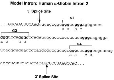
One sequence element frequently found in close proximity to mammalian 5′ splice sites is the sequence GGG (26, 30, 35). The presence of a G triplet in the vicinity of a 5′ splice site has been demonstrated to be of predictive value for identification of exons in sequenced genomic DNA, which suggests that the recognition of such sequences by splicing factors may play an important role in 5′ splice site selection in a large number of pre-mRNAs. Indeed, several intron enhancer sequences that function to activate an upstream exon contain G triplets (2, 6, 18, 25, 34). We have modeled the function of G triplets in 5′ splice site recognition using an intron containing multiple G triplets (26). This intron, the second intron of the human alpha-globin gene, contains multiple G triplets, which we arbitrarily grouped into three elements designated G1, G2, and G4 (Fig. 1). Maximal in vitro and in vivo activity of the intron is dependent upon the presence of these G triplets, and we therefore refer to these elements as intronic splicing enhancers. When the G1, G2, and G4 elements were mutated, in vivo splicing of the alpha-globin intron was reduced from near 100% to about 45% (26).
FIG. 1.
Sequence of the second intron of the human alpha-globin gene. G elements are denoted, as well as mutants used in this and previous studies (26).
The alpha-globin G triplet elements also affect splice site selection. In precursor RNAs containing two identical 5′ splice sites separated by wild-type G1 and G2 elements, the distal or 5′-most splice site was used almost exclusively. Progressive mutation of the G triplets in these elements resulted in a switch from the distal to the proximal 5′ splice site (26). The elements act additively to increase activity and define 5′ splice sites, suggesting that only a single element need be occupied by a trans-acting factor (26).
In the present study, we provide evidence that the alpha-globin G-rich elements interact directly with the RNA component of the U1 snRNP by base pairing with nucleotides 8 to 10 of U1 RNA. This interaction is novel in that it does not require nucleotides 2 to 7 of U1 RNA, which are generally believed to interact with 5′ splice site sequences. Replacement of one or more G triplets in alpha-globin substrates containing duplicated 5′ splice sites with mutant sequences complementary to altered U1 RNAs permitted substantial, but not total, rescue of the 5′ splice site selection phenotype, suggesting that the ability of the G triplets to bind U1 RNA can account for most, but not all, of the selectivity afforded by G triplets. Interestingly, increased concentrations of SR proteins were unable to modulate 5′ splice site choice in alpha-globin substrates containing duplicated 5′ splice sites. Given the prevalence of G triplets in the region immediately downstream of vertebrate 5′ splice sites, it is likely that interaction between G-rich elements and U1 RNA represents a common mechanism for recognizing 5′ splice sites in vertebrate pre-mRNAs.
MATERIALS AND METHODS
Plasmids, in vitro transcription, and HeLa cell transfection.
Alpha-globin sequences were inserted into pCDNA3 (Invitrogen) for in vitro and in vivo transcription as described previously (26). The wild-type and mutant G1-G2 RNA expression plasmids used to generate psoralen cross-linking probes were constructed by ligating annealed complementary oligonucleotides into pUC19. The T7 promoter used for in vitro transcription of these RNAs was positioned so that transcription initiated at the first G nucleotide of the G1 element. The wild-type U1 RNA expression vector was kindly provided by Alan Weiner (University of Washington) and has been previously described (42). Mutations were introduced into this U1 plasmid by PCR-mediated mutagenesis. Mammalian expression vectors coding for human ASF-SF2 and SC35 were kindly provided by James Manley (Columbia University) and have been described previously (38). HeLa cell maintenance, transfection, RNA isolation, and reverse transcription (RT)-PCR analysis were done as described previously (26). Cloned RT-PCR products were sequenced to confirm splice site usage. Capped [32P]GTP-labeled in vitro transcripts were prepared from linearized templates as described previously (26).
Psoralen cross-linking.
For psoralen cross-linking, 150,000 cpm of 32P-labeled RNA was incubated for 5 min at 30°C under splicing conditions described previously (26) with the addition of psoralen (HRI Associates) to a final concentration of 0.02 μg/μl. Reactions were illuminated at 365 nm for 15 min to generate RNA-RNA cross-links, deproteinated, and analyzed by electrophoresis on 5 or 8% polyacrylamide–8.3 M urea gels as indicated in the figure legends. For immunoprecipitation, cross-linking reaction mixtures were added to beads bound by an antibody to the U1 70,000-molecular-weight protein (U1 70K) in 500 μl of cold NET buffer (150 mM NaCl, 50 mM Tris[pH 7.5], 0.05% NP-40), rocked at 4°C for 12 h, and washed extensively with NET buffer. The bound RNAs were purified from the washed beads and analyzed by electrophoresis on 5% polyacrylamide–8.3 M urea gels.
Oligonucleotide-mediated cleavage and blocking of U1 snRNA.
The 5′ end of U1 snRNA was removed by preincubating reaction mixtures with 2 μg of a DNA oligonucleotide complementary to U1 snRNA nucleotides 1 to 11 and 2 U of RNase H (Promega) at 30°C for 15 min prior to the addition of substrate RNA. Loop 2 of U1 snRNA was removed after cross-linking and deproteinization by incubating the cross-linked RNAs with 2 U of RNase H and 2 μg of a DNA oligonucleotide complementary to U1 RNA nucleotides 64 to 75 for 30 min at 37°C. RNAs were purified by proteinase K digestion and phenol-chloroform extraction, precipitated with ethanol, and analyzed by denaturing gel electrophoresis and primer extension.
The 5′ end of U1 snRNA was blocked with a 2′-O-methyl RNA oligonucleotide (Oligos Etc.) complementary to U1 positions 1 to 11 which was added to cross-linking reactions just prior to substrate addition. A similar oligonucleotide complementary to U3 RNA was used as a control.
Primer extension mapping of psoralen cross-links.
To generate enough cross-linked RNA for primer extension analysis, psoralen cross-linking reactions were scaled up to 200 μl (80 μl of nuclear extract) and the labeled G1-G2 substrate was supplemented with unlabeled substrate. The cross-linked RNAs were purified from 5% polyacrylamide denaturing gels and used for primer extension as described previously (16). The sequence of the U1 loop 2 primer used in this analysis was 5′ CGGAGTGCAATG 3′. The primer extension products were separated on 7% polyacrylamide–8.3 M urea DNA sequencing gels and visualized by autoradiography.
Glycerol gradient fractionation.
Psoralen cross-linking reactions were performed in a 200-μl volume using 80 μl of nuclear extract. The reaction mixtures were layered onto 10 to 30% glycerol gradients prepared in a buffer which contained 2 mM MgCl2, 20 mM Tris (pH 7.9), and 80 mM KCl. The gradients were centrifuged for 16 h at 33,000 rpm in an SW40Ti rotor. Fractions (0.5 ml) were collected, treated with proteinase K, and phenol-chloroform extracted. RNAs were recovered by ethanol precipitation and separated on 8% polyacrylamide–8.3 M urea gels. The distribution of nuclear RNAs across the gradients was monitored by silver staining, and the free and cross-linked 32P-labeled G1-G2 RNAs were visualized by autoradiography.
RESULTS
SR proteins are ineffective in regulating 5′ splice site usage in human alpha globin.
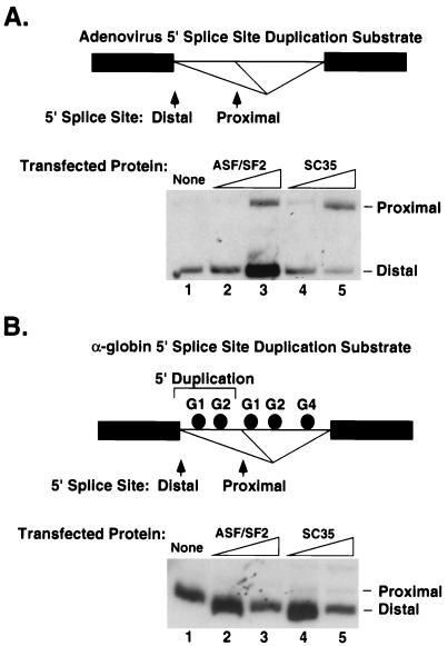
As shown in Fig. 1, the second intron of the human alpha-globin gene contains multiple G triplets, four of which are close to the 5′ splice site and which we have grouped into two elements termed G1 and G2. Introduction of a single mutant G into either of these elements both reduces splicing efficiency in vivo and alters 5′ splice site usage when substrates are created in which the region around the 5′ splice site (including the G1 and G2 elements) is duplicated (the duplication construct is diagramed in Fig. 2B). When the elements are wild type, over 90% of the spliced RNA product arises from use of the upstream distal 5′ splice site. Mutation of both G1 and G2 drops usage of this site to 25%, indicating the importance of these sequences for splice site specificity (26). The observed additivity of the ability of the elements to dictate distal usage suggested recognition of individual G triplets by a trans-acting factor.
FIG. 2.
SR proteins are unable to affect 5′ splice site usage in duplication constructs containing the alpha-globin 5′ splice sites and neighboring G elements. The minigenes utilized for in vivo transfections contained duplicated 5′ splice sites and proximal intron sequences from either adenovirus (A) or alpha globin (B), diagramed at the top of each panel. Transfections with expression plasmids coding for either ASF-SF2 or SC35 used no (A and B, lane 1) or 0.5 (A and B, lanes 2 and 4) or 1.0 (A and B, lanes 3 and 6) μg of expression plasmid per 10-cm-diameter dish of HeLa cells. Whole-cell RNA was analyzed by RT-PCR using primers for flanking exons. RNAs resulting from usage of the proximal or distal sites (see diagrams) are indicated.
Our previous observation that the G-rich elements in the alpha-globin intron promote early spliceosome assembly and affect 5′ splice site selection (26) suggests that these elements might be recognized by a factor involved in the early recognition of 5′ splice sites. Because ample evidence existed in the literature that SR proteins influence recognition of 5′ splice sites and can recognize purine-rich short enhancer sequences, we initially investigated the effects of SR proteins on this system by transfection studies using a reporter plasmid containing duplicated alpha-globin 5′ splice sites and their naturally accompanying G-triplet sequences (the minigene diagrammed in Fig. 2B) compared to a similar reporter containing duplicated 5′ splice sites from the second intron of the adenovirus major late transcription unit. As shown in Fig. 2A, coexpression of the adenovirus-based plasmid with expression plasmids coding for either human ASF-SF2 or SC35 altered 5′ splice site utilization towards the proximal site, as has been observed in other studies (5, 38). When the adenovirus minigene was expressed in HeLa cells, the proximal splice site was used in 6% of the spliced transcripts (standard error [SE], 1.4). Proximal-site usage increased to 19% (SE, 3) in the presence of exogenous ASF-SF2 (1 μg of expression plasmid) or 57% (SE, 2.7) in the presence of exogenous SC35. In contrast, no activation of proximal-site usage was observed with the alpha-globin construct (Fig. 2B). In the absence of exogenous SR protein, the proximal alpha-globin 5′ splice site was used 5% of the time (SE, 0.33). This usage was unchanged in the presence of endogenous SF2 (3%; SE, 1.0) or SC35 (4%; SE, 0.58). Other SR proteins, including SRp75, SRp55, SRp54, and SRp20, also failed to alter the 5′ splice site usage of the alpha-globin construct (data not shown). Even when we used mutant alpha-globin minigenes that demonstrated about equal usage of the two 5′ splice sites because of introduced mutations into the G elements separating the splice sites, we were unable to detect an effect of SR proteins on splice site usage. Although these experiments cannot rule out involvement of SR proteins, they did suggest that it would be profitable to examine other mechanisms.
An RNA containing the G1 and G2 elements interacts with the U1 snRNP.
Previous experiments had indicated that the G-rich enhancer sequences operated prior to the formation of the spliceosomal A complex, suggesting that they might interact with a factor important for initial complex formation and which could participate in 5′ splice site specificity. This possibility led us to ask if these elements interact directly with U1 snRNA. The base pairing of U1 RNA to substrate RNAs can be monitored by RNA-RNA psoralen cross-linking (39). In this assay, 32P-labeled substrate RNAs are incubated with nuclear extract under splicing conditions in the presence of the RNA cross-linking agent psoralen. Samples are illuminated with 365-nm-wavelength light to induce RNA-RNA cross-linking and analyzed on denaturing polyacrylamide RNA gels. Cross-linked RNAs appear as radiolabeled species migrating more slowly than substrate RNA.
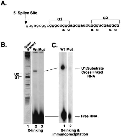
We applied this assay to short RNAs containing either wild-type or mutant G1 and G2 elements, but no splice sites (referred to here as the G1-G2 RNA, representing nucleotides 10 to 36 of the second intron of the human alpha-globin gene [Fig. 3]). We noticed a prominent band migrating above wild-type but not mutant substrate after cross-linking (Fig. 3B, lanes 2 and 3). This band was both psoralen and extract dependent (data not shown), suggesting that it represented cross-linking of the wild-type G1-G2 RNA to a nuclear RNA and not inter- or intramolecular cross-linking of substrate RNA. It was also ATP dependent (not shown). The gel migration of the band was severely retarded in the gel compared to the migration of free substrate, immediately suggesting that it represented an adduct between substrate RNA and a nuclear RNA. The migration of the band was also retarded with respect to the position of free U1 snRNA (Fig. 3B, compare the cross-link position in lane 2 to the stained U1 RNA position in lane 1), suggesting that it could have resulted from cross-linking between substrate and U1 RNAs.
FIG. 3.
U1 snRNA binds to GGG enhancer elements in the human alpha-globin intron. (A) Sequence of the alpha-globin intron 2 5′ splice site and adjacent G1 and G2 intron elements. The sequence of the short alpha-globin wild-type G1-G2 RNA used for psoralen cross-linking is 5′ GGGCCGGGAGCGAUCUGGGUCGAGGGGG 3′. The mutant G1-G2 RNA sequence is 5′ GGGCCAGCAGCGAUCUAGCUCGAUGCG 3′. (B) Psoralen cross(X)-linking reactions using the wild-type (Wt) or mutant (Mut) 32P-labeled G1-G2 RNA were prepared as described in Materials and Methods and were analyzed on an 8% polyacrylamide–8.3 M urea denaturing gel. The gel was silver stained to identify nuclear RNAs (lane 1), and 32P-labeled G1-G2 RNA was visualized by autoradiography (lanes 2 and 3). The positions of the U1 and U2 snRNAs, free G1-G2 RNA, and the cross-linked species are indicated. (C) Psoralen cross-linking reactions were analyzed by immunoprecipitation with an anti-U1 70K antibody. RNAs precipitated by this antibody were separated on a 5% polyacrylamide–8.3 M urea denaturing gel and visualized by autoradiography. The film in panel B was exposed overnight, while that in panel C was exposed for 7 days.
Several lines of evidence indicated that this band did indeed represent a U1 snRNA-substrate RNA adduct. The cross-linked species could be detected by Northern blot analysis using a U1 snRNA probe (not shown), indicating the presence of U1 RNA. In addition, the cross-linked RNA resided in a complex that was immunoprecipitated with antibodies that recognize the U1 snRNP-specific 70K protein (Fig. 3C), indicating that the cross-linked species is in a U1 snRNP. The appearance of a cross-linked and immunoprecipitable band required wild-type G elements (Fig. 3C, compare lanes 1 and 2). A significant amount of free wild-type (5% of input) but not mutant substrate was also recovered in the U1 70K immunoprecipitation. This RNA most likely represents substrate that was bound to U1 snRNP but not cross-linked because of the overall low efficiency of psoralen cross-linking. These experiments demonstrated that RNA containing the wild-type G1 and G2 elements interacts with the U1 snRNP and forms direct contacts with U1 snRNA.
Interaction of U1 snRNA and G1-G2 RNA does not require the complete 5′ end of U1 snRNA.
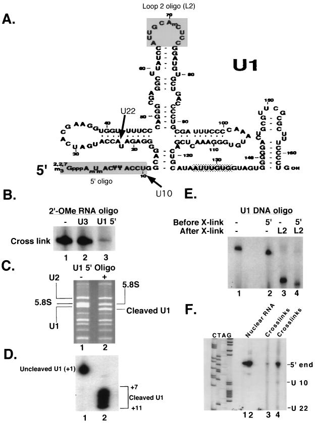
The 5′ end of U1 snRNA (nucleotides 1 to 10 [Fig. 4A]) is known to interact with 5′ splice sites through base pairing (29, 42), and removal or sequestration of these nucleotides inhibits the in vitro splicing of most substrates (1, 3, 37). To determine if this 5′ sequence is required for the interaction of U1 RNA with the G1-G2 RNA, we performed the psoralen cross-linking assay using nuclear extracts in which the 5′ end of U1 had been sequestered with a 2′-O-methyl RNA oligonucleotide complementary to U1 nucleotides 1 to 11. This treatment effectively inhibited the assembly of spliceosomal complexes on an adenovirus splicing substrate, presumably by preventing U1 snRNA from interacting with the 5′ splice site (data not shown). The same concentration of a control oligonucleotide complementary to U3 RNA had no effect (data not shown). The anti-U1 but not the anti-U3 2′-O-methyl RNA inhibited formation of the psoralen cross-link between U1 RNA and G1-G2 RNA (Fig. 4B). This result supports the observation that U1 snRNA can be psoralen cross-linked to the G1-G2 RNA. Furthermore, it implicates U1 nucleotides 1 to 11 as critical for the interaction.
FIG. 4.
Alpha-globin G triplets psoralen cross-link to nucleotides U10 and U22 of U1 snRNA. (A) Sequence of U1 snRNA. The sequences targeted by the U1 5′ and loop 2 oligonucleotides are shaded. The Sm protein binding region is indicated by a stippled box. The positions of mapped cross-links (U10 and U22) are indicated. (B) Psoralen cross-linking reactions using the wild-type G1-G2 RNA were carried out in nuclear extracts treated with 2′-O-methyl RNA oligonucleotides complementary to U3 RNA or U1 RNA nucleotides 1 to 11. The cross-linking products were analyzed on a 5% polyacrylamide–8.3 M urea denaturing gel and visualized by autoradiography. The labeled band corresponds to the cross-linked species in Fig. 1. (C) Analysis of U1 snRNA in nuclear extract after incubation with RNase H (lane 1) or RNase H plus a DNA oligonucleotide complementary to U1 nucleotides 1 to 11 (lane 2). RNAs were separated on a 10% polyacrylamide–8.3 M urea denaturing gel and visualized by ethidium bromide staining. The positions of U2 snRNA, 5.8S RNA, and cleaved and uncleaved U1 are indicated. (D) Primer extension analysis of U1 snRNA in nuclear extract after incubation with RNase H (lane 1) or RNase H plus a DNA oligonucleotide complementary to U1 nucleotides 1 to 11 (lane 2). The U1 loop 2 oligonucleotide was used to prime RT, and the products were analyzed on a 6% polyacrylamide–8.3 M urea sequencing gel and visualized by autoradiography. The positions of uncleaved and cleaved U1 molecules are indicated. Nucleotide numbers were assigned by comparison to an adjacent U1 sequencing reaction (not shown). (E) Analysis of the G1-G2–U1 cross(X)-linked species by DNA oligonucleotide-directed RNase H cleavage using the U1 5′ oligonucleotide (lane 2), loop 2 oligonucleotide (lane 3), or both (lane 4). Treatment with the 5′ oligonucleotide was done before cross-linking, and treatment with the loop 2 oligonucleotide was done after cross-linking and RNA purification. RNAs were analyzed on 5% polyacrylamide–8.3 M urea denaturing gels and visualized by autoradiography. (F) Primer extension analysis of cross-linked RNAs. Cross-linked RNAs were purified and analyzed by primer extension using the U1 loop 2 oligonucleotide. The positions of two transcription stops (U10 and U22) and the 5′ end of U1 RNA are indicated. In lanes 1 and 2, 5 or 10 μg of nuclear RNA was analyzed by primer extension. The U10 and U22 stops were not observed when RNAs were analyzed following psoralen cross-linking in the absence of substrate RNA and therefore are not intramolecular cross-links (data not shown).
To further characterize the interaction of U1 with the G1-G2 RNA, psoralen cross-linking reactions were performed using nuclear extracts in which the terminal 5′ sequences of U1 had been removed by DNA oligonucleotide-directed RNase H cleavage prior to the addition of substrate. Cleavage of U1 was monitored by gel electrophoresis of nuclear RNAs (Fig. 4C). The faster migration of U1 snRNA from the oligonucleotide-treated extract (Fig. 4C, lane 2) compared to mock-treated extract (lane 1) indicated that almost all of the U1 in the extract had been cleaved. Primer extension analysis indicated, however, that RNase H cleavage was incomplete and produced multiple species containing residual nucleotides downstream of nucleotide A7 (Fig. 4D).
In contrast to the 2′-O-methyl sequestration experiments described above (Fig. 4B), psoralen cross-linking with G1-G2 RNA was still observed following oligonucleotide-directed RNase treatment (Fig. 4E, lane 2). However, the cross-linked RNA was reduced in size, reflecting the shortened U1 snRNA (compare the bands in Fig. 4E, lanes 1 and 2). The observation of a shorter cross-linked species following U1 cleavage indicates that the intact 5′ end of U1, which is known to interact with nucleotides +1 to +6 of 5′ splice sites, is not required for the interaction of G1-G2 RNA with U1 snRNA. The ability of U1 cleavage to increase the migration of the cross-linked species also provides further evidence that the cross-link involves U1 snRNA. Both the RNase H cleavage experiment and the RNA antisense experiments used oligonucleotides complementary to nucleotides 1 to 11 of U1 RNA. As described above, however, nucleotides 7 to 10 of U1 were inefficiently cleaved by RNAse H digestion and therefore were available for base pairing with the substrate (Fig. 4D). The observation that cross-linking was prevented when U1 nucleotides 1 to 11 were sequestered, but not when U1 nucleotides 1 to 6 were cleaved with RNase H, suggests that the G1-G2 RNA interacts with U1 nucleotides 7 to 11. In a likely scenario, a G triplet base pairs with the U1 sequence CCU (nucleotides 8 to 10).
Additional RNase H mapping was performed to further characterize the cross-linked species. U1 RNA loop 2 was targeted for cleavage using an oligonucleotide complementary to U1 RNA nucleotides 64 to 75 (Fig. 4A). In intact U1 snRNPs, the U1A protein protects this loop from cleavage; therefore, these reactions were done after cross-linking and deproteinization. Cleavage within loop 2 of U1 RNA generates diagnostic 5′ and 3′ U1 RNA fragments, only one of which would likely be linked to the substrate via a cross-link and therefore radiolabeled. As shown in Fig. 4E, cleavage of loop 2 significantly increased the migration of the cross-linked product RNA (compare control lane 1 with lane 3). The ability of a second U1-specific oligonucleotide to direct cleavage of the cross-linked species further proves that the observed cross-linking involves U1 snRNA.
Only one of the two U1 fragments was detected by autoradiography, indicating that only one of the fragments was cross-linked to 32P-labeled substrate. To identify this cross-linked fragment, we performed loop 2 cleavage analysis of cross-linked product RNA from reactions using extract in which the 5′ end of U1 snRNA had been cleaved prior to cross-linking. In this case (Fig. 4E, lane 4), the 32P-labeled band was slightly smaller than the product resulting from cross-linking to U1 RNA still containing its 5′ sequences, reflecting removal of the 5′ end of U1 RNA (compare lanes 3 and 4). Observation of a smaller band upon cleavage of the 5′ end of U1 RNA positions the cross-link to the 5′-most U1 RNA fragment generated by loop 2 cleavage (nucleotides 7 to 64).
The site of cross-linking to G triplets maps to uridine residues 10 and 22 in U1 snRNA.
To exactly determine the position of the cross-link(s), we performed primer extension using purified cross-linked product RNA and an oligonucleotide primer complementary to U1 loop 2 (U1 nucleotides 64 to 75 [Fig. 4A]). This assay is based on the observation that reverse transcriptase pauses at cross-linked nucleotides. Using this approach, we identified two RT stops at uridine 10 (U10) and U22 in U1 snRNA (Fig. 4F, lanes 3 and 4, and 4A) that were not observed when total nuclear RNA was extended (Fig. 4F, lanes 1 and 2). These stops likely represent cross-linked nucleotides. The U10 and U22 cross-links were not observed when the G1-G2 RNA was omitted form the psoralen cross-linking reaction, suggesting that the stops do not result from intramolecular cross-links in the U1 snRNA (not shown). The U10 cross-link is consistent with our 2′-O-methyl and RNase H cleavage data and supports a model in which a G triplet in the substrate RNA interacts with U1 nucleotides 8 to 10 (CCU). The cross-link involving U22 might result from additional, novel interactions of the substrate with U1, or perhaps the unpaired U22 is spatially positioned so that it is cross-linked to substrate bound near nucleotides 8 to 10.
The G1-G2 RNA exists in a large complex.
The U1 70K immunoprecipitation data (Fig. 3C) indicated that the G1-G2 RNA interacts with U1 snRNA in a complex which contains the U1 70K protein. We were interested in knowing the size of this complex and if it was likely to contain intact U1 snRNP. To address this issue, cross-linking reaction products were fractionated on 10 to 30% glycerol gradients, and the fractionated RNAs were analyzed by electrophoresis on denaturing gels. The position of U1 snRNA in the gradient was identified by silver staining (Fig. 5A), and the cross-link was detected in the same gel by autoradiography (Fig. 5B). This analysis indicated that the cross-linked species containing the G1-G2 RNA and the U1 RNA resides in a complex large enough to contain a complete U1 snRNP. In fact, the observed complex peak is slightly larger than the peak of bulk U1 snRNA, suggesting the involvement of additional yet-unidentified components.
FIG. 5.
An alpha-globin enhancer element containing G elements forms a complex that sediments larger than a U1 snRNP. Psoralen cross-linking reaction mixtures using the G1-G2 RNA were layered onto 10 to 30% glycerol gradients and centrifuged for 16 at 33,000 rpm in an SW40Ti rotor. Fractions (0.5 ml) were collected, and RNAs were recovered by ethanol precipitation and separated on 8% polyacrylamide–8.3 M urea gels. (A) The distribution of nuclear RNAs across the gradients was monitored by silver staining. (B) The free and cross-linked 32P-labeled G1-G2 RNAs were visualized by autoradiography.
Targeting modified U1 snRNAs to a mutant G1 element.
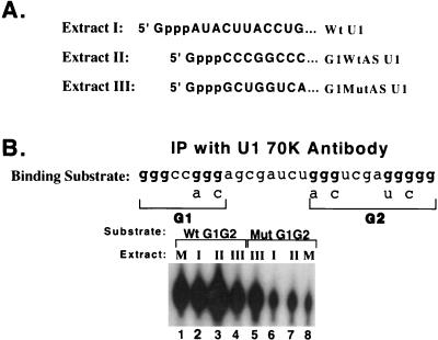
The experiments described above indicated that U1 snRNPs bind to G-rich intronic elements in the alpha-globin intron. We next wanted to determine if U1 binding to these elements accounts for their effects on splicing. Our approach was to attempt to mimic the binding of U1 RNA to a G element by creating a mutant G element and a mutant U1 RNA capable of base pairing with the created element. The modified U1 snRNAs generated in this study are summarized in Table 1. A mutant alpha-globin in vivo splicing substrate was created in which the wild-type G1 element GGGCCGGG was altered to UGACCAGC (the G1 mutant diagrammed in Fig. 1). This alteration depressed both activity and 5′ splice site specificity to an extent sufficient to reproducibly observe complementation should it occur. The sequence of the 5′ end of U1 was changed to be complementary to the mutant G1 element (denoted G1 MutASU1) and coexpressed in HeLa cells along with the mutant alpha-globin gene. We also generated a modified U1 complementary to the wild-type G1 element as a control (denoted G1WtAs U1 snRNA). The sequences of the 5′ ends of these U1 RNAs are shown in Fig. 6A. This approach, which bypassed the natural mechanism for bringing U1 to the enhancer, allowed us to ask if U1 binding alone is sufficient to mimic the activity of a G1 element.
TABLE 1.
Potential base pairing of the G1 and G2 elements with the U1 snRNAs used in this study
| Base pairinga | RNAb | ΔGc (kcal/mol) | Figured |
|---|---|---|---|
| 5′ G G G C C G G G 3′ | Wt G1 | −1.5 | 7C(1) |
| ····· | |||
| 3′ U C C A U U C A U A 5′ | Wt U1 | ||
| 5′ G G G U C G A G G G G 3′ | Wt G2 | −3.1 | 7C(1) |
| ······ | |||
| 3′ U C C A U U C A U A 5′ | Wt U1 | ||
| 5′ U G A C C A G C 3′ | Mutant G1 | >0 | 7C(2), 7E(1), 8B(2) |
| ··· | |||
| 3′ U C C A U U C A U A 5′ | Wt U1 | ||
| 5′ A G C U C G A U G C G 3′ | Mutant G2 | >0 | 7C(2 and 6), 7D (plot) |
| ······ | |||
| 3′ U C C A U U C A U A 5′ | Wt U1 | ||
| 5′ U G A C C A G C 3′ | Mutant G1 | −12.6 | 7C(4), 8B(3) |
| ········ | |||
| 3′ A C U G G U C G A 5′ | G1MutAS U1 | ||
| 5′ U G A C C A G C 3′ | Mutant G1 | −1.3 | 7D (plot) |
| ····· | |||
| 3′ A C U A U U C A U A 5′ | G1-1 rescue U1 | ||
| 5′ A G C U C G A U G C G 3′ | Mutant G2 | −18.1 | 7E(5 and 8) |
| ··········· | |||
| 3′ U C G A G C U A C G C A 5′ | G2MutAS U1 |
Aligned to allow for base pairing (periods) between U1 nucleotides 8 to 10 and a GGG element. Potential base pairing of the G1 and G2 elements with U1 nucleotides in addition to 8 to 10 is also indicated. Experimental evidence, however, indicated that these positions are not essential for the interaction of the wild-type elements with U1 (Fig. 4).
Identity of each G element and U1 snRNA. Wt, wild type.
ΔG values were calculated as described previously (13).
Figure and lane numbers (in parentheses) where the indicated element and U1 combination analyzed for in vivo effects on splicing are shown.
FIG. 6.
A mutant G element will bind in extract to a U1 snRNP containing a complementary mutated U1 snRNA. (A) Sequences of the 5′ ends of wild-type (Wt) U1 RNA and U1 RNAs modified to complement wild-type or mutant G1 elements. (B) Wild-type and mutant G1-G2 RNAs were incubated in nuclear extracts prepared from cells transfected with the U1 snRNA genes shown in panel A and analyzed by immunoprecipitation (IP) with an anti-U1 70K antibody. Precipitated RNAs were separated on a 5% polyacrylamide–8.3 M urea denaturing gel and visualized by autoradiography.
Before assaying the G1 MutASU1 for splicing effects, we wanted to verify that it was expressed in cells and that it could bind to the mutant G1 element. Therefore, we expressed this RNA in HeLa cells under the control of the U1 promoter and prepared nuclear extracts from transfected cells in which to assay for the presence of the transfected mutant U1 RNA. To differentiate endogenous U1 from transfected mutant U1 RNA, transfected extracts were incubated with RNase H and DNA oligonucleotides complementary to the 5′ end of either the wild-type or mutant U1 RNA. This treatment allowed us to differentially cleave these RNAs and distinguish them by denaturing gel electrophoresis. Using this method we determined that the G1MutAS U1 accumulated in transfected HeLa cells, albeit at a level significantly lower than that of endogenous wild-type U1 (not shown).
We next asked if the G1MutAS U1 snRNA present in this nuclear extract could bind the mutant G1 substrate. To investigate this, wild-type or mutant G1-G2 RNA was incubated in nuclear extracts prepared from mock-transfected cells or cells transfected with wild-type, G1WtAS, or G1MutAS U1, and the binding of substrate to each U1 was assayed by U1 70K immunoprecipitation. If U1 is bound to the substrate, the substrate should be immunoprecipitated by the U1 snRNP antibody. If U1 does not associate with the substrate, little substrate should be recovered by this immunoprecipitation. This assay is similar to the precipitation of non-cross-linked substrate by U1-specific antibodies shown in Fig. 3C. As shown in Fig. 6B, the mutant substrate was efficiently precipitated from the extract expressing U1 RNA with a 5′ sequence complementary to the mutant G1 element (lane 5) while it was inefficiently precipitated from the control extracts expressing either normal U1 RNA or U1 RNA with a 5′ end complementary to wild-type G1-G2 RNA (lanes 6, 7, and 8). Phosphorimager analysis indicated that the mutant G1-G2 RNA was immunprecipitated five times more efficiently from the G1MutAS U1 extract than from the other three extracts tested.
In this experiment, the wild-type substrate was immunoprecipitated from each extract (Fig. 6B, lanes 1 to 4). Immunoprecipitation was greatest with the extract expressing a U1 RNA complementary to the wild-type G1 element in the wild-type substrate (extract II, lane 3). Phosphorimager analysis indicated that the wild-type RNA was immunoprecipitated 2.5 times more efficiently from this extract than from the standard extract. Considerable precipitation, however, was observed with standard extract or extract expressing wild-type U1, reflecting the ability of wild-type U1 to bind to the wild-type sequence.
These experiments indicated that the G1MutAS U1 RNA accumulates in transfected cells, associates with the U1 70K protein, and is targeted to RNAs containing the mutant G1 element. We therefore proceeded to ask if this modified U1 could rescue the splicing defects associated with the mutant G1 element.
Rescue of distal splice site selection by U1 snRNAs complementary to mutant G elements.
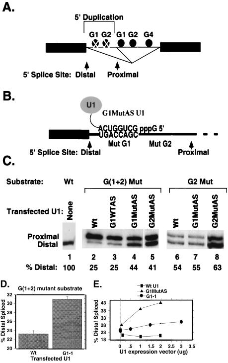
Our first functional test of the G1MutAS U1 was to determine its effect on 5′ splice site utilization using a 5′ splice site duplication substrate similar to that described in the legend to Fig. 2 in which both of the wild-type G1 or G2 elements between the 5′ splice sites were mutated as described in the previous section. The role of U1 RNA binding to these elements was tested by attempting to mimic distal splice site utilization in the mutant duplication minigene by expression of complementary mutant U1 RNAs (Fig. 7). The potential base pairing of these modified U1 snRNAs and substrate elements is shown in Table 1. HeLa cells were cotransfected with vectors expressing the mutant duplication minigene and either the wild-type G1WtAS, or G1MutAS U1 snRNAs shown in Fig. 6A and Table 1. The potential base-pairing interactions between the G1MutAS U1 RNA and the mutant G1 element in this substrate are modeled in Fig. 7B and in Table 1. As shown in the RT-PCR assay in Fig. 7C, the proximal site was preferred when either wild-type U1 (25%; SE, 1.9) or the G1WtAs U1 was cotransfected with the G(1+2) mutant duplication substrate (lanes 2 and 3). This is the splicing pattern previously observed for this substrate in the absence of mutant U1 RNA expression (26). In contrast, the distal site was activated in a dose-dependent manner when the G1MutAS U1 RNA that can bind mutant G1 elements was coexpressed (Fig. 7C, lane 4, and E). When 2 μg of G1MutAS DNA was used for transfection, the distal splice site was utilized in 44% (SE, 1.2%) of the spliced transcripts. This is comparable to what was observed for a substrate with a wild-type G1 element in this position (26). Thus, expression of a suppressor U1 RNA complementary to a mutant G element restored the 5′ splice site specificity of the element.
FIG. 7.
A complementary mutant U1 snRNA rescues 5′ splice site specificity when G triplets are mutated in a construct containing duplicated 5′ splice sites. (A) The 5′ splice site duplication substrates contained two identical 5′ splice sites separated by 58 nucleotides containing either wild-type (solid circles) or mutant (cross-hatched circles) G1 and G2 elements. The G1 and G2 mutations were identical to those indicated in Fig. 1A. The sequences downstream of the proximal 5′ splice site were wild type in each construct. Potential splicing pathways using either the distal or proximal 5′ splice sites are illustrated. (B) Model of potential base pairing between the G1MutAS U1 snRNA and a mutant (Mut) G1 element in the G(1+2) mutant duplication substrate. (C) RT-PCR (15-cycle) analysis of splicing products produced in transiently transfected HeLa cells. The positions of PCR products corresponding to RNAs spliced using the distal or proximal 5′ splice site are indicated. The substrates and cotransfected U1 genes used are indicated above the lanes. In the G(1+2) mutant, the G1 and G2 elements between the duplicated 5′ splice sites are both mutant. In the G2 mutant, the G1 element is wild type (Wt). The percentage of products resulting from usage of the distal site is indicated below each lane. (D) Rescue of distal 5′ splice site usage by expression of a U1 snRNA complementary to the first G triplet of the mutant G1 element (designated G1-1) in the G(1+2) mutant 5′ splice site duplication substrate. Means and standard errors were calculated from three experiments. (E) Graphical representation of distal splice site activation in the G(1+2) mutant substrate in HeLa cells cotransfected with increasing amounts of wild-type, G1MutAS, or G1-1 U1 expression plasmids.
The G1 element that was targeted by the altered U1 in the above experiment begins at +9 within the intron, or immediately adjacent to the 5′ splice site. To see if we could alter utilization by targeting a more distal G element, we asked if a complementary U1 targeted to G2 (the G2 element begins at +26 of the intron) could rescue distal 5′ splice site utilization in a duplication construct in which both the G1 and G2 elements were mutated [the G(1+2) Mut construct in Fig. 7] or in a construct in which only the G2 element was mutated (the G2 Mut construct in Fig. 7). The latter construct has a basal-level usage of the distal 5′ splice site of 50% (Fig. 7C, right). Co-transfection of the G2MutAS U1 (2 μg) was also able to rescue distal 5′ splice site usage in the G(1+2) mutant duplication substrate (Fig. 7C, lane 5). The specificity of this activation is illustrated by the ability of the G2MutAS, but not G1MutAS, to rescue distal splice site usage in a G2 duplication substrate containing only the G2 mutation (Fig. 7C, lanes 6 to 8). This substrate has a mutant G2 element and a wild-type G1 element in its duplicated 5′ end and therefore contains the target sequence for the G2MutAS, but not the G1MutAS, U1 snRNA.
The rescue U1 snRNAs described above had considerable complementarity to the mutant G1 or G2 elements (Table 1). To determine if a degree of complementarity similar to that proposed for the interaction of wild-type U1 with wild-type elements (Fig. 4) could rescue distal splicing, we tested a U1 snRNA complementarity to only the first 3 nucleotides of the mutant G1 element (denoted G1-1 [Table 1]). In the G1-1 U1 snRNA, nucleotides 8 to 10 have been changed to 5′ UCA 3′ to complement the first three nucleotides of the mutant G1 element, 5′ UGA 3′. When this U1 was cotransfected with the G(1+2) mutant construct described above (Fig. 7A), the distal 5′ splice site was used in 31% of the accumulated spliced transcripts (SE, 0.58) compared to 23% (SE, 0.68) for controls transfected with wild-type U1 (Fig. 7D). Distal 5′ splice site usage in cells transfected with the G1-1 U1 was significantly above that observed in the wild-type U1 control when compared by analysis of variance (P = 0.0003) and responded to the amount of G1-1 U1 DNA used (Fig. 7E).
These experiments indicate that a U1 snRNP bound to a mutant G element is sufficient to activate distal 5′ splice site utilization. This alteration in 5′ splice site utilization in the presence of a complementing U1 RNA indicates that the G-rich elements modulate 5′ splice site selection, at least in part, by recruiting the U1 snRNP.
Rescue of mutant alpha-globin splicing efficiency with a U1 snRNA complementary to the mutant G1 element.
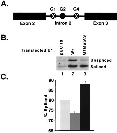
We next asked if the in vivo splicing efficiency of a substrate with a single 5′ splice site and a mutant G1 element could be rescued by cotransfection with the G1MutAS U1 snRNA. A previously characterized alpha-globin splicing substrate containing mutant G1 and G4 elements (Fig. 8A), which is incompletely spliced after transfection because of the mutated G elements (26), was expressed in HeLa cells cotransfected with either PUC 19 DNA or plasmids expressing wild-type or G1MutAS U1 snRNA. As shown in Fig. 8C, the percentage of accumulated transcripts that were spliced was higher in RNA from cells expressing the G1MutAS U1 snRNA (88%; SE, 1.2 [lane 3]) than in the PUC- (80%; SE, 1.1 [lane 1]) or wild-type U1 (74%; SE, 0.9 [lane 2])-transfected cells. Therefore, the splicing defect attributed to the mutant G1 element was reversed by experimentally restoring its interaction with U1 snRNP.
FIG. 8.
A complementary mutant U1 snRNA rescues alpha-globin splicing activity when G triplets are mutated in a construct containing a single 5′ splice site. (A) The alpha-globin in vivo expression construct contained intron 2 and exons 2 and 3 of the human alpha-globin 2 gene. This intron contained a wild-type G2 element (solid circle) and G1 and G4 elements (cross-hatched circles) mutated as indicated in Fig. 1A. (B) RT-PCR (15-cycle) analysis of splicing products produced in transiently transfected HeLa cells. The positions of PCR products corresponding to unspliced and spliced RNAs are indicated. The cotransfected U1 expression vector is indicated above each lane. Wt, wild type. (C) Means and standard errors calculated from three independent cotransfection experiments. The percentage spliced was defined as spliced/(unspliced + spliced).
These experiments provide strong evidence that the binding of U1 snRNP to wild-type G-rich elements is functionally important. Mutation of these elements abolished U1 binding, reduced splicing efficiency, and altered splice site selection. A modified U1 capable of binding mutant elements was, however, capable of rescuing both splicing defects.
DISCUSSION
We have previously identified G-rich elements in the second intron of the human alpha-globin gene that are important for efficient splicing in vivo and in vitro. These elements also promote distal 5′ splice site selection when positioned between duplicated, identical 5′ splice sites and affect in vitro spliceosome assembly (26). The minimal discernible element is the simple sequence GGG, and these elements function additively (26).
Interestingly, despite their purine content, G-triplet elements appear not to be binding sites for SR proteins (Fig. 2). In repeated attempts to alter either activity or splice site specificity in the alpha-globin intron through the addition of SR proteins, we were unable to alter either by increasing the concentration of a number of SR proteins, including the SR proteins ASF-SF2 and SC35 that dominantly cause recognition of proximal 5′ splice sites in a number of systems (5, 38). Given the intronic location of the elements, the preference of the G triplets for activating 5′ splice sites residing upstream of the elements, and the observed preference of SR proteins for binding to exonic rather than intronic enhancer sequences, it is perhaps not surprising that G-triplet elements do not operate via SR-mediated pathways. Furthermore, the observed refractory response to SR proteins suggests a difference between recognition of G triplets and recognition of the similar sequence intron enhancer (UAGAGU) from the hnRNP A1 gene that is recognized by hnRNP A1 (7).
Because these elements affect early spliceosome assembly and 5′ splice site selection, we postulated that they might bind a factor that recruits U1 snRNP to the 5′ splice site to initiate commitment complex formation. Surprisingly, we found that the G-rich elements themselves interact with the U1 snRNP and that the U1 snRNA can be cross-linked to these elements in the presence of the RNA cross-linking agent psoralen (Fig. 3). The interaction of U1 snRNP with these elements is different from other U1-substrate interactions in that it does not require nucleotides 2 to 7 of U1 snRNA (Fig. 4). The nature of this interaction remains under investigation, and the contributions of additional factors are possible.
We tested the functional significance of this interaction by targeting modified U1 snRNAs to mutant G-rich elements. Expression of these modified U1 RNAs in transfected HeLa cells was able to rescue both the splicing efficiency and splice site selection properties of the G-rich element. The rescue U1 increased the splicing efficiency of a substrate with a single 5′ splice site and a mutant G-rich element (Fig. 8) and restored distal splice site utilization in a substrate with duplicated 5′ splice sites separated by mutant G-rich elements (Fig. 7). The degree of rescue imparted by these engineered U1 genes was significant. Mutation of the G1 element positioned between two alpha-globin 5′ splice sites reduced distal 5′ splice site use by 48% (26), while expression of a U1 complementary to this mutant G1 element (G1MutAS U1) restored distal splice site use by 44% (Fig. 7C). This represents a 90% rescue of G1 activity. A similar rescue efficiency was seen when a complementary U1 RNA was targeted to a mutant G2 element (Fig. 7C). These rescue U1s, however, had extensive complementarity to their target elements. In contrast, the G1-1 U1 RNA complemented only the first 3 nucleotides of a mutant G1 element. Mutating these 3 nucleotides of the G1 element in an otherwise wild-type alpha-globin intron reduced distal 5′ splice site usage by 18% (26). The G1-1 U1 was able to rescue distal 5′ splice site usage by 8% (Fig. 7D). This represents a rescue efficiency of about 50%. Given the low ΔG associated with a 3-bp interaction (Table 1), the degree of compensation observed with the G1-1 U1 strongly supports the functionality of the U1 RNA–G-triplet interaction proposed in this study.
These results indicate that the interaction of U1 with G-rich elements is functionally relevant and represents a mechanism for loading U1 snRNP onto splicing substrates. Thus, G triplets bind U1 snRNPs similarly to the SR-induced binding of U1 snRNPs to regions near 5′ splice sites; in the former case, however, binding favors usage of a 5′ splice site upstream of the U1 binding site, suggesting fundamental differences between the binding of U1 snRNPs to either exon enhancers or 5′ splice sites and the binding to an intronic GGG-containing enhancer.
The interaction of U1 with these elements likely initiates commitment complex assembly and nucleates spliceosome formation. We do not know if the U1 snRNPs that interact with an intronic G-rich element also base pair with the 5′ splice site during splicing. We do know that such base pairing is not required, because the rescue U1 RNAs used in our cotransfection experiments have modified 5′ ends that are unable to base pair with the 5′ splice site. It is plausible that the U1 bound to the intronic elements recruits a second U1 via protein-protein interactions, but it is more likely that a second U1 is not required. There is ample evidence indicating that U1 base pairing with the 5′ splice site is not required for efficient, accurate splicing. First, U1 is not necessarily required for splicing. Its function can be replaced by high concentrations of SR proteins which subsequently recruit U4, -5, and -6 snRNPs (9, 37). In some introns, the U4, -5, and -6 tri-snRNP can recognize the 5′ splice site in the absence of U1 or high concentrations of SR proteins (10). Furthermore, Cohen and coworkers have shown that U1 can act at a distance from the 5′ splice site to promote recognition of the 5′ splice site in the absence of base pairing (8). In their experiments, a weakened 5′ splice site unable to interact with U1 was rescued by targeting U1 snRNPs (termed shift U1) to introduced sequences upstream or downstream of the mutant 5′ splice site. This effect was dependent upon the ability of U6 snRNA to base pair with the remaining nonmutated sequences of the 5′ splice site (21). It is unclear if U1 snRNPs affect splicing by a similar mechanism in the natural alpha-globin pre-mRNA. There are, however, fundamental differences between alpha globin and the shift U1 system. Most prominently, U1 binding to G elements in alpha globin affects the activities of strong, wild-type splice sites while the shift U1s activated incapacitated sites via binding of U6 snRNA.
We suggest that our observations with alpha globin can be extended to other introns with abundant G triplets (26, 30, 35). In small introns, G-rich elements are proposed to interact with U1 snRNPs as an initial step in intron definition, maximizing mRNA production. In large introns, we propose that G-rich elements downstream of 5′ splice sites serve to load U1 snRNP onto the 5′ end of the intron and promote recognition of the 5′ splice site. Such a mechanism would increase the information content of authentic 5′ splice sites and help the splicing machinery accurately identify legitimate exons. The G-rich sequences at the 5′ and 3′ ends of large introns have also been reported to bind hnRNP A1 (4). Blanchette and Chabot have proposed that interactions between hnRNP A1 molecules bound at these sites function to bring the ends of the intron together for splicing (4). The interaction of G-rich elements with U1 and hnRNP A1 are most likely temporally separated and compatible.
The binding of U1 snRNPs to G-rich sequences is not fully understood. It is unclear if the base-pairing interaction detected by psoralen cross-linking is the basis of the interaction or if it represents a footprint that allowed us to visualize the interaction. Our observation that U1 remains bound to the G1-G2 RNA throughout lengthy immunoprecipitation experiments (precipitated, free substrate in Fig. 3C, lane 1) in the absence of cross-linking demonstrates that the interaction of U1 snRNP and the G1-G2 RNA is quite stable. This suggests that interactions in addition to the brief base pairing documented here may be involved. If an additional factor is involved, however, such a factor may be U1 snRNP associated, because purified U1 snRNP recognizes and binds to the G1-G2 RNA (unpublished results). The involvement of an additional component is supported by the fact the cross-linking reported here is ATP dependent. It is reasonable to speculate that this interaction is mediated by a phosphorylation event, or perhaps a helicase activity.
The U1 snRNP has several roles in RNA maturation beyond base pairing with the 5′ splice site. The U1 snRNP is found in complexes assembled on exonic splicing enhancers (36, 40), and it is a component of a polyadenylation enhancer in the human calcitonin gene (23). U1 binding to sequences upstream of polyadenylation signals in terminal exons can inhibit polyadenylation (15, 17), and the binding of U1 to pseudo-5′ splice sites contributes to tissue specific splicing of Drosophila P element transcripts (33). U1 is also a component of a complex that assembles on the negative regulator of splicing element in the Rous sarcoma virus, where it is proposed to compete for binding with U11 snRNA (27). Finally, we provide evidence that U1 snRNP interacts with intronic G-rich elements to promote efficient splicing and accurate splice site selection. Considering this ever-expanding list of functions, it is not surprising that U1 is the most abundant of the spliceosomal snRNPs.
ACKNOWLEDGMENTS
We thank the members of the Berget laboratory, especially Leslie Landree and Hua Lou, for valuable discussions throughout the course of this project. We recognize Valerija Vitkauskas, an undergraduate summer student, for performing several transfection experiments, and we acknowledge the dedicated technical assistance of Wade Wilson. We thank James Manley (Columbia University) for providing us with ASF-SF2 and SC35 expression plasmids, and we acknowledge Alan Weiner (University of Washington) for providing the wild-type U1 snRNA gene used in these studies.
This work was supported by grant RO1 GM38526 to S.M.B.
REFERENCES
- 1.Berget S M, Robberson B L. U1, U2, and U4/U6 small nuclear ribonucleoproteins are required for in vitro splicing but not polyadenylation. Cell. 1986;46:691–696. doi: 10.1016/0092-8674(86)90344-2. [DOI] [PubMed] [Google Scholar]
- 2.Black D L. Activation of c-src neuron-specific splicing by an unusual RNA element in vivo and in vitro. Cell. 1992;69:795–807. doi: 10.1016/0092-8674(92)90291-j. [DOI] [PubMed] [Google Scholar]
- 3.Black D L, Chabot B, Steitz J A. U2 as well as U1 small nuclear ribonucleoproteins are involved in premessenger RNA splicing. Cell. 1985;42:737–750. doi: 10.1016/0092-8674(85)90270-3. [DOI] [PubMed] [Google Scholar]
- 4.Blanchette M, Chabot B. Modulation of exon skipping by high-affinity hnRNP A1-binding sites and by intron elements that repress splice site utilization. EMBO J. 1999;18:1939–1952. doi: 10.1093/emboj/18.7.1939. [DOI] [PMC free article] [PubMed] [Google Scholar]
- 5.Caceres J F, Stamm S, Helfman D M, Krainer A R. Regulation of alternative splicing in vivo by overexpression of antagonistic splicing factors. Science. 1994;265:1706–1709. doi: 10.1126/science.8085156. [DOI] [PubMed] [Google Scholar]
- 6.Carlo T, Sterner D A, Berget S M. An intron splicing enhancer containing a G-rich repeat facilitates inclusion of a vertebrate micro-exon. RNA. 1996;2:342–353. [PMC free article] [PubMed] [Google Scholar]
- 7.Chabot B, Blanchette M, Lapierre I, La Branche H. An intron element modulating 5′ splice site selection in the hnRNP A1 pre-mRNA interacts with hnRNP A1. Mol Cell Biol. 1997;17:1776–1786. doi: 10.1128/mcb.17.4.1776. [DOI] [PMC free article] [PubMed] [Google Scholar]
- 8.Cohen J B, Broz S D, Levinson A D. U1 small nuclear RNAs with altered specificity can be stably expressed in mammalian cells and promote permanent changes in pre-mRNA splicing. Mol Cell Biol. 1993;13:2666–2676. doi: 10.1128/mcb.13.5.2666. [DOI] [PMC free article] [PubMed] [Google Scholar]
- 9.Crispino J D, Blencowe B J, Sharp P A. Complementation by SR proteins of pre-mRNA splicing reactions depleted of U1 snRNP. Science. 1994;265:1866–1869. doi: 10.1126/science.8091213. [DOI] [PubMed] [Google Scholar]
- 10.Crispino J D, Mermoud J E, Lamond A I, Sharp P A. Cis-acting elements distinct from the 5′ splice site promote U1- independent pre-mRNA splicing. RNA. 1996;2:664–673. [PMC free article] [PubMed] [Google Scholar]
- 11.Elrick L L, Humphrey M B, Cooper T A, Berget S M. A short sequence within two purine-rich enhancers determines 5′ splice site specificity. Mol Cell Biol. 1998;18:343–352. doi: 10.1128/mcb.18.1.343. [DOI] [PMC free article] [PubMed] [Google Scholar]
- 12.Eperon I C, Ireland D C, Smith R A, Mayeda A, Krainer A R. Pathways for selection of 5′ splice sites by U1 snRNPs and SF2/ASF. EMBO J. 1993;12:3607–3617. doi: 10.1002/j.1460-2075.1993.tb06034.x. [DOI] [PMC free article] [PubMed] [Google Scholar]
- 13.Freier S M, Kierzek R, Jaeger J A, Sugimoto N, Caruthers M H, Neilson T, Turner D H. Improved free-energy parameters for predictions of RNA duplex stability. Proc Natl Acad Sci USA. 1986;83:9373–9377. doi: 10.1073/pnas.83.24.9373. [DOI] [PMC free article] [PubMed] [Google Scholar]
- 14.Fu X D. Specific commitment of different pre-mRNAs to splicing by single SR proteins. Nature. 1993;365:82–85. doi: 10.1038/365082a0. [DOI] [PubMed] [Google Scholar]
- 15.Furth P A, Choe W T, Rex J H, Byrne J C, Baker C C. Sequences homologous to 5′ splice sites are required for the inhibitory activity of papillomavirus late 3′ untranslated regions. Mol Cell Biol. 1994;14:5278–5289. doi: 10.1128/mcb.14.8.5278. [DOI] [PMC free article] [PubMed] [Google Scholar]
- 16.Grabowski P J. Characterization of RNA. In: Hames B D, Higgins S J, editors. RNA processing: a practical approach. Vol. 1. Oxford, England: IRL Press; 1994. pp. 31–55. [Google Scholar]
- 17.Gunderson S I, Polycarpou-Schwarz M, Mattaj I W. U1 snRNP inhibits pre-mRNA polyadenylation through a direct interaction between U1 70K and poly(A) polymerase. Mol Cell. 1998;1:255–264. doi: 10.1016/s1097-2765(00)80026-x. [DOI] [PubMed] [Google Scholar]
- 18.Haut D D, Pintel D J. Intron definition is required for excision of the minute virus of mice small intron and definition of the upstream exon. J Virol. 1998;72:1834–1843. doi: 10.1128/jvi.72.3.1834-1843.1998. [DOI] [PMC free article] [PubMed] [Google Scholar]
- 19.Heinrichs V, Ryner L C, Baker B S. Regulation of sex-specific selection of fruitless 5′ splice sites by transformer and transformer-2. Mol Cell Biol. 1998;18:450–458. doi: 10.1128/mcb.18.1.450. [DOI] [PMC free article] [PubMed] [Google Scholar]
- 20.Hong W, Bennett M, Xiao Y, Feld Kramer R, Wang C, Reed R. Association of U2 snRNP with the spliceosomal complex E. Nucleic Acids Res. 1997;25:354–361. doi: 10.1093/nar/25.2.354. [DOI] [PMC free article] [PubMed] [Google Scholar]
- 21.Hwang D Y, Cohen J B. U1 snRNA promotes the selection of nearby 5′ splice sites by U6 snRNA in mammalian cells. Genes Dev. 1996;10:338–350. doi: 10.1101/gad.10.3.338. [DOI] [PubMed] [Google Scholar]
- 22.Jamison S F, Pasman Z, Wang J, Will C, Luhrmann R, Manley J L, Garcia-Blanco M A. U1 snRNP-ASF/SF2 interaction and 5′ splice site recognition: characterization of required elements. Nucleic Acids Res. 1995;23:3260–3267. doi: 10.1093/nar/23.16.3260. [DOI] [PMC free article] [PubMed] [Google Scholar]
- 23.Lou H, Neugebauer K M, Gagel R F, Berget S M. Regulation of alternative polyadenylation by U1 snRNPs and SRp20. Mol Cell Biol. 1998;18:4977–4985. doi: 10.1128/mcb.18.9.4977. [DOI] [PMC free article] [PubMed] [Google Scholar]
- 24.Mayeda A, Krainer A R. Regulation of alternative pre-mRNA splicing by hnRNP A1 and splicing factor SF2. Cell. 1992;68:365–375. doi: 10.1016/0092-8674(92)90477-t. [DOI] [PubMed] [Google Scholar]
- 25.McCarthy E M, Phillips J A., III Characterization of an intron splice enhancer that regulates alternative splicing of human GH pre-mRNA. Hum Mol Genet. 1998;7:1491–1496. doi: 10.1093/hmg/7.9.1491. [DOI] [PubMed] [Google Scholar]
- 26.McCullough A J, Berget S M. G triplets located throughout a class of small vertebrate introns enforce intron borders and regulate splice site selection. Mol Cell Biol. 1997;17:4562–4571. doi: 10.1128/mcb.17.8.4562. [DOI] [PMC free article] [PubMed] [Google Scholar]
- 27.McNally L M, McNally M T. U1 small nuclear ribonucleoprotein and splicing inhibition by the Rous sarcoma virus negative regulator of splicing element. J Virol. 1999;73:2385–2393. doi: 10.1128/jvi.73.3.2385-2393.1999. [DOI] [PMC free article] [PubMed] [Google Scholar]
- 28.Michaud S, Reed R. An ATP-independent complex commits pre-mRNA to the mammalian spliceosome assembly pathway. Genes Dev. 1991;5:2534–2546. doi: 10.1101/gad.5.12b.2534. [DOI] [PubMed] [Google Scholar]
- 29.Mount S M, Pettersson I, Hinterberger M, Karmas A, Steitz J A. The U1 small nuclear RNA-protein complex selectively binds a 5′ splice site in vitro. Cell. 1983;33:509–518. doi: 10.1016/0092-8674(83)90432-4. [DOI] [PubMed] [Google Scholar]
- 30.Nussinov R. Conserved quartets near 5′ intron junctions in primate nuclear pre-mRNA. J Theor Biol. 1988;133:73–84. doi: 10.1016/s0022-5193(88)80025-0. [DOI] [PubMed] [Google Scholar]
- 31.Sawa H, Shimura Y. Association of U6 snRNA with the 5′-splice site region of pre-mRNA in the spliceosome. Genes Dev. 1992;6:244–254. doi: 10.1101/gad.6.2.244. [DOI] [PubMed] [Google Scholar]
- 32.Sharp P A. Split genes and RNA splicing. Cell. 1994;77:805–815. doi: 10.1016/0092-8674(94)90130-9. [DOI] [PubMed] [Google Scholar]
- 33.Siebel C W, Fresco L D, Rio D C. The mechanism of somatic inhibition of Drosophila P-element pre-mRNA splicing: multiprotein complexes at an exon pseudo-5′ splice site control U1 snRNP binding. Genes Dev. 1992;6:1386–1401. doi: 10.1101/gad.6.8.1386. [DOI] [PubMed] [Google Scholar]
- 34.Sirand-Pugnet P, Durosay P, Brody E, Marie J. An intronic (A/U)GGG repeat enhances the splicing of an alternative intron of the chicken beta-tropomyosin pre-mRNA. Nucleic Acids Res. 1995;23:3501–3507. doi: 10.1093/nar/23.17.3501. [DOI] [PMC free article] [PubMed] [Google Scholar]
- 35.Solovyev V V, Salamov A A, Lawrence C B. Predicting internal exons by oligonucleotide composition and discriminant analysis of spliceable open reading frames. Nucleic Acids Res. 1994;22:5156–5163. doi: 10.1093/nar/22.24.5156. [DOI] [PMC free article] [PubMed] [Google Scholar]
- 36.Staknis D, Reed R. SR proteins promote the first specific recognition of pre-mRNA and are present together with the U1 small nuclear ribonucleoprotein particle in a general splicing enhancer complex. Mol Cell Biol. 1994;14:7670–7682. doi: 10.1128/mcb.14.11.7670. [DOI] [PMC free article] [PubMed] [Google Scholar]
- 37.Tarn W Y, Steitz J A. SR proteins can compensate for the loss of U1 snRNP functions in vitro. Genes Dev. 1994;8:2704–2717. doi: 10.1101/gad.8.22.2704. [DOI] [PubMed] [Google Scholar]
- 38.Wang J, Manley J L. Overexpression of the SR proteins ASF/SF2 and SC35 influences alternative splicing in vivo in diverse ways. RNA. 1995;1:335–346. [PMC free article] [PubMed] [Google Scholar]
- 39.Wassarman D A, Steitz J A. Interactions of small nuclear RNA's with precursor messenger RNA during in vitro splicing. Science. 1992;257:1918–1925. doi: 10.1126/science.1411506. [DOI] [PubMed] [Google Scholar]
- 40.Watakabe A, Tanaka K, Shimura Y. The role of exon sequences in splice site selection. Genes Dev. 1993;7:407–418. doi: 10.1101/gad.7.3.407. [DOI] [PubMed] [Google Scholar]
- 41.Wyatt J R, Sontheimer E J, Steitz J A. Site-specific cross-linking of mammalian U5 snRNP to the 5′ splice site before the first step of pre-mRNA splicing. Genes Dev. 1992;6:2542–2553. doi: 10.1101/gad.6.12b.2542. [DOI] [PubMed] [Google Scholar]
- 42.Zhuang Y, Weiner A M. A compensatory base change in U1 snRNA suppresses a 5′ splice site mutation. Cell. 1986;46:827–835. doi: 10.1016/0092-8674(86)90064-4. [DOI] [PubMed] [Google Scholar]
- 43.Zuo P, Manley J L. The human splicing factor ASF/SF2 can specifically recognize pre-mRNA 5′ splice sites. Proc Natl Acad Sci USA. 1994;91:3363–3367. doi: 10.1073/pnas.91.8.3363. [DOI] [PMC free article] [PubMed] [Google Scholar]


