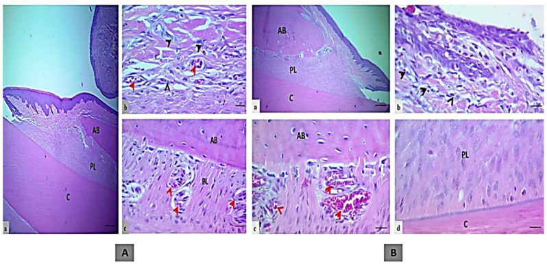
Figure 12.
A histological section of a rat incisor tooth and its surrounding periodontal tissues. Treatment control group (A): mild inflammatory cells (black arrows) with intact junctional epithelium and a stable bony surface with dense, well-formed bone and multiple blood vessels (red arrows) (H&E, scale bar 10 μm in section (a), and 20 μm in section (b,c)). Testing group (B): mild inflammatory cells (black arrowheads) with intact junctional epithelium and well-formed, dense bone (H&E, scale bar 10 μm in section (a), and 20 μm in section (b–d)) (AB; alveolar bone, PL; periodontal ligament and C; cementum) [415].

