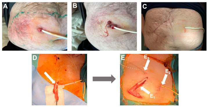
Figure 1.
The appearance of the wound before, during, and after surgery. (A) The wound in October 2020 before antibacterial treatment. (B) The wound in March 2021 before antibacterial treatment. (C) The wound in October 2021 with no local signs of infection, four months after phage treatment and surgery. (D) Removal of the velour from the LVAD driveline during surgery; velour indicated with an arrow. (E) In a picture of surgery, arrow E1 is pointing at the previous driveline canal, arrow E2 indicates the exit site of the new driveline canal, and arrow E3 indicates an 8-Fr catheter for local phage infusion in the new canal.

