

Abstract
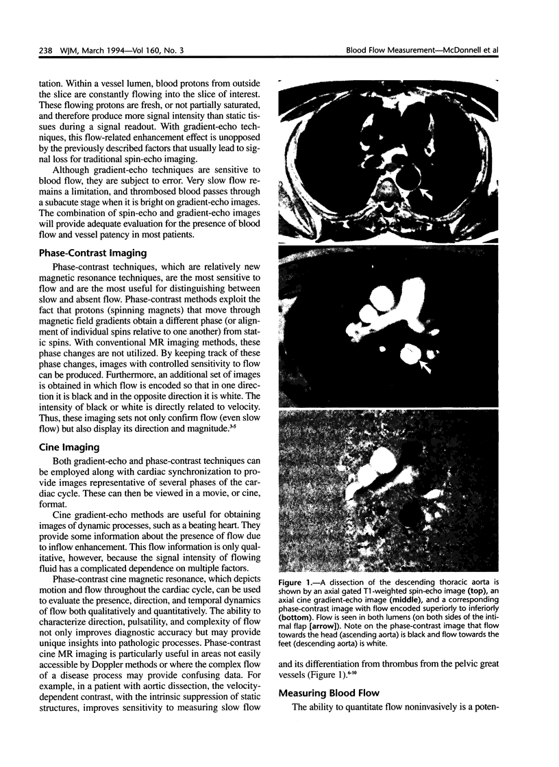
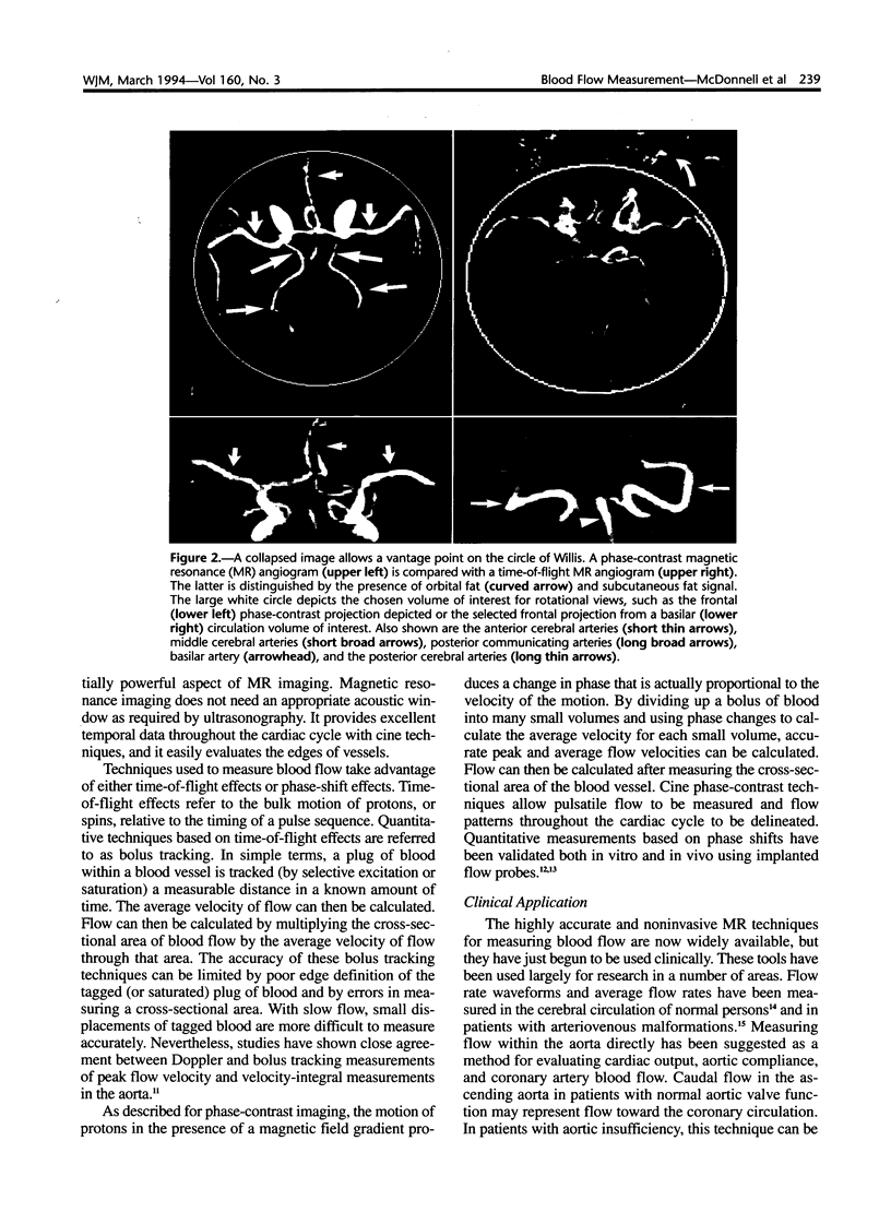
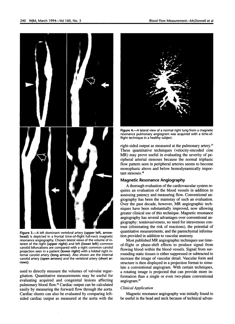
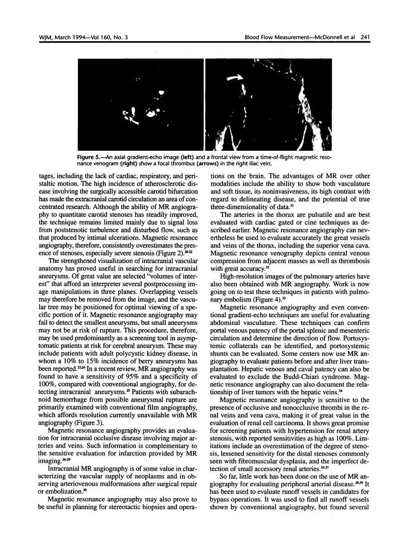
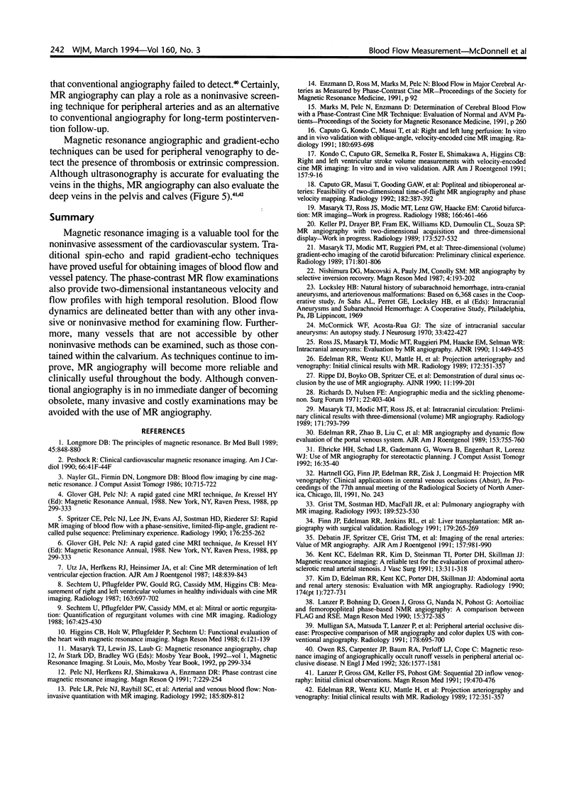
Blood flow can be shown as a negative image with magnetic resonance spin-echo techniques or as a positive image with gradient-echo techniques. Phase contrast refers to techniques where structures can be seen because of flow-induced phase shifts. These techniques can show the presence (slow flow) and also the direction of flow. Gradient-echo techniques--including phase-contrast versions--can be used with cardiac synchronization to obtain multiple images during the cardiac cycle. These images can be viewed in a movie or cine format to provide dynamic information about blood flow. Blood flow can be measured by using contrast media in boluses or even more elegantly with phase-contrast methods. Clinical applications of flow measurements are growing rapidly. Phase-contrast or gradient-echo techniques can be used to create magnetic resonance angiography, which was first used to study the carotid bifurcation and intracranial circulation and is now being used throughout the body.
Full text
PDF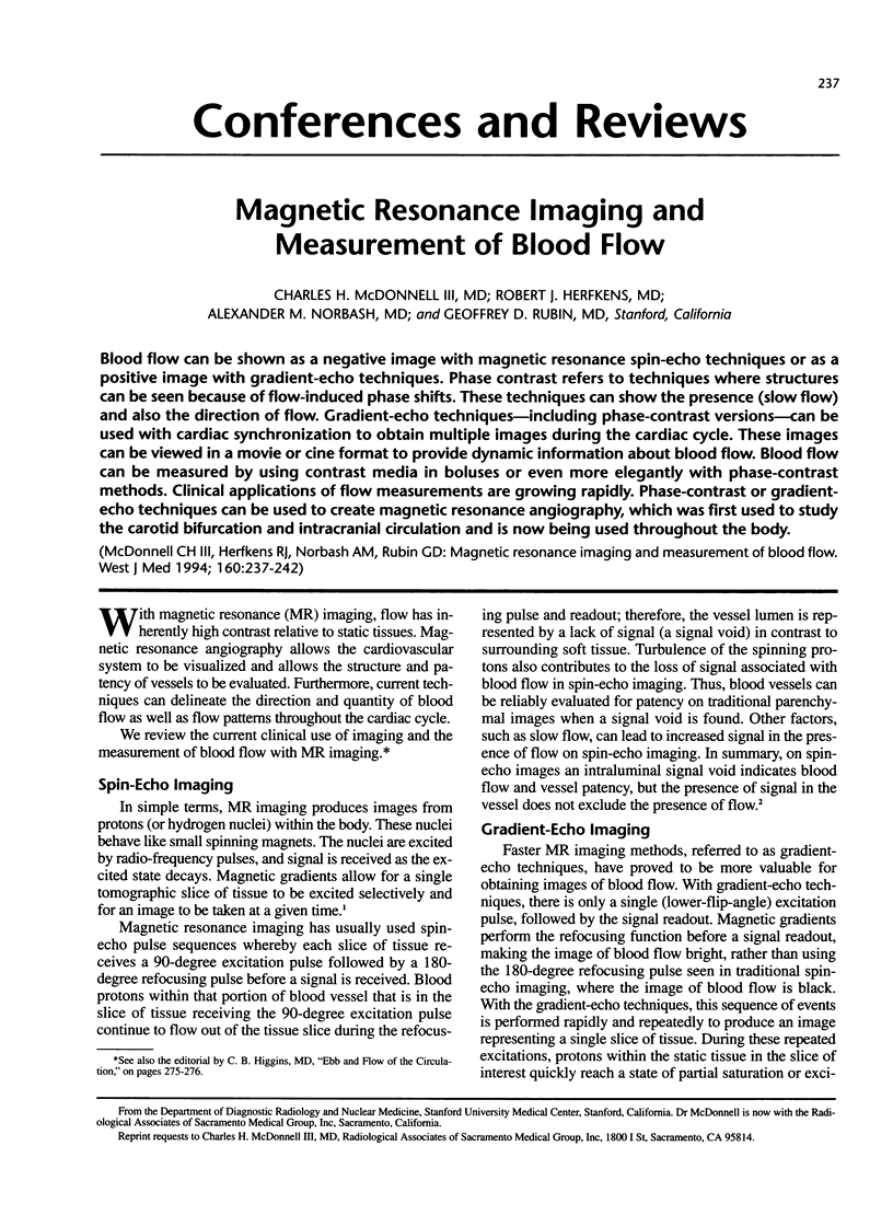
Images in this article
Selected References
These references are in PubMed. This may not be the complete list of references from this article.
- Caputo G. R., Kondo C., Masui T., Geraci S. J., Foster E., O'Sullivan M. M., Higgins C. B. Right and left lung perfusion: in vitro and in vivo validation with oblique-angle, velocity-encoded cine MR imaging. Radiology. 1991 Sep;180(3):693–698. doi: 10.1148/radiology.180.3.1871279. [DOI] [PubMed] [Google Scholar]
- Caputo G. R., Masui T., Gooding G. A., Chang J. M., Higgins C. B. Popliteal and tibioperoneal arteries: feasibility of two-dimensional time-of-flight MR angiography and phase velocity mapping. Radiology. 1992 Feb;182(2):387–392. doi: 10.1148/radiology.182.2.1732954. [DOI] [PubMed] [Google Scholar]
- Debatin J. F., Spritzer C. E., Grist T. M., Beam C., Svetkey L. P., Newman G. E., Sostman H. D. Imaging of the renal arteries: value of MR angiography. AJR Am J Roentgenol. 1991 Nov;157(5):981–990. doi: 10.2214/ajr.157.5.1927823. [DOI] [PubMed] [Google Scholar]
- Edelman R. R., Wentz K. U., Mattle H., Zhao B., Liu C., Kim D., Laub G. Projection arteriography and venography: initial clinical results with MR. Radiology. 1989 Aug;172(2):351–357. doi: 10.1148/radiology.172.2.2748814. [DOI] [PubMed] [Google Scholar]
- Edelman R. R., Wentz K. U., Mattle H., Zhao B., Liu C., Kim D., Laub G. Projection arteriography and venography: initial clinical results with MR. Radiology. 1989 Aug;172(2):351–357. doi: 10.1148/radiology.172.2.2748814. [DOI] [PubMed] [Google Scholar]
- Edelman R. R., Zhao B., Liu C., Wentz K. U., Mattle H. P., Finn J. P., McArdle C. MR angiography and dynamic flow evaluation of the portal venous system. AJR Am J Roentgenol. 1989 Oct;153(4):755–760. doi: 10.2214/ajr.153.4.755. [DOI] [PubMed] [Google Scholar]
- Ehricke H. H., Schad L. R., Gademann G., Wowra B., Engenhart R., Lorenz W. J. Use of MR angiography for stereotactic planning. J Comput Assist Tomogr. 1992 Jan-Feb;16(1):35–40. doi: 10.1097/00004728-199201000-00007. [DOI] [PubMed] [Google Scholar]
- Finn J. P., Edelman R. R., Jenkins R. L., Lewis W. D., Longmaid H. E., Kane R. A., Stokes K. R., Mattle H. P., Clouse M. E. Liver transplantation: MR angiography with surgical validation. Radiology. 1991 Apr;179(1):265–269. doi: 10.1148/radiology.179.1.2006289. [DOI] [PubMed] [Google Scholar]
- Glover G. H., Pelc N. J. A rapid-gated cine MRI technique. Magn Reson Annu. 1988:299–333. [PubMed] [Google Scholar]
- Glover G. H., Pelc N. J. A rapid-gated cine MRI technique. Magn Reson Annu. 1988:299–333. [PubMed] [Google Scholar]
- Grist T. M., Sostman H. D., MacFall J. R., Foo T. K., Spritzer C. E., Witty L., Newman G. E., Debatin J. F., Tapson V., Saltzman H. A. Pulmonary angiography with MR imaging: preliminary clinical experience. Radiology. 1993 Nov;189(2):523–530. doi: 10.1148/radiology.189.2.8210385. [DOI] [PubMed] [Google Scholar]
- Higgins C. B., Holt W., Pflugfelder P., Sechtem U. Functional evaluation of the heart with magnetic resonance imaging. Magn Reson Med. 1988 Feb;6(2):121–139. doi: 10.1002/mrm.1910060202. [DOI] [PubMed] [Google Scholar]
- Keller P. J., Drayer B. P., Fram E. K., Williams K. D., Dumoulin C. L., Souza S. P. MR angiography with two-dimensional acquisition and three-dimensional display. Work in progress. Radiology. 1989 Nov;173(2):527–532. doi: 10.1148/radiology.173.2.2798885. [DOI] [PubMed] [Google Scholar]
- Kent K. C., Edelman R. R., Kim D., Steinman T. I., Porter D. H., Skillman J. J. Magnetic resonance imaging: a reliable test for the evaluation of proximal atherosclerotic renal arterial stenosis. J Vasc Surg. 1991 Feb;13(2):311–318. [PubMed] [Google Scholar]
- Kim D., Edelman R. R., Kent K. C., Porter D. H., Skillman J. J. Abdominal aorta and renal artery stenosis: evaluation with MR angiography. Radiology. 1990 Mar;174(3 Pt 1):727–731. doi: 10.1148/radiology.174.3.2305057. [DOI] [PubMed] [Google Scholar]
- Kondo C., Caputo G. R., Semelka R., Foster E., Shimakawa A., Higgins C. B. Right and left ventricular stroke volume measurements with velocity-encoded cine MR imaging: in vitro and in vivo validation. AJR Am J Roentgenol. 1991 Jul;157(1):9–16. doi: 10.2214/ajr.157.1.2048544. [DOI] [PubMed] [Google Scholar]
- Lanzer P., Bohning D., Groen J., Gross G., Nanda N., Pohost G. Aortoiliac and femoropopliteal phase-based NMR angiography: a comparison between FLAG and RSE. Magn Reson Med. 1990 Sep;15(3):372–385. doi: 10.1002/mrm.1910150304. [DOI] [PubMed] [Google Scholar]
- Lanzer P., Gross G. M., Keller F. S., Pohost G. M. Sequential 2D inflow venography: initial clinical observations. Magn Reson Med. 1991 Jun;19(2):470–476. doi: 10.1002/mrm.1910190240. [DOI] [PubMed] [Google Scholar]
- Longmore D. B. The principles of magnetic resonance. Br Med Bull. 1989 Oct;45(4):848–880. doi: 10.1093/oxfordjournals.bmb.a072371. [DOI] [PubMed] [Google Scholar]
- Masaryk T. J., Modic M. T., Ross J. S., Ruggieri P. M., Laub G. A., Lenz G. W., Haacke E. M., Selman W. R., Wiznitzer M., Harik S. I. Intracranial circulation: preliminary clinical results with three-dimensional (volume) MR angiography. Radiology. 1989 Jun;171(3):793–799. doi: 10.1148/radiology.171.3.2717754. [DOI] [PubMed] [Google Scholar]
- Masaryk T. J., Modic M. T., Ruggieri P. M., Ross J. S., Laub G., Lenz G. W., Tkach J. A., Haacke E. M., Selman W. R., Harik S. I. Three-dimensional (volume) gradient-echo imaging of the carotid bifurcation: preliminary clinical experience. Radiology. 1989 Jun;171(3):801–806. doi: 10.1148/radiology.171.3.2717755. [DOI] [PubMed] [Google Scholar]
- Masaryk T. J., Ross J. S., Modic M. T., Lenz G. W., Haacke E. M. Carotid bifurcation: MR imaging. Work in progress. Radiology. 1988 Feb;166(2):461–466. doi: 10.1148/radiology.166.2.3336721. [DOI] [PubMed] [Google Scholar]
- McCormick W. F., Acosta-Rua G. J. The size of intracranial saccular aneurysms. An autopsy study. J Neurosurg. 1970 Oct;33(4):422–427. doi: 10.3171/jns.1970.33.4.0422. [DOI] [PubMed] [Google Scholar]
- Mulligan S. A., Matsuda T., Lanzer P., Gross G. M., Routh W. D., Keller F. S., Koslin D. B., Berland L. L., Fields M. D., Doyle M. Peripheral arterial occlusive disease: prospective comparison of MR angiography and color duplex US with conventional angiography. Radiology. 1991 Mar;178(3):695–700. doi: 10.1148/radiology.178.3.1994405. [DOI] [PubMed] [Google Scholar]
- Nayler G. L., Firmin D. N., Longmore D. B. Blood flow imaging by cine magnetic resonance. J Comput Assist Tomogr. 1986 Sep-Oct;10(5):715–722. doi: 10.1097/00004728-198609000-00001. [DOI] [PubMed] [Google Scholar]
- Nishimura D. G., Macovski A., Pauly J. M., Conolly S. M. MR angiography by selective inversion recovery. Magn Reson Med. 1987 Feb;4(2):193–202. doi: 10.1002/mrm.1910040214. [DOI] [PubMed] [Google Scholar]
- Owen R. S., Carpenter J. P., Baum R. A., Perloff L. J., Cope C. Magnetic resonance imaging of angiographically occult runoff vessels in peripheral arterial occlusive disease. N Engl J Med. 1992 Jun 11;326(24):1577–1581. doi: 10.1056/nejm199206113262428. [DOI] [PubMed] [Google Scholar]
- Pelc L. R., Pelc N. J., Rayhill S. C., Castro L. J., Glover G. H., Herfkens R. J., Miller D. C., Jeffrey R. B. Arterial and venous blood flow: noninvasive quantitation with MR imaging. Radiology. 1992 Dec;185(3):809–812. doi: 10.1148/radiology.185.3.1438767. [DOI] [PubMed] [Google Scholar]
- Pelc N. J., Herfkens R. J., Shimakawa A., Enzmann D. R. Phase contrast cine magnetic resonance imaging. Magn Reson Q. 1991 Oct;7(4):229–254. [PubMed] [Google Scholar]
- Peshock R. M. Clinical cardiovascular magnetic resonance imaging. Am J Cardiol. 1990 Oct 26;66(14):41F–44F. doi: 10.1016/0002-9149(90)90641-d. [DOI] [PubMed] [Google Scholar]
- Richards D., Nulsen F. E. Angiographic media and the sickling phenomenon. Surg Forum. 1971;22:403–404. [PubMed] [Google Scholar]
- Rippe D. J., Boyko O. B., Spritzer C. E., Meisler W. J., Dumoulin C. L., Souza S. P., Heinz E. R. Demonstration of dural sinus occlusion by the use of MR angiography. AJNR Am J Neuroradiol. 1990 Jan-Feb;11(1):199–201. [PMC free article] [PubMed] [Google Scholar]
- Ross J. S., Masaryk T. J., Modic M. T., Ruggieri P. M., Haacke E. M., Selman W. R. Intracranial aneurysms: evaluation by MR angiography. AJNR Am J Neuroradiol. 1990 May;11(3):449–455. [PMC free article] [PubMed] [Google Scholar]
- Sechtem U., Pflugfelder P. W., Cassidy M. M., White R. D., Cheitlin M. D., Schiller N. B., Higgins C. B. Mitral or aortic regurgitation: quantification of regurgitant volumes with cine MR imaging. Radiology. 1988 May;167(2):425–430. doi: 10.1148/radiology.167.2.3357951. [DOI] [PubMed] [Google Scholar]
- Sechtem U., Pflugfelder P. W., Gould R. G., Cassidy M. M., Higgins C. B. Measurement of right and left ventricular volumes in healthy individuals with cine MR imaging. Radiology. 1987 Jun;163(3):697–702. doi: 10.1148/radiology.163.3.3575717. [DOI] [PubMed] [Google Scholar]
- Spritzer C. E., Pelc N. J., Lee J. N., Evans A. J., Sostman H. D., Riederer S. J. Rapid MR imaging of blood flow with a phase-sensitive, limited-flip-angle, gradient recalled pulse sequence: preliminary experience. Radiology. 1990 Jul;176(1):255–262. doi: 10.1148/radiology.176.1.2353099. [DOI] [PubMed] [Google Scholar]
- Utz J. A., Herfkens R. J., Heinsimer J. A., Bashore T., Califf R., Glover G., Pelc N., Shimakawa A. Cine MR determination of left ventricular ejection fraction. AJR Am J Roentgenol. 1987 May;148(5):839–843. doi: 10.2214/ajr.148.5.839. [DOI] [PubMed] [Google Scholar]