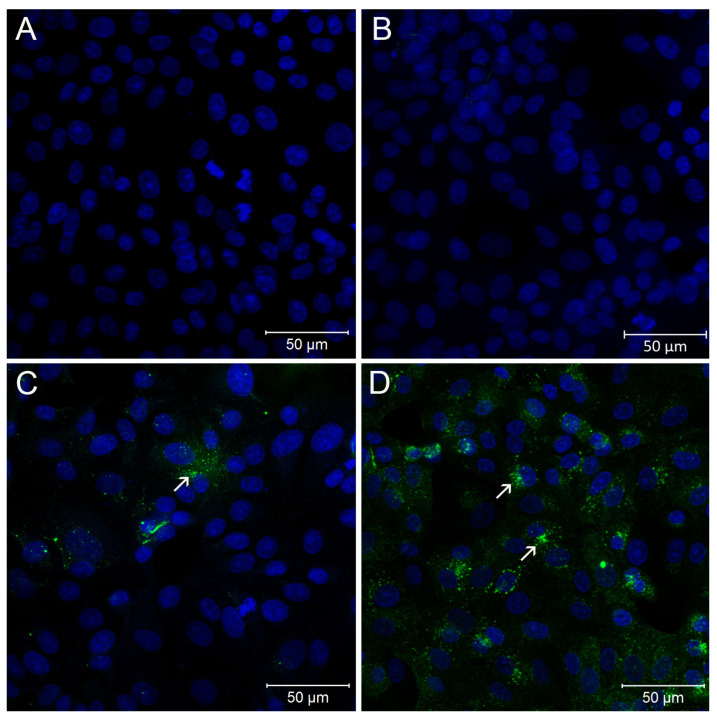
Figure 4.
Immunofluorescence assay of Vero cells inoculated with rectal swab samples. (A) Mock-infected Vero cells. (B) Rectal swab sample (id_31386). (C) Rectal swab sample (id_30771). (D) Second passage of sample id_30771. Nucleus—Blue; SARS-CoV-2 Spike—Green. In MOCK infected and sample id_31386, staining for spike protein was not observed (A,B). Rectal swab sample (id_30771) positive staining for spike in cytoplasmic dots (C). Supernatant of second passage of the sample id_30771 showing the immunofluorescence in a higher number of cells (D). Representative areas of SARS-CoV-2 infected cells are indicated by white arrows.

