Abstract
We report a young male patient who presented with chest and shoulder tip pain with spontaneous intraperitoneal haemorrhage (haemoperitoneum) due to gastric vessel rupture. Point‐of‐care ultrasound detected abdominal free fluid, which led to a CT scan of the abdomen and reached the diagnosis. Intra‐abdominal bleeding can present as referred chest or shoulder tip pain, as more commonly seen in females with pelvic pathologies. Point‐of‐care ultrasound may add diagnostic value with the detection of a haemoperitoneum in this context.
Keywords: abdominal apoplexy/intra‐abdominal haemorrhage, gastric vessel rupture, haemoperitoneum, point‐of‐care ultrasound (POCUS), referred pain, ultrasonography
Introduction
The spontaneous rupture of gastric vessels is a rare condition that can be life‐threatening. 1 It has been reported to occur in adulthood at any age, typically presenting in shock in conjunction with abdominal pain. 2 The diagnosis is usually made with cross‐sectional imaging, such as a CT scan with contrast. However, it can present more insidiously in a younger person. 3 This article describes how point‐of‐care ultrasound (POCUS) detected abdominal free fluid in a young male patient presenting to the emergency department (ED) with chest and shoulder tip pain, which expedited CT imaging of the abdomen and the ultimate diagnosis of left gastric vessel rupture with haemoperitoneum.
Case report
A 25‐year‐old male presented to the ED with 14 hours of atraumatic left‐sided chest pain radiating to his left shoulder tip. He reported that the pain was severe with sudden onset that morning at 01:00 while walking home. The pain was pleuritic in nature and worse with coughing and in a supine position. He had consumed a few alcoholic drinks and inhaled several electronic cigarettes the evening before. He denied using any recreational drugs. There was no significant past medical history apart from appendicectomy as a child. Despite adequate analgesia, which included intravenous fentanyl, he remained symptomatic.
On examination, he appeared uncomfortable, but alert. His vital signs included a temperature of 36.8°C, heart rate of 82 beats/min, blood pressure of 150/67 mmHg, respiratory rate of 18 breaths/min and oxygen saturation of 98% in room air. There were no localising features or abnormalities on systems review, including a normal cardiovascular, respiratory and abdominal examination. ECG, chest x‐ray and blood tests were unremarkable (haemoglobin 158 g/L, platelets 326/L, red blood cell 5.24 × 10¹²/L and high sensitivity cTroponin I 2 ng/L). Subsequent coagulopathy and vasculitis screening were normal.
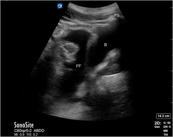
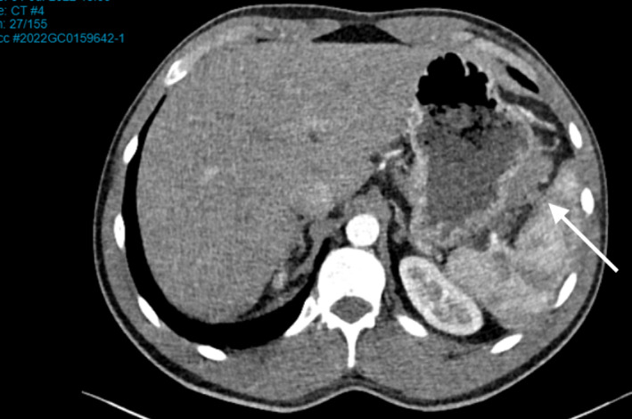
Point‐of‐care ultrasound was performed immediately after the initial assessment, which excluded a pneumothorax and pericardial effusion. Given the lack of a cause for his symptoms, the abdomen was then scanned using a focussed assessment with sonography for trauma (FAST) protocol, which identified a large volume of pelvic free fluid (Figures 1, S1). CT abdomen with contrast (portal venous phase) confirmed a large volume haemoperitoneum, predominately in the pelvis, with extension between the spleen and the greater curvature of the stomach (Figure 2). Based on these findings, the source of haemorrhage was thought to be from the spontaneous rupture of the left gastric vessel. However, given the stability of the patient, a further CT angiogram was not performed.
Figure 1.

Ultrasound image demonstrating a large volume of free fluid in the pelvis. B, bladder; FF, free fluid; PS, pubic symphysis.
Figure 2.

CT image (abdominal axial slice) demonstrating the gastric vessel haemorrhage, measuring 65 × 22 mm, visualised between the greater curvature of the stomach and the left gastroepiploic artery (arrow).
He was admitted to the intensive care unit under the surgical team with haematology consultation. He was managed conservatively as he remained haemodynamically stable with static serial haemoglobin levels and was discharged 4 days later without any complication. Consent to publish the case report was obtained from the patient and approved by the Gold Coast Hospital and Human Research Ethics Committee (EC00160).
Discussion
This article reports a rare presentation of spontaneous intraperitoneal haemorrhage (haemoperitoneum) of a young man from left gastric vessel rupture, detected with POCUS. He was managed conservatively as an inpatient. However, his presentation with referred chest and shoulder tip pain was atypical and could have easily been misdiagnosed and discharged from the ED with possible deterioration. The detection of abdominal free fluid on POCUS was key to his diagnosis and highlights how it can be employed to rapidly assess patients with this presentation.
Spontaneous intraperitoneal haemorrhage (abdominal apoplexy) was first described by Barber in 1901. 4 Spontaneous intraperitoneal haemorrhage usually arises from one of the smaller abdominal arteries or veins and is diagnosed after haemorrhage from an aortic aneurysm or aortic dissection has been excluded. Spontaneous intraperitoneal haemorrhage is rare with its true incidence unknown. 5 , 6 It mostly occurs due to rupture of aneurysmal disease, which can be predisposed by vasculitis, hypertension, dyslipidaemia or tumour. 7 , 8 There have also been a few cases reported as a result of medical procedures, such as endoscopy. 9
In prior case reports, patients with rupture of the gastric vessels typically present with abdominal pain after vomiting or gagging and haemodynamic instability, which were absent in this case. 10 , 11 In all of these cases, the diagnosis was made on a CT scan of the abdomen with contrast, 12 with the majority occurring as a result of the rupture of left gastric vessels, including short gastric, gastroepiploic or splenic arteries. 13 It is a condition that typically requires surgical intervention or an endovascular procedure to achieve haemostasis, with a good prognosis with timely intervention. 14
Left shoulder tip pain, otherwise known as Kehr's sign, is due to the presence of blood or other irritants in the peritoneal cavity. 15 Classically, this is due to a ruptured spleen but, besides left gastric vessel rupture, this may result from other pathologies around the left hemidiaphragm, such as ruptured ectopic pregnancy (females), ruptured ovarian cysts (females), pancreatitis/pseudocyst rupture, renal calculi, gastrointestinal perforation or abscesses. 16 , 17
Point‐of‐care ultrasound was instrumental in the diagnosis of this patient, without which he may have been discharged home from the ED with subsequent deterioration. Given his presentation and demographics, abdominal pathology was not initially considered as a cause of his chest pain. POCUS was instrumental in shifting the cognitive bias to consider other differential diagnoses. Many ED clinicians are already familiar with using POCUS to detect abdominal free fluid (i.e. FAST scan protocol), usually conducted in the setting of trauma. Consideration should be given to performing POCUS looking for abdominal free fluid in patients presenting with unexplained severe chest pain radiating to the shoulder tip.
Authorship statement
We confirm that (i) the authorship listing conforms with the journal's authorship policy and (ii) that all authors are in agreement with the content of the submitted manuscript.
Funding statement
No funding information is provided.
Conflict of interest
None declared.
Author contributions
KY, PS and SW conceptualised the report. KY and PS wrote the initial draft, which was reviewed and revised by JX, RX and SW. Supervision was provided by PS and SW. All authors read and approved the final manuscript, and KY takes responsibility for the paper as a whole. KY contributed to writing – original draft preparation and writing – review and editing. PS contributed to writing – review and editing and supervision. JN contributed to writing – review and editing. RM and SW contributed to supervision. All authors critically revised the report, commented on draft of the manuscript and approved the final report.
Supporting information
Figure S1. Ultrasound image series of (a) the right upper quadrant, (b) the liver tip and (c) the left upper quadrant with no free fluid demonstrated.
References
- 1. Abdelrazeq AS, Saleem TB, Nejim A, Leveson SH. Massive hemoperitoneum caused by rupture of an aneurysm of the marginal artery of Drummond. Cardiovasc Intervent Radiol 2008; 31(Suppl 2): S108–10. [DOI] [PubMed] [Google Scholar]
- 2. Harbour LN, Koch MS, Louis TH, Fulmer JM, Guileyardo JM. Abdominal apoplexy: two unusual cases of hemoperitoneum. Proc (Bayl Univ Med Cent) 2012; 25(1): 16–9. [DOI] [PMC free article] [PubMed] [Google Scholar]
- 3. Gregory CR, Proctor VK, Thomas SM, Ravi K. Spontaneous haemorrhage from a left gastric artery aneurysm as a cause of acute abdominal pain. Ann R Coll Surg Engl. 2017; 99(2): e49‐e51. [DOI] [PMC free article] [PubMed] [Google Scholar]
- 4. Barber MC. Intra‐abdominal haemorrhage associated with labour. Br Med J 1909; 2(2534): 203–4. [DOI] [PMC free article] [PubMed] [Google Scholar]
- 5. Saeed Y, Farkas Z, Azeez S. Idiopathic spontaneous intraperitoneal hemorrhage due to rupture of short gastric artery presenting as massive gastrointestinal bleeding: a rare case presentation and literature review. Cureus 2020. Nov 16; 12(11): e11499. [DOI] [PMC free article] [PubMed] [Google Scholar]
- 6. Carmeci C, Munfakh N, Brooks JW. Abdominal apoplexy. South Med J 1998; 91(3): 273–4. [DOI] [PubMed] [Google Scholar]
- 7. Kohara Y, Fujimoto K, Katsura H, Komatsubara T, Ichikawa K, Higashiyama H. Biphasic clinical course of a ruptured right gastric artery aneurysm caused by segmental arterial mediolysis: a case report. BMC Surg 2020; 20(1): 191. [DOI] [PMC free article] [PubMed] [Google Scholar]
- 8. Yamaguchi M, Kumada K, Sugiyama H, Okamoto E, Ozawa K. Hemoperitoneum due to a ruptured gastroepiploic artery aneurysm in systemic lupus erythematosus. A case report and literature review. J Clin Gastroenterol 1990; 12(3): 344–6. [DOI] [PubMed] [Google Scholar]
- 9. Pricolo R, Cipolletta L, Rossi GP, Bianco MA, Cuccurullo A. Rara complicanza in corso di esofago‐gastro‐duodenoscopia [A rare complication of esophagogastroduodenoscopy]. Minerva Dietol Gastroenterol. 1985; 31(2): 339–42. [PubMed] [Google Scholar]
- 10. Shimpi TR, Shikhare S, Chan DY, Peh WC, Chawla A. Vomiting‐induced short gastric artery apoplexy. BJR Case Rep 2016; 3(1): 20150216. [DOI] [PMC free article] [PubMed] [Google Scholar]
- 11. Jabr FI, Skeik N. Short gastric artery apoplexy after gagging. J Med Liban 2012; 60(3): 173–5. [PubMed] [Google Scholar]
- 12. Lavoipierre AM, Vellar ID. Gastroepiploic artery pseudoaneurysm as a cause of abdominal apoplexy. Role of CT Scanning Australas Radiol 1984; 28(1): 23–5. [DOI] [PubMed] [Google Scholar]
- 13. Sandstrom A, Jha P. Ruptured left gastric artery aneurysms: three cases managed successfully with open surgical repair. Ann Vasc Surg 2016; 36: 296.e9‐e12. [DOI] [PubMed] [Google Scholar]
- 14. Park SJ, Kwon SW, Oh JH, Park HC. Glue embolization of a ruptured celiac trunk aneurysm. Vascular 2009; 17: 112–5. [DOI] [PubMed] [Google Scholar]
- 15. Lowenfels AB. Kehr's sign‐‐a neglected aid in rupture of the spleen. N Engl J Med 1966; 274(18): 1019. [DOI] [PubMed] [Google Scholar]
- 16. Wu CE, Liu CH. A man with pain in the left upper quadrant and left shoulder. Gastroenterology 2020; 3(20): 30274–2. [DOI] [PubMed] [Google Scholar]
- 17. Nyhsen C, Mahmood SU. Life‐threatening haemoperitoneum secondary to rupture of simple ovarian cyst. BMJ Case Rep 2014; 2014: bcr2014205061. [DOI] [PMC free article] [PubMed] [Google Scholar]
Associated Data
This section collects any data citations, data availability statements, or supplementary materials included in this article.
Supplementary Materials
Figure S1. Ultrasound image series of (a) the right upper quadrant, (b) the liver tip and (c) the left upper quadrant with no free fluid demonstrated.


