
Figure 1.
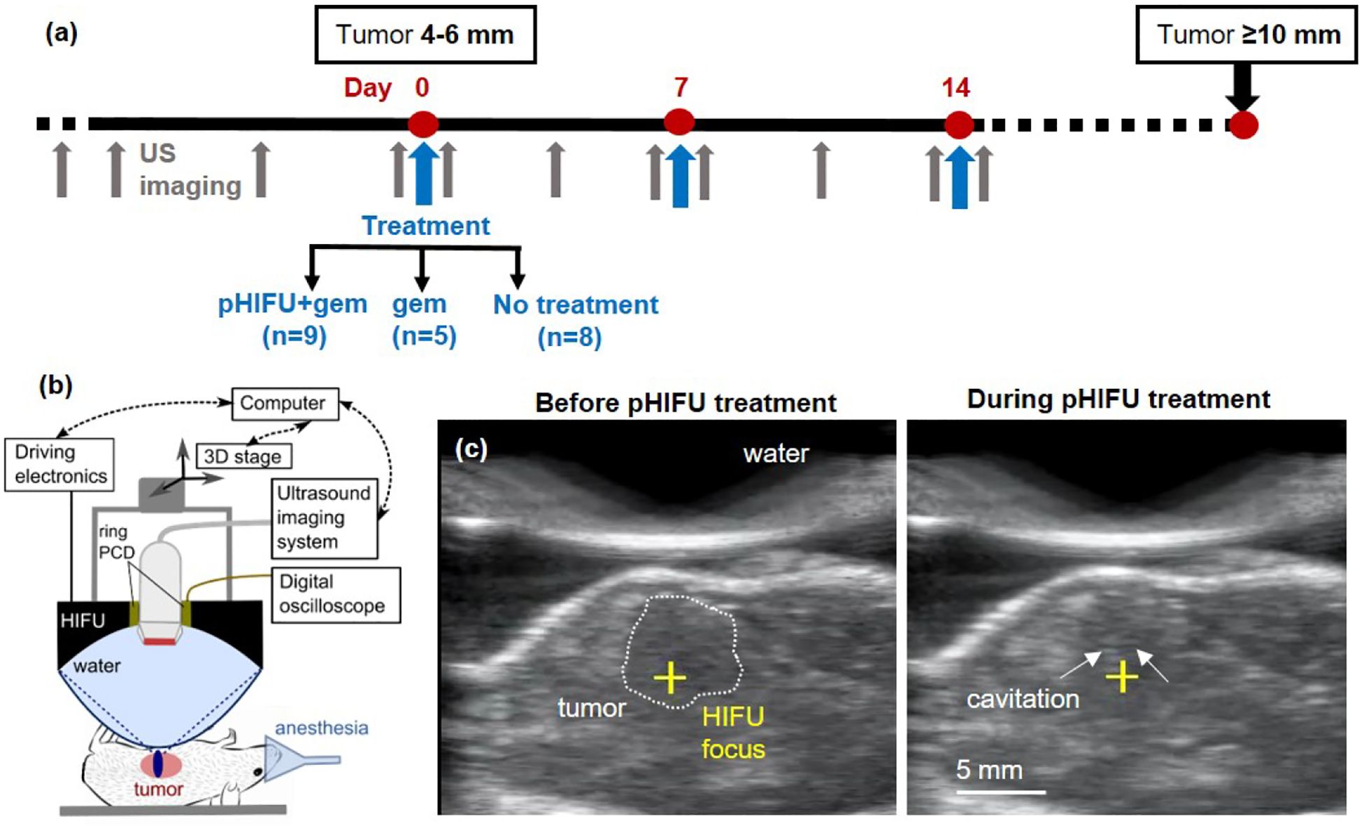
The study timeline and pHIFU methods. (a) Mice were enrolled when the tumor reached 4–6 mm in size, per US imaging, and treated weekly until tumor size reached 10 mm – the study endpoint. Mice were randomly assigned to three groups: pHIFU followed by IP administration of gemcitabine (gem), gem only or no treatment. (b) Experimental setup for pHIFU treatment of KPC mouse tumors (Alpinion VIFU 2000 dry system). (c) B-mode ultrasound (US) images from the inline probe obtained immediately prior to (left) and during (right) pHIFU treatment. Note the faint hyperechoic region (arrows) appearing prefocally and corresponding to inertial cavitation (see also Supplemental Video 1).