ABSTRACT
Three critical aspects that define high concentration antibody products (HCAPs) are as follows: 1) formulation composition, 2) dosage form, and 3) primary packaging configuration. HCAPs have become successful in the therapeutic sector due to their unique advantage of allowing subcutaneous self-administration. Technical challenges, such as physical and chemical instability, viscosity, delivery volume limitations, and product immunogenicity, can hinder successful development and commercialization of HCAPs. Such challenges can be overcome by robust formulation and process development strategies, as well as rational selection of excipients and packaging components. We compiled and analyzed data from US Food and Drug Administration-approved and marketed HCAPs that are ≥100 mg/mL to identify trends in formulation composition and quality target product profile. This review presents our findings and discusses novel formulation and processing technologies that enable the development of improved HCAPs at ≥200 mg/mL. The observed trends can be used as a guide for further advancements in the development of HCAPs as more complex antibody-based modalities enter biologics product development.
KEYWORDS: Container closure components, excipients, formulation, high concentration, immunoglobulin, monoclonal antibody, protein concentration
Introduction
Monoclonal antibodies (mAbs) are therapeutic proteins derived from a single clone or line of cloned cells that have been successfully used to treat a wide array of debilitating and life-threatening diseases in immunology, infectious diseases, and oncology. Since their introduction as drugs, mAbs therapies have become a tremendous therapeutic and commercial success, and more than 120 mAbs therapies have been approved by the US Food and Drug Administration (FDA) and introduced in the market.1–3 Because of their success in treatment over the past 2 decades, the global therapeutic market for mAbs was valued at approximately $115.2 billion in 2018 and surpassed over $150 billion in revenue by the end of 2019 and is projected to grow to about $300 billion by 2025.4 Currently, mAbs are also the most-marketed biologic, and nearly 4000 mAbs are currently undergoing preclinical and clinical developments, which further validate their prevalence and potential for growth.5
Detailed descriptions of the antibody structure, its instabilities and formulation approaches are included in a review by Wang et al.6 Briefly, mAbs are Y-shaped proteins composed of two light chains and two heavy chains linked by a disulfide bridge. In addition to high specificity, mAbs therapies have the ability to target and interact with various antigens, receptors, and immune checkpoints, giving them the ability to treat a wide variety of disease states.7 The variable regions on the heavy chain and the light chain are composed of 110 amino acids that make up the antigen-binding fragments (Fabs), which includes the complementarity-determining region (CDR) that accounts for the target specificity of the antibody. The remaining protein sequence comprises the constant regions, which culminate to form the Fc (fragment crystallizable) regions. All these varying regions of the mAbs work together to give the antibody its effector and receptor-binding capabilities. Antibodies, also referred to as immunoglobulins (Ig), can be further divided into five classes: IgA, IgD, IgE, IgM, and IgG, which are named based on their constant regions. As discussed below, most mAbs therapies are of the IgG subtype.
High concentration antibody products (HCAPs) can be broadly defined as injectable mAbs or polyclonal antibody therapies, which typically have an overall product concentration of ≥100 mg/mL. These high protein concentrations often present additional physical stability challenges. Therefore, to enable therapeutic use of HCAPs, they must be formulated, stabilized, developed, and manufactured using robust processes. The art and science of antibody formulation and stabilization evolves over time, and several reviews6,8–12 and books13–16 have described the basic concepts in antibody drug product formulation development and manufacturing.17,18 Other reviews have highlighted issues associated with protein aggregation, including possible mechanisms and how manufacturing processes for antibody drug products should be designed to overcome these challenges.18–25 Similarly, several studies have addressed challenges like viscosity, phase separation, and opalescence, which are particularly prevalent in HCAPs.26–30
With the introduction of computational predictive techniques31 coupled with experimental methods,32 developability assessment of antibody candidates in early discovery is now commonly included in the pharmaceutical development process.33–35 Biophysical characterization is a key component of the developability assessment that should be done before a candidate enters biopharmaceutical development. The use of such techniques has enabled smart design of biopharmaceuticals, wherein any potential aggregation or instability is identified and can be mitigated in the early stages of development.
HCAPs have become immensely popular and widely successful in the therapeutic space for the following reasons.
Subcutaneous delivery of <2 mL dose volume
Without the use of enabling technologies or novel devices, the maximum volume of products delivered via subcutaneous (SC) administration is often considered to be 2 mL.36,37 HCAPs enable the delivery of a larger amount of protein in this <2 mL dose volume, which presents a unique advantage over intravenous (IV) administration options, which often require hospitalization or a visit to an infusion center for administration by a health-care professional, as the SC administration can be performed in a doctor’s clinic or even at home.
Self-administration
The advantages of self-administration are coupled with those of SC delivery. With SC administration, certain patients can self-administer the dose using a prefilled syringe (PFS) or autoinjector (AI) pen. Such administration gives more flexibility and freedom to the patient in managing their dosing schedule and living a normal life while managing a chronic health condition.
Management of chronic diseases
Providing HCAPs as drug–device combination products for self-administration presents a unique advantage with managing chronic diseases that require long-term drug administration and ensure patient compliance.
Manufacturing and logistics cost
During the manufacturing of HCAPs, the drug substance is typically brought to a high concentration, frozen and shipped to the drug product fill finish site. Since HCAPs have a high protein concentration per unit volume of the drug substance, the cost of shipping, storage, and inventory management is significantly lower than lower concentration solutions.
Life-cycle management approaches that later introduce a more patient-centric dosage form post-approval are becoming increasingly popular. In some cases, a liquid dosage form is introduced as a follow-on to an initially approved lyophilized product. In other cases, the introduction of an SC route of administration (ROA) as opposed to the IV ROA has offered some of the advantages discussed above.38–43 Further, introducing a PFS or AI as a life cycle management strategy for an initially approved ready-to-use product (packaged in vial and delivered using a syringe and needle) has become a notable trend in the industry.44
Several recent reviews have summarized trends in liquid formulations of therapeutic proteins,45 formulations of commercially available antibodies in the US,1 approved parenteral protein formulations within the European Union,46 and US FDA-approved therapeutic antibodies with high concentration formulations.2 However, the field of high concentration antibody formulation has greatly evolved, as summarized in several studies.47–50 While these reviews have highlighted the significance of HCAP development and challenges with the development, manufacturing and testing of HCAPs, a systematic review of the HCAPs approved and commercialized in the US has been missing. To fill this gap, in this review we summarize the prescription information from individual package inserts of all HCAPs (monoclonal and polyclonal antibodies) that were granted an FDA approval or emergency use authorization (EUA). We further identify common trends and strategies in HCAP development, including formulation, stabilization, dosage form development, and packaging of commercial HCAPs. We also discuss novel formulation and processing technologies that enable the development of improved HCAPs at ≥200 mg/mL. The observed trends in this review can guide future development of HCAPs.
Scope
Here, high-concentration antibody product (HCAP) is defined as a solution or a lyophilized product upon reconstitution with a protein concentration ≥100 mg/mL. The term HCAP is relative and subjective in nature and a cutoff of 100 mg/mL protein concentration cannot be universally applied to all mAbs or mAb-like modalities. Some reviews recommend that the term “high concentration protein formulation” should rather refer to concentrations at which protein–protein interactions as a result of factors, such as crowding, solution properties, and high viscosity are significant and can affect the stability and delivery of such products.50,51 Several mAb products are formulated at concentrations <100 mg/mL. For example, Evenit® (romosozumab-aqqg), SkyriziTM (risankizumab-rzaa) and StelaraTM (ustekinumab) are all formulated as solutions and have a protein concentration of 90 mg/mL, only slightly missing the >100 mg/mL cutoff. Taltz® (ixekizumab), XgevaTM (denosumab) and ProliaTM (denosumab) are all formulated as solutions and have a protein concentration of 80 mg/mL, 70 mg/mL, and 60 mg/mL, respectively. While the unique formulation and stabilization trends in such products (<100 mg/mL but >50 mg/mL) can be studied and reviewed, the focus of this review is limited to only antibody products that are ≥ 100 mg/mL.
We referred to the prescription information, including the Description (Section 11) and How Supplied/Storage and Handling (Section 16), of all HCAPs approved in the US in compiling the information in this review. Data on all 46 (n = 46) approved high concentration mAbs (≥100 mg/mL) were compared, contrasted, and sorted based on 13 different identifiers. These identifiers were as follows: International nonproprietary name, brand name, initial FDA approval date, antibody target, antibody isotype and subtype if applicable, indication, conc. (mg/mL), ROA, dosage form (lyophilized solid (LYO) or solution (SOL)), primary container (PFS, AI/pen or vial), pH range, formulation, and storage condition Table S1.
After the data from each product’s individual package insert were collected, the information was analyzed and compiled into either a bar graph, pie chart, or table. For the formulation composition, we performed an in-depth review of the excipients used in stabilization of these HCAPs, their probable role in formulation and stabilization of these HCAPs and tried to identify common trends. The compiled data, observed trends, and our findings are further discussed in the subsequent sections of this review.
REVIEW of HCAPs
Approvals of antibody products (1998–2021)
Since we have referred to the prescription information for each of the 46 approved HCAP products in this review, we recorded the year for their initial FDA approval as provided in the prescription information. In several instances, the year of initial FDA approval was substantially earlier than the approval date for an HCAP product. For example, the Orencia® HCAP liquid product in the ClickJect™ AI was approved and launched in 2016, after an initial FDA approval of the LYO product for IV infusion in 2005. As shown in Figure 1, there has been a consistent increase in the number of approved antibody products since 1998. Among the 46 products included in this review, the greatest annual number of initial FDA approvals has occurred since 2015. Among the 46 reviewed HCAPs, six new antibody products were approved each year in years 2015, 2017, and 2018 (initial approvals).
Figure 1.
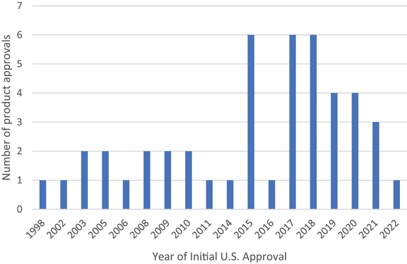
Number of product approvals each year among HCAPs between 1998–2021. (n = 46).
A consistent increase in the number of approved antibody products since 1998 with the greatest annual number of initial US approvals have occurred since 2015.
Route of administration
As shown in Figure 2, the SC ROA is the most used for HCAPs (n = 34), followed by IV (n = 6), both SC and IV (n = 2), intravitreal (n = 3), and intramuscular (IM) (n = 1). The SC route is the most prevalent due to advantages, such as home self-administration and rapid onset of action.52 SC injections are popular for peptides, proteins, certain hormones (insulin), and antibody products because they are often usually used to treat chronic diseases in which at-home administration is preferred for reasons such as patient convenience, decreased burden on health-care professionals, ease of use, reduction in hospitalization or in-patient costs, and overall reduction in treatment costs when compared to the IV administration.51,53,54 However, the SC ROA does suffer from a few drawbacks, including patient variability (e.g., substantially different body mass index), protein aggregation upon administration, incomplete absorption, and potential differences in pharmacokinetic profile and bioavailability compared to an IV route.55 Predicting the bioavailability of antibodies following SC administration can be challenging; unlike IV administrations, which are 100% bioavailable, SC administrations can have variable bioavailability, with values in the range of 60–80%.53
Figure 2.
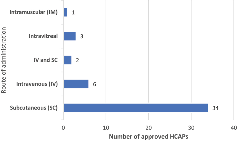
Routes of administration among approved HCAPs (n = 46).
Among the 46 HCAP, Subcutaneous (SC) route of administration is the most used for HCAPs (n = 34), followed by intravenous (IV) (n = 6), both SC and IV (n = 2), intravitreal (n = 3) and intramuscular (IM) (n = 1).
Typically, dose volumes of 2 mL or less can be conveniently administered via SC administration. Antibody solutions, therefore, need to be formulated at high concentrations (>100 mg/mL) to deliver effective doses of 200 mg or more. Concentrating protein solutions at such elevated levels can lead to crowding due to reduced intermolecular distance and the formation of a network of reversible protein–protein interactions or self-associations. These interactions do not manifest at low concentrations and can lead to protein aggregation and instability in HCAPs.56–61 Despite these drawbacks, SC administration is simpler and more practical than IV administration due to its reduced burden on health-care staff, ease of patient administration, and overall reduction of health-care costs.54
Protein solutions can also be administered via IM injection. Since muscles are more vascularized than the SC matrix of the skin, IM injections can lead to higher systemic exposure, thereby making IM injections much less desirable. 5252 Synagis® (palivizumab injection) is used to prevent serious lung infection in children and babies caused by respiratory syncytial virus (RSV) and is currently the only HCAP administered by IM injection by a health-care provider. Palivizumab injection is prescribed for infants and children 24 months and younger at the start of RSV season. A dose of 15 mg per kg of body weight is administered intramuscularly prior to commencement of the RSV season, and the remaining doses are administered monthly throughout the RSV season.
Three HCAPs are administered via intravitreal injection: 1) SusvimoTM (ranibizumab injection 100 mg/L for ocular implant), 2) Beovu® (brolucizumab-dbll), and 3) VabysmoTM (faricimab-svoa). These products are indicated for the treatment of patients with neovascular (wet) age-related macular degeneration. One probable reason HCAP is developed for intravitreal injection is due to the intravitreal dose volume limitation of about 20–50 µL. Overall, very few intravitreal injection products have been approved by the FDA (Macugen®, Lucentis®, Eylea®, and Beovu®, VabysmoTM). Lucentis® (same active ingredient as SusvimoTM) and Eylea® (aflibercept) are not HCAPs (<100 mg/mL concentration) as defined in this review. All intravitreal injection products have a dose volume of about 50 µL. As noted herein, while Beovu® (brolucizumab-dbll) is formulated at 120 mg/mL, the dose per injection is 6 mg that can be delivered in 50 µL dose volume. Similarly, Susvimo® (ranibizumab injection) is supplied for intravitreal use via implant. Each single-dose vial contains 10 mg of ranibizumab in 0.1 mL of solution (100 mg/mL). The implant is designed to contain approximately 0.02 mL (2 mg) of ranibizumab solution when filled. Similarly, VabysmoTM (faricimab-svoa) is a 120 mg/mL solution supplied in a vial that can deliver a 0.05 mL dose solution containing 6 mg of the drug.
Therapeutic area
We classified the approved HCAPs in eight disease areas, based on the indications for which they were approved. The therapeutic areas include immunology, infectious disease, cardiology, respiratory, neurology, ophthalmology, hematology, and oncology. As shown in Figure 3, among the 46 HCAP products, 20 products are prescribed in immunology, 4 each for infectious diseases and cardiology, 5 for respiratory, 6 for neurology, 3 for ophthalmology, 1 for hematology, and 3 for oncology. Not surprisingly, the largest percentage of approved HCAPs is indicated for immunology indications because of the chronic nature of these diseases. Because of the need for long-term management of immunological diseases, such as psoriasis, rheumatoid arthritis (RA), psoriatic arthritis, and ankylosing spondylitis, self-administration or at home administration by a caregiver via SC administration is the desired form of care. Such SC therapies are typically administered by prefilled syringes, AI, or injection pens, which are helpful when considering geriatric populations who may have low dexterity.54,54Across all approved antibody therapeutics, which include both high and low concentration products, about 40% of the products are indicated for use in immunology disease areas.4
Figure 3.
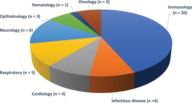
Different therapeutic areas for which HCAPs are approved and marketed (n = 46).
The number of products (n) in parentheses, and the total number of products is 46.
Among the 46 HCAP products, a majority of approved HCAPs are indicated for immunology indications, with 20 products prescribed in Immunology, 4 each for Infectious diseases and Cardiology, 5 for Respiratory, 6 for Neurology, 3 for Ophthalmology, 1 for Hematology, and 3 for Oncology.
Antibody types, isotypes, and formats
Among the 46 HCAPs reviewed herein, two products are fixed dose combinations (FDCs) containing two types of antibodies each. REGEN-COV™ contains casirivimab (human IgG1κ) and imdevimab (human IgG1λ) and Phesgo® contains pertuzumab (humanized IgG1) and trastuzumab (human IgG1κ) as active ingredients. The various categories of antibodies included among the 46 HCAPs are shown in Figure 4. As noted, four products are polyclonal antibodies. Most antibody products are immunoglobulin G 1 (IgG1) (30 products). Among the IgG1 antibodies, 2 are chimeric, 9 are human, pertuzumab is a humanized IgG1, 13 human IgG1κ (kappa subtype), tocilizumab is a humanized IgG1κ (kappa subtype), faricimab-svoa is a humanized bispecific IgG1 and 3 human IgG1λ (lambda subtype). There is a total of 5 human IgG2 antibodies, among which fremanezumab is a human IgG2 Δa/kappa antibody. There are four human IgG4, while emicizumab-kxwh is a bispecific humanized IgG4.
Figure 4.
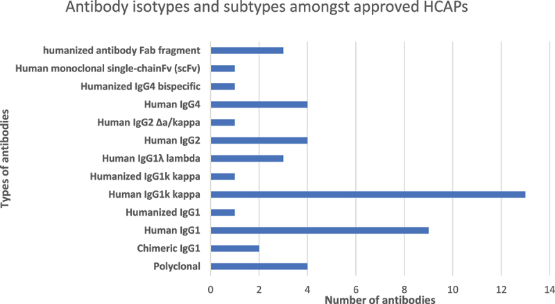
Antibody isotypes and subtypes amongst approved HCAPs.
Most antibody products are Immunoglobulin G 1 (IgG1) (30 products). Among the IgG1 antibodies, there are 2 chimeric IgG1, 9 human IgG1, 13 human IgG1k (kappa subtype), and 3 human IgG1λ (lambda subtype).
In addition to the full-length antibodies described above, brolucizumab-dbll, the active ingredient of Beovu®, is a human monoclonal single-chain Fv. Three products, Cimzia® (certolizumab pegol), liquid and lyophilized dosage forms, and Susvimo™ (ranibizumab) contain a humanized antibody Fab as the active ingredient. It is worth noting that the four HCAPs, Gammagard® (100 mg/mL), Hyqvia® (100 mg/mL), Hizentra® (200 mg/mL), and Xembify® (200 mg/mL) are derived from serum and are thus polyclonal, meaning that they are composed of antibodies with multiple specificities and isotypes. Our findings are in line with an observed industry trend wherein the most commonly manufactured antibody isotype for clinical and therapeutic use is human IgG1, which includes IgG1k and IgG1λ.7
Dosage form (lyophilized solid vs. Ready to use solution)
As shown in Figure 5, among the 46 products reviewed here, the most prominent type of dosage form is the Ready-to-Use Solution (SOL) (n = 41). Only four products are marketed as Lyophilized solid (LYO) (n = 4). It is important to note that all 4 LYO products in this list (Cimzia®, Cosentyx®, Nucala®, and Xolair®), are also approved and marketed as SOL dosage forms. This observed trend clearly demonstrates that the most desired form of the mAb formulation is a solution that can be readily administered without any further preparation,7 while the lyophilized solid is not the preferred dosage form and product presentation for HCAP. The lyophilization process, including freezing and primary and secondary drying, can sometimes be harsh on the proteins. The lyophilization process can lead to physicochemical and interfacial instabilities in proteins as a result of buffer or sugar crystallization and the associated pH shift.62–67 According to an early estimate in 2006, only about half of all commercial antibody products were considered stable enough to be formulated in a liquid form, hence the lyophilized form was preferable to inhibit protein aggregation.6 LYO products were developed initially because of their short development times. This trend has changed considerably in the past 15 y and as noted herein, about 90% of currently approved HCAPs are ready-to-use liquid dosage form. These SOL dosage forms are usually preferred over LYO products since they can be readily supplied for self-administration using a pen or AI, thereby providing additional treatment flexibility to patients and ensuring treatment adherence and compliance. Further, the LYO dosage form may require longer processing times and may cost more compared to the SOL dosage form, due to the additional steps of freezing, primary and secondary drying typically involved in the lyophilization process.
Figure 5.
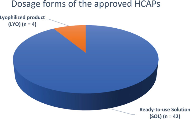
Dosage forms of the approved HCAPs (n = 46).
Among the 46 HCAPs reviewed here, the most prominent type of dosage form is the Ready-to-Use Solution (SOL) (n = 41), and only four products are marketed as Lyophilized solid (LYO) (n = 4).
The four products mentioned above, Cimzia® (certolizumab pegol), Cosentyx® (secukinumab), Nucala® (mepolizumab) and Xolair® (omalizumab), can technically be classified as both LYO and SOL, since both forms of these molecules are approved. The formulation compositions of the SOL dosage forms for each of these four products are unique and slightly different from their corresponding LYO products. This observation signifies the importance of rational formulation development and excipient selection based on the dosage form being developed. A Cimzia® for injection (LYO) vial upon reconstitution provides 200 mg/mL certolizumab pegol and contains lactic acid, polysorbate and sucrose. Cimzia® injection (SOL), supplied as a solution in a single-dose prefilled syringe, delivers 200 mg/mL certolizumab pegol and contains sodium acetate and sodium chloride. It is noteworthy that lactic acid is used as a buffering agent for the LYO product, whereas the SOL product is buffered with sodium acetate. Likewise, sucrose is used as a lyoprotectant and tonicity agent in the LYO product, whereas the SOL product uses sodium chloride as a tonicity agent and does not contain a sugar stabilizer. While the LYO product contains polysorbates, the SOL product does not contain surfactants.
Cosentyx® injection (SOL), supplied in a Sensoready pen or a prefilled syringe, contains 150 mg of secukinumab formulated with L-histidine/histidine hydrochloride monohydrate, L-methionine, polysorbate 80, trehalose dihydrate, and sterile water for injection. Cosentyx® injection is also available as a 75 mg/0.5 mL prefilled syringe and has an identical composition to the 150 mg dose prefilled syringe. Cosentyx® for injection (LYO) contains 150 mg of secukinumab formulated with L-histidine/histidine hydrochloride monohydrate, polysorbate 80, and sucrose. The formulation compositions of the SOL and LYO dosage forms of Cosentyx® are different. While both dosage forms are buffered with a histidine buffer and contain polysorbate 80 as a surfactant, sucrose is the lyoprotectant in LYO and SOL includes trehalose as a stabilizer. The SOL contains L-methionine, which is a common antioxidant in liquid protein formulations.
Nucala® (mepolizumab) for injection LYO is supplied in a single-dose vial and contains 100 mg mepolizumab, polysorbate 80, sodium phosphate dibasic heptahydrate, and sucrose. Whereas Nucala® (mepolizumab) injection SOL dosage form is supplied in a PFS or AI and contains 100 mg/mL mepolizumab, citric acid monohydrate, ethylenediaminetetraacetic acid (EDTA), disodium dihydrate, polysorbate 80, sodium phosphate dibasic heptahydrate, and sucrose. While the LYO product is buffered with phosphate buffer (pH = 7.0), the SOL product is buffered with citrate-phosphate buffer (pH = 6.3). Unlike the LYO product, the SOL product also contains a chelator (EDTA).
Xolair® (omalizumab) for injection of LYO dosage form is stabilized with L-histidine, L-histidine hydrochloride monohydrate, polysorbate 20, and sucrose. Xolair® injection SOL dosage form is stabilized with L-arginine hydrochloride, L-histidine, L-histidine hydrochloride monohydrate, and polysorbate 20. While sucrose is used as a lyoprotectant in the LYO dosage form, the SOL dosage form does not contain any sugar and is stabilized using L-arginine hydrochloride, which is an effective viscosity reducing agent.
It should be noted that all the dosage forms in this review are single use products and do not contain antimicrobial preservatives. Antimicrobial preservatives are known to have deleterious effects on protein stability and potentially induce aggregation.68–70
Protein concentration
The observed protein concentrations for the 46 HCAPs are shown in Table S1. As discussed above, Cimzia®, Cosentyx®, Nucala®, and Xolair® are all approved and marketed as both SOL and LYO products. In Table S1, the protein concentration (mg/mL) for both LYO and SOL dosage forms of these four products is shown only once since the protein concentration for both SOL and LYO products is identical. Dupixent®, Emgality®, Kevzara®, and Repatha® HCAPs are marketed and approved in two different product strengths (protein concentrations), hence in Table S1, the protein concentration for each of these strengths is taken into consideration. Dupixent® has two approved product strengths (150 and 175 mg/mL), Emgality® has two approved product strengths (110 and 120 mg/mL), Kevzara® has two approved product strengths (131.6 and 175 mg/mL), Repatha® has two approved product strengths (120 and 140 mg/mL). Among the 46 total products reviewed here, 25 products had protein concentration in the range of 100–125 mg/mL, 17 products were in the 126–150 mg/mL range, 2 products were in the 151–175 mg/mL range, 1 product was in the 176–199 mg/mL range, and 5 products had protein concentration of 200 mg/mL. Benlysta®, Cimzia® LYO and SOL, Hizentra® and Xembify® all have a protein concentration of 200 mg/mL. Based on our findings, 200 mg/mL is the highest protein concentration achieved among currently commercialized HCAPs.
Table 1.
List of excipients generally used in protein formulations and their functions.
| Excipients | Function | Examples |
|---|---|---|
| Buffer | Maintain stable pH environment | Histidine, Phosphate, Acetate, Citrate, etc. |
| Sugar and polyol stabilizers | Conformational stabilizer for the antibody | Disaccharides – Sucrose, Trehalose, Maltose. Polyols – Sorbitol, Mannitol |
| Tonicity Agent | Maintain iso-tonicity | Sucrose, NaCl, Trehalose, Mannitol |
| Surfactant | Reduce interfacial stress | Polysorbate 20 and 80, Poloxamer 188 |
| Amino acid stabilizers | Stabilize the antibody by buffering or charge interactions or blocking hydrophobic interactions | Arginine, Proline, Glycine, Histidine, Methionine |
| Chelating agent | Prevent metal induced degradation | DTPA, EDTA |
| Viscosity Modifiers | Reduce solution viscosity | Arginine, Proline, Glycine, NaCl |
| Antioxidants | Inhibit oxidation | Methionine |
Formulation composition and key aspects of stabilization
The different types of excipients typically used in protein formulation, their function, and some representative examples of each class of excipients are shown in Table 1. We identified seven important classes of excipients, namely tonicity-modifying agents, sugar and polyol stabilizers, buffer, surfactant, amino acid stabilizers, viscosity modifiers, chelators, and antioxidants. These classes of excipients and observed trends for their use in stabilization of HCAPs are discussed below.
Tonicity-modifying agents
Before discussing tonicity agents, it is important to first understand the definition of, and the difference between, tonicity and osmolality. As described by Strickley and Lambert,1 “Tonicity involves non-permeable molecules and is a measure of the osmotic pressure gradient between two solutions separated by a permeable membrane. Osmolality involves both non-permeable molecules and permeable molecules and is a measure of the osmotic pressure of a solution.” To make a solution isotonic with human blood before injection, tonicity agents, such as disaccharides (sucrose, trehalose), polyols (sorbitol, mannitol), and sodium chloride, are added to the formulation to achieve tonicity close to the osmolality of blood (300 mOsm/kg).1 As seen in Figure 6, sucrose, which is in 18 of 46 products, is the most frequently used tonicity agent for HCAP formulation. Trogarzo® and VabysmoTM contain sodium chloride, in addition to sucrose. Trehalose, another disaccharide, is found in five products. The polyols sorbitol and mannitol were found in four and two products, respectively. Sodium chloride is found in four products. Thirteen of the products do not contain a disaccharide, polyol, or sodium chloride as tonicity agent, but some of these products contain amino acids L-arginine, glycine, and L-proline, which might function as tonicity agents. Xolair® (omalizumab SOL dosage form), Enspryng® (satralizumab), Aduhelm® (aducanumab-avwa), Evkeeza® (evinacumab-dgnb), Actemra® (tocilizumab), and Hemlibra® (emicizumab-kxwh) do not contain disaccharide, polyol, or sodium chloride, but instead contain L-arginine or L-arginine hydrochloride. Arginine is a basic amino acid that is widely used in biopharmaceutical processing, formulation, and stabilization of biotherapeutics. It finds application as a stabilizer during refolding of proteins and as an inhibitor of protein aggregation.71 While the most common application of arginine in formulating HCAPs is inclusion as a viscosity reducing agent, it can also act as a tonicity agent. Polyclonal antibody products Gammagard Liquid®, Hyqvia®, Hizentra® and Xembify® do not contain sugars or sodium chloride as tonicity adjusting agents, but contain the amino acids glycine or proline, which are known to act as tonicity agents and stabilizers.72,73 Gammagard Liquid®, Hyqvia®, and Xembify® contain glycine, whereas Hizentra® contains proline. Siliq™ (rodalumab) and Repatha® (evolocumab) contain proline as a tonicity agent. Siliq™ is formulated in glutamate buffer with proline and polysorbate 20, while Repatha® is formulated in acetate buffer with proline and polysorbate 80.
Figure 6.
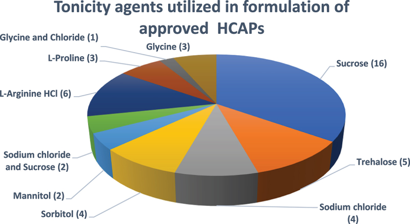
Tonicity agents utilized in formulation of approved HCAPs (n = 46).
The number of products that contain the tonicity agent (n) in parenthesis and the total number of products is 46.
Among the 46 HCAPs, sucrose is the most frequently utilized tonicity agent for HCAP formulation and found in 18 of 46 products. Trehalose can be found in five products. The polyols sorbitol and mannitol were found in four and two products, respectively. Sodium chloride is found in 4 products. Thirteen of the products do not contain a disaccharide, polyol, or sodium chloride as tonicity agent, but some of these products contain amino acids L-arginine, glycine, and L-proline.
Sugar and polyol stabilizer
As seen in Figure 7, among the 46 HCAPs we reviewed, 18 products contain sucrose, 5 contain trehalose, 4 contain sorbitol and 2 contain mannitol. Seventeen products do not contain a sugar or polyol stabilizer. Sucrose is the most commonly used excipient because it is not only a tonicity agent but also a protein conformational stabilizer.74–77 Disaccharides (sucrose, trehalose, and maltose) and polyols (sorbitol, mannitol) are commonly employed in protein stabilization since they act as conformational stabilizers, primarily stabilizing the protein by the preferential exclusion mechanism.78,79 For example, sucrose, sorbitol and trehalose are increasingly used as stabilizers in HCAP development.80–83 While these disaccharides and polyols can be used in HCAPs, their physicochemical properties, crystallization, and their behavior in protein solutions during typical biopharmaceutical process unit operations should be carefully assessed. One such example of biopharmaceutical unit operation is freezing of biological drug substances at large scale, which enables manufacturing flexibility and storage of the material for a longer duration, thereby allowing scheduling of manufacturing campaigns and improved management of the drug product supply chain. Trehalose,63,64,84 sorbitol,82 and mannitol65,66 are known to crystallize in protein solutions during freezing and can adversely affect the stability of proteins; therefore, their use in an HCAP formulation and stabilization should be carefully evaluated. Connolly and colleagues identified an optimal range of trehalose-mAb (w/w) ratio of 0.2–2.4%, capable of physically stabilizing mAb formulations during long-term frozen storage.63 Sundaramurthi and Suryanarayanan studied the crystallization behavior of trehalose in the presence of 1) a crystallizing (mannitol) solute, 2) a non-crystallizing (sucrose) solute, and 3) a combination of mannitol and a model protein.67 Their findings indicate that mannitol, by readily crystallizing as a hemihydrate, accelerates trehalose dehydrate crystallization. However, by remaining amorphous, sucrose completely inhibits trehalose dehydrate crystallization. These studies indicate that while crystallization of disaccharides or polyols can be a concern, this can be easily mitigated by using higher protein to sugar ratios or by incorporating amorphous excipients like sucrose in the formulation. One of the major advantages of sucrose is that it does not crystallize and remains amorphous during freezing, making it the most widely accepted and used stabilizer in both LYO and SOL dosage forms of HCAPs.
Figure 7.
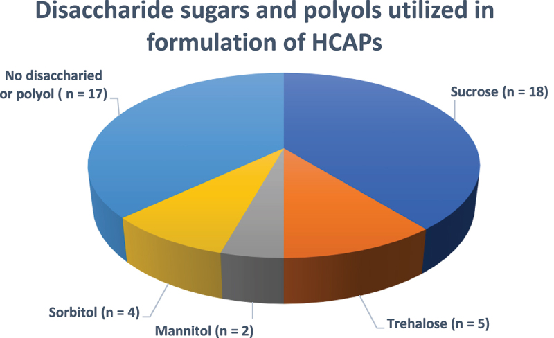
Disaccharide sugars and polyols utilized in formulation of approved HCAPs (n = 46).
The number of products that contain the disaccharide or polyol (n) in parenthesis and the total number of products is 46.
Among the 46 reviewed HCAPs, 18 products contain sucrose, 5 contain trehalose, 4 contain sorbitol and 2 contain mannitol. Seventeen products do not contain a sugar or polyol stabilizer.
Buffer and formulation pH
When considering any mAb formulation, the pH range of the solution is vital to formulation since proteins are stable against chemical modifications and aggregation over a narrow pH range.7 For example, a lower pH could lead to protein degradation and cleavage, while a high pH could cause protein deamidation. It has been shown that even brief exposure of an IgG1 protein outside of optimal pH can cause small amounts of aggregation to occur.25 Stabilizing buffers for mAbs should be rationally selected, as reactions leading to physicochemical instability are primarily driven by pH and ionic strength of the buffer.6 Buffers play a vital role in mAb formulation because they not only affect storage stability of the mAb but also the length of SC absorption after administration. Additionally, both the type and the concentration of the buffer can affect protein stability, thereby signifying the importance of choosing the most optimal buffer composition.6 For example, one study showed that an increase in buffer tonicity greatly increased the level of SC absorption, especially for neutral excipient such as mannitol. Other tonicity agents, such as sodium chloride, had a very minimal effect on SC absorption due to fast distribution upon SC injection.85 Among commercial antibody formulations, the buffer concentration is usually between 0.003 M and 0.10 M for SC administration, with the highest buffer concentrations administered via the IM route in Synagis, at 0.025 M (histidine buffer).1
Figure 8 shows the prevalence of different buffers in the formulation of the 46 HCAPs we reviewed. Histidine, individually or in combination with other amino acids or inorganic buffers, is the most prevalent buffer system used to stabilize HCAPs (n = 32). Among these 32 products, 25 use only histidine as the buffer system. Enspryng® (satralizumab) and Hemlibra® (emicizumab-kxwh) are buffered using a combination of histidine and aspartic acid. Takhzyro® is buffered using citrate phosphate and histidine, while VabysmoTM (faricimab-svoa) and Dupixent® (dupilumab) use a histidine and acetate buffer system. Cimzia® (SOL dosage form of certolizumab pegol), Aimovig® (erenumab-aooe), and Repatha® (evolocumab) are buffered using acetate. Beovu® (brolucizumab-dbll) is buffered with sodium citrate. Orencia® (abatacept) and Nucala® (mepolizumab) LYO dosage forms use a phosphate buffer, and Nucala® (mepolizumab) SOL dosage form is buffered with citrate phosphate. Lactic acid buffers the LYO dosage form of Cimzia® (certolizumab pegol), whereas glutamate buffers Siliq™ (rodalumab). As noted in the section on tonicity agents, the polyclonal antibody products Gammagard Liquid®, Hyqvia®, and Xembify® are buffered with glycine, whereas Hizentra® contains proline. It is important to note that Humira® (adalimumab) does not contain any buffer and hence can be termed the only “buffer-free” mAb product. This product contains mannitol, polysorbate 80, and sterile water for injection besides the active adalimumab and is distinctly different from the initial launch version of Humira®. The initially approved and marketed Humira® was a lower concentration product (50 mg/mL) that used a citrate phosphate buffer. Buffer-free or self-buffering of high concentration antibody solutions has been extensively studied,86–88 and findings suggest that at high concentration, the antibody itself can buffer the solution, thereby eliminating the need for inorganic or amino acid-based buffers.
Figure 8.
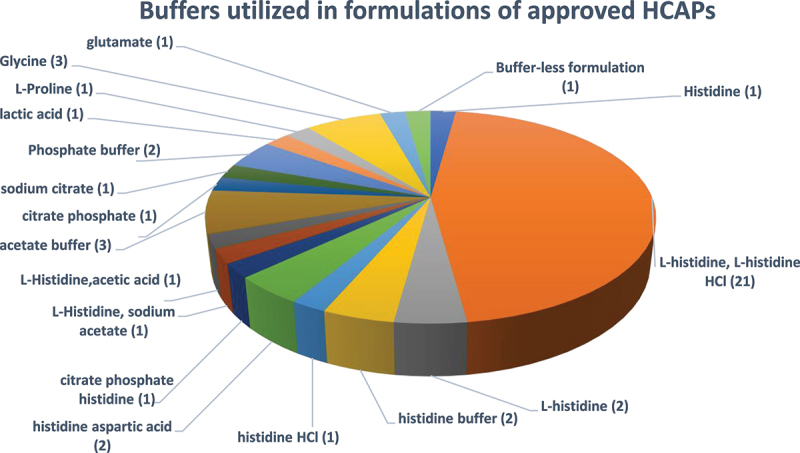
Buffers utilized in formulations of approved HCAPs (n = 46).
The number of products that contain a buffering agent (n) in parenthesis and the total number of HCAPs is 46.
Figure shows the prevalence of different buffers in the formulation of the 46 HCAPs, where Histidine, individually or in combination with other amino acids or inorganic buffers, is the most prevalent buffer system used to stabilize HCAPs (n = 32). In addition, glycine (n = 3), glutamate (n = 1), L-Proline (n = 1), lactic acid (n = 1), phosphate buffer (n = 2), acetate buffer (n = 3), citrate buffer, and its combination (n = 2) have been used as alternate buffers. Formulation of 1 product is bufferless.
Histidine is the most commonly used buffer because it has a buffering capacity in the pH range of 5.5 to 7.0 and does not have destabilizing effects on mAbs. Unlike other biopharmaceutical buffers, such as sodium phosphate and tris-hydrochloride, histidine buffers do not exhibit significant pH shifts during freezing,62 making it equally suitable for both SOL and LYO dosage forms. Moreover, histidine is an inexpensive and cost-effective buffer.89 While histidine is an inert buffer, some studies report its degradation in a biologics formulation buffer. Wang and colleagues report the investigation of a degradant in a biologics formulation buffer containing L-histidine.90,91 As described in this study, histidine can degrade to urocanic acid. Histidine degradation was slightly increased by Mn+2. The chelating agents, EDTA, and diethylenetriaminepentaacetic acid (DTPA) counteracted the Mn+2 effects. This degradation is thought to be caused by microbial contamination of the formulation buffer. Adding alanine or cysteine as an excipient was found to reduce this degradation by 97% and 98%, respectively. Although histidine is a widely used buffer for HCAP, its stability in development and manufacturing should be appropriately controlled.
The pH range of the HCAP formulations is depicted in Figure 9. Among the 46 reviewed HCAPs, 6 products have pH in the 4.5–5.0 range, 12 products have pH in the range of 5.1–5.5, 21 products have pH in the range of 5.6–6.0, four products have pH in the range of 6.1–6.5, one product has pH in the range of 6.6–7.0, and two have pH in the 7.1–7.5 range. As expected, most antibodies are stabilized in the pH range of 5–6. One of the critical aspects of stabilization of antibodies in liquid formulations is the control of asparagine deamidation and aspartate isomerization, especially if the residues are in the CDRs of mAbs.92,93 The rate limiting step in both deamidation and isomerization reactions at physiological pH is the formation of the Asp intermediate, which involves the attack by nitrogen of the peptide backbone on the carbonyl carbon of the asparagine or the aspartate side chain. Asparagine deamidation is promoted at neutral pH, whereas aspartate isomerization is promoted at slightly acidic pH ≤ 5.0. Hence, most antibody formulations are stabilized in the pH range of 5–6 to control these chemical modifications, thereby achieving optimal stability and a marketable product with a shelf life of at least 24 months.87
Figure 9.
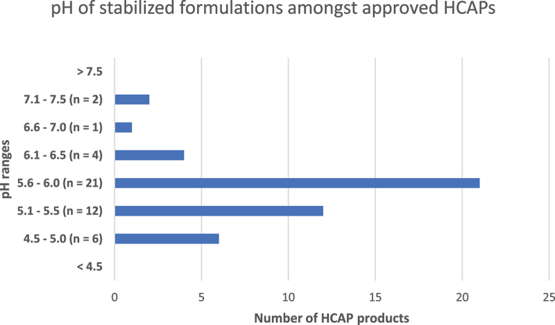
pH of stabilized formulations amongst approved HCAPs (n = 46).
Among the 46 reviewed HCAPs, 6 products have pH in the range of 4.5–5.0, 12 products have pH in the range of 5.1–5.5, 21 products have pH in the range of 5.6–6.0, four products have pH in the range of 6.1–6.5, one product has pH in the range of 6.6–7.0, and two have pH in the range of 7.1–7.5.
Surfactants
The main function of surfactants is to minimize surface adsorption and adhesion to packaging surfaces or containers, reducing aggregation and particle formation. Surfactants primarily act by reducing the interfacial stress that the antibody would typically undergo during various drug product manufacturing unit operations, such as filtration, mixing, filling, and during shipping of the finished drug product between manufacturing site, storage site, and the health-care sites (e.g., pharmacies, hospitals, and home). Additionally, surfactants can be used to reduce mechanical stress and decrease protein–protein interactions, which could lead to aggregation.7 Only three surfactants are currently used in the marketed antibody formulations: polysorbate 20, polysorbate 80, and poloxamer 188.1 As seen in Figure 10, among the 46 HCAPs reviewed here, polysorbate 80 was the most used surfactant (n = 28), followed by polysorbate 20 (n = 10) and poloxamer 188 (n = 3). Cimzia® (LYO dosage form of certolizumab-pegol) contains polysorbate (the type of polysorbate is not described in prescription information). Four products, Synagis®, Cimzia® (SOL dosage form of certolizumab-pegol) and the polyclonal antibody products Gammagard Liquid® and Hyqvia®, do not contain any added surfactant.
Figure 10.
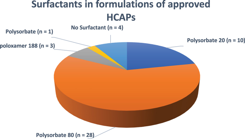
Surfactants in formulations of approved HCAPs (n = 46).
The number of products that contain a surfactant (n) in parenthesis and the total number of HCAPs is 46.
Among the 46 HCAPs reviewed here, polysorbate 80 was the most used surfactant (n = 28) followed by polysorbate 20 (n = 10) and poloxamer 188 (n = 3). Four products have no surfactant.
While polysorbates 20 and 80 are widely used in stabilizing HCAPs, they are chemically diverse mixtures and may degrade through oxidation and hydrolysis pathways.94 The hydrolysis pathway can be either chemically induced or enzymatically catalyzed. Several recent reviews have discussed the chemical structure of polysorbates, their degradation,95,96 analytical techniques for identifying degradation products,97 and control strategies for polysorbates in biopharmaceuticals.98 Since polysorbate degradation may inadvertently affect the quality, efficacy, safety, and stability of the protein formulation, comprehensive development and characterization studies should be performed during pharmaceutical development. In a high-throughput screening study of excipients, poloxamers were shown to be superior when compared to polysorbates at protecting Antigen 18A aggregation at air–liquid interface.99 A recent study compared polysorbates and poloxamer 188 in their abilities to stabilize several mAb formulations; while poloxamer 188 was found to offer protection against interfacial stress in liquid formulations, visible protein-polydimethylsiloxane (silicone oil) particles were observed in some protein formulations using poloxamer 188 during long-term storage at 2–8°C.100 The results of this study reiterate the importance of comprehensive development and characterization studies to be performed during screening and final selection of surfactant during pharmaceutical development of each product.
Amino acids as stabilizers
In addition to the widespread use of histidine as a buffer, amino acids can also act as tonicity agents (e.g., glycine and proline), protein stabilizers (e.g., arginine), antioxidants (e.g., methionine), and viscosity reducing agents (e.g., arginine) in antibody formulations.1,72,73 Figure 11 depicts the 81 instances when an amino acid was used in stabilizing the 46 reviewed HCAPs. As shown, L-histidine was used in 30 products, L-histidine hydrochloride monohydrate was used in 23 products, L-methionine was used in 8 products, L-arginine hydrochloride was used in 6 products, glycine and L-proline were both used in 4 products each, L-arginine was used in 3 products, L-aspartic acid in 2 products (Enspryng® and Hemlibra®) and L-lysine hydrochloride was used in Saphnelo®.
Figure 11.
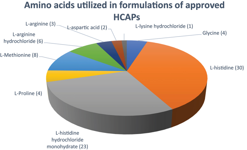
Amino acids utilized in formulations of approved HCAPs (n = 46).
Number of products that contain an amino acid (n) in parenthesis.
Among 46 HCAPs, L-histidine was used in 30 products, L-histidine hydrochloride monohydrate was used in 23 products, L-methionine was used in 8 products, L-arginine hydrochloride was used in 6 products, glycine and L-proline both were used in 4 products each, L-arginine was used in 3 products, L-aspartic acid in 2 products, and L-lysine hydrochloride was used in 1 product.
We further evaluated how many amino acids, or their salts, are used in each of the 46 HCAPs. As shown in Figure 12 among the 46 HCAPs, 9 products do not use any amino acid for stabilization of the formulation. These products are Orencia®, Cimzia® (LYO and SOL dosage forms), Praluent®, Nucala® (LYO and SOL dosage forms), Beovu®, Humira®, and Aimovig®. These products are stabilized by sugars, non-amino acid buffers (e.g., acetate and phosphate), and surfactants. Another nine products use one amino acid to stabilize the formulation; the polyclonal antibody products GammagardLiquid®, Hyqvia®, and Xembify® contain glycine as the only amino acid, whereas Hizentra® contains proline as the only amino acid stabilizer. Based on the prescription information, glycine and proline are claimed as a buffer and stabilizer in the polyclonal antibody products. Repatha® and Siliq™ contain proline as the only amino acid stabilizer, Anthim® and Takhzyro® contain histidine as the only amino acid stabilizer and Susvimo™ contains histidine hydrochloride as the only amino acid stabilizer.
Figure 12.
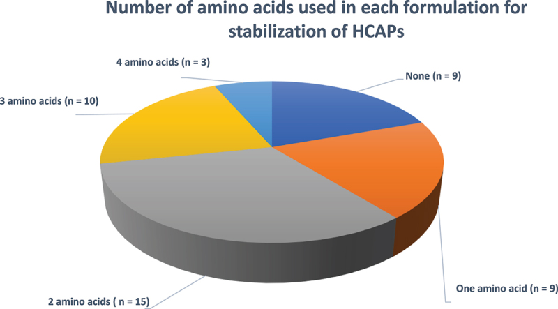
Number of amino acids used in each formulation for stabilization of HCAPs (n = 46).
Among the 46 HCAPs, 9 products do not utilize any amino acid for stabilization of the formulation. These products are stabilized using sugars, non-amino acid buffers (e.g., acetate and phosphate), and surfactants. Another nine products utilize one amino acid to stabilize the formulation
A total of 15 products include two amino acids (and their respective salts) in the formulation composition, wherein the most common combination of amino acids and their salt used is L-histidine (base) and L-histidine hydrochloride (11 products). Synagis® uses the combination of glycine and histidine amino acids, Dupixent® contains L-histidine and L-arginine hydrochloride, Kevzara® includes the combination of histidine and arginine, and VabysmoTM contains L-histidine and L-methionine. It should be noted that VabysmoTM also contains acetic acid as a buffering agent besides L-histidine. Ten products use two amino acids (and their respective salts) in the formulation composition to stabilize the product. Cosentyx® (SOL dosage form), Rituxan Hycela®, Darzalex Faspro®, Herceptin Hylecta®, and Phesgo® utilize L-methionine in addition to the histidine–histidine HCl buffer. The probable role of L-methionine in these formulations is of an antioxidant. Xolair® (SOL dosage form) and Benlysta® include L-arginine HCl in addition to the histidine–histidine HCL combination, wherein the arginine likely acts as a viscosity reducing agent. Enspryng® and Hemlibra® use L-arginine in addition to the histidine-aspartic acid buffer combination. It is important to note that neither of these products contain any sugar stabilizer. Saphnelo® includes L-lysine hydrochloride in addition to the histidine–histidine HCl buffer.
Finally, three products, Aduhelm®, Evkeeza®, and Actemra®, use a total of 4 amino acids (and their respective salts) as stabilizers in the HCAP formulation. Aduhelm® and Actemra® contain L-methionine and L-arginine HCl in addition to the histidine and histidine HCl combination. Evkeeza® contains L-proline and L-arginine HCl in addition to the histidine base and histidine HCl combination. It is important to note that all three products contain only amino acids (and their respective salts) with a surfactant; they do not contain sodium chloride or sugar (or polyol) as stabilizer. This indicates that a stable HCAP can be achieved by using a rationally selected amino acid combination.
Viscosity reducing agents
Viscosity is a critical attribute to consider when developing an HCAP. When the antibodies are formulated as concentrated solutions (>100 mg/mL), the viscosity of the solutions is expected to increase exponentially beyond the generally pharmaceutically acceptable limit of 20cP for SC injections due to the short-range protein–protein interactions. Weakening of such protein–protein interactions by using co-solutes, thereby enhancing the stability and reducing viscosity of a high concentration mAb solution, is an acceptable development strategy.101 Factors affecting the viscosity in high concentration solutions of mAbs have been extensively studied.102–110 Amino acids in their salt forms and several common salts, such as arginine HCl, histidine HCl, lysine HCl, sodium chloride, sodium sulfate, and sodium acetate, could potentially be used as viscosity-lowering excipients in high concentration mAb formulations.111 Amino acids arginine and lysine were found to reduce the high viscosity of serum albumin solutions for pharmaceutical injection.112 Similarly, the viscosities of the bovine gamma globulin solution (250 mg/mL) and the human gamma globulin solution (292 mg/mL) at physiological pH were reduced to <50 cP by using 1 M arginine HCl.113 In one study, proline was found to be more effective than a combination of glycine and trehalose at reducing the viscosity of high concentration antibody formulation at a protein concentration of ~225 mg/mL.114 Uncharged amino acids, such as glycine, alanine, phenylalanine, and tryptophan can modify the hydrophobic interactions and possibly reduce viscosity.115,116 Among the reviewed 46 HCAPs, we found a total of 23 instances wherein viscosity reducing agents (i.e., arginine and its hydrochloride salt, proline, lysine hydrochloride, glycine, and sodium chloride) were used (Figure 13). The most prevalent viscosity reducing agent was arginine (n = 9), used as arginine HCl (six products) and arginine (three products). Sodium chloride is used in four products (Cimzia® SOL dosage form, Emgality®, Trogarzo®, and Benlysta®). It should be noted that, while sodium chloride in protein formulations serves primarily as a tonicity agent, it can also function as a viscosity reducing agent. The definite role of sodium chloride in each of these compositions is not disclosed in the product insert, but we infer that it may also function as a viscosity lowering agent. In addition to sodium chloride, Benlysta® also contains arginine HCl. Proline is used in four products (Hizentra®, Repatha®, Siliq™, and Evkeeza®). Besides proline, Evkeeza® contains arginine HCl, Saphnelo® contains L-lysine HCl. Finally, glycine is used as a buffering and stabilizing agent in four products (Synagis®, and three polyclonal antibodies Gammagard Liquid®, Hyqvia®, and Xembify). While it cannot be verified if glycine acts as a viscosity reducing agent in these products, we have listed it here since prior reviews have identified glycine as a probable viscosity lowering agent.1
Figure 13.
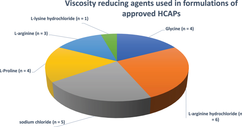
Viscosity reducing agents used in formulations of approved HCAPs (n = 23).
The number of products that contain a viscosity reducing agent (n) in parenthesis.
Among the reviewed 46 HCAPs, there are 23 instances wherein viscosity reducing agents (i.e., arginine and its hydrochloride salt, proline, lysine hydrochloride, glycine, and sodium chloride) were used.
Chelators and antioxidants
When considering the long-term stability of antibody solutions, chemical modifications, such as oxidation, must be considered. Oxidation is prevalent mainly because it can occur with or without a stimulus, the latter being referred to as auto-oxidation.7 The most commonly oxidizable amino acids in antibodies include Met, Tyr, Trp, His, and Cys.6 To mitigate oxidation, chelators, such as EDTA or DTPA,117,118 and amino acids, such as methionine,119–121have been added to stabilize protein solutions. The mechanism of stabilization differs, where chelators chelate metal ions in solution, and methionine acts as a sacrificial antioxidant. Among the HCAP formulations reviewed here, Nucala® (SOL dosage form) and Ajovy® use disodium EDTA as a chelator. A total of seven products contain L-methionine, including Cosentyx® (SOL dosage form), Aduhelm®, Actemra®, and four antibody products co-formulated with recombinant human hyaluronidase PH20 enzyme (rHuPH20; Halozyme Therapeutics, Inc.). These four products are Rituxan Hycela® (rituximab and hyaluronidase), Darzalex Faspro® (daratumumab and hyaluronidase-fihj), Herceptin Hylecta® (trastuzumab and hyaluronidase) and Phesgo® (pertuzumab, trastuzumab, and hyaluronidase-zzxf). It is worth mentioning that Nucala® (SOL dosage form) and Cosentyx® (SOL dosage form) contain EDTA and methionine, respectively, which are not found in their respective LYO dosage form products.
Primary containers (vial vs prefilled syringe vs auto injector or prefilled pen)
The three types of primary packaging configurations that are typically used for mAb drug products are as follows: 1) glass vials, 2) glass PFS, and 3) glass cartridges. Non-glass polymeric materials, such as cyclic olefin polymer, have also been recently used for biologics requiring ultra-low temperature storage. For example, West’s Daikyo Crystal Zenith® (CZ) vials (cyclic olefin polymer) are used for packaging of Imlygic™ (talimogene laherparepvec). CZ vials offer a break-resistant, high-performance alternative to glass for complex drugs. Biologics manufacturers have effectively used PFS as a primary packaging configuration.122 The PFS offers some unique advantages compared to vials, wherein the drug can be administered directly by the patient or the caregiver with minimal drug preparation and manipulation prior to administration. PFS also offers the advantage of requiring fewer drug overages as compared to vials and minimizing drug wastage. Several PFS products also have an added safety device that protects the caregiver from accidental needle stick injury. Additionally, PFS has been further developed into AI, thereby offering some unique administration advantages. While the AI or PFS offer convenient administration and delivery options for the drug, the primary packaging in such systems is still a glass container. Glass vials are typically made of USP Type 1 glass. Although glass containers are inert and generally provide a stable environment for biologics storage, there have been issues with glass vials during long-term storage, resulting in product recalls due to glass delamination that leads to the appearance of glass particles in solution.123 Glass delamination can occur when thin layers of glass become detached from their container and leach into solution. Since certain formulation buffers and their pH can accelerate glass delamination, the selection of primary packaging components becomes an extremely critical aspect of successful parenteral drug product development.
One final aspect when considering primary packaging components for biologics drug product is the type of rubber stopper used. Generally, polymer-coated rubber stoppers, which are known to be inert toward biologics and do not induce instability or protein aggregation, are used. While this may seem unlikely, the material used by the rubber stopper can leach into the drug solution and contribute toward product immunogenicity. The above discussion highlights the importance of rational selection of primary packaging components for storage of parenteral product.
Two main types of prefilled syringes that can be used for biologic drug products are the staked needle syringe in which the injection cannula is already pre-attached, and the Luer Lock syringes in which the user attaches a hypodermic needle prior to injection. Staked needle syringes are popular, as the needle is attached and does not require much preparation prior to dosage administration. To efficiently deliver the drug solution, the PFS barrels are siliconized. As silicone oil-inducing protein aggregation and subvisible particle formation in biologics solutions have been reported,124–127 appropriate development studies should be performed to evaluate the sensitivity of each biologic drug to silicone oil.127,128
Of the 46 HCAP products reviewed here, the most prevalent primary packaging configuration was glass vials (n = 25) (Figure 14). One product is marketed in both a vial and PFS presentation, and a total of 20 products are marketed in PFS presentations. While vials might be most prevalent, PFS packaging has become increasingly popular in recent times. Among the 20 products marketed in PFS presentation, 5 products are marketed only as PFS, 10 products are marketed as both PFS and AI presentations, and 4 products are marketed as both PFS and prefilled pen. For both AI and prefilled pens, the primary packaging is still a PFS. Repatha® is marketed as PFS, AI, and a prefilled cartridge, which is administered using an on-body infuser (Pushtronex® system). The higher prevalence of glass vials as primary packaging components for HCAPs is logical because glass vials can be used for both LYO and SOL formulations. PFS are certainly gaining popularity as primary packaging components because of the unique at-home self-administration advantage they offer, thereby ensuring treatment flexibility, patient compliance, and positive treatment outcomes.
Figure 14.
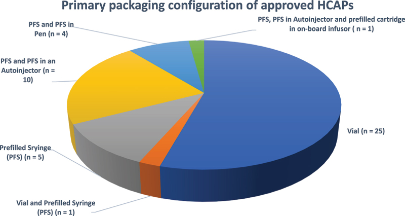
Primary packaging configuration of approved HCAPs (n = 46).
The primary packaging configuration for each of the HCAP, number of products (n) in parentheses, and the total number of HCAPs is 46.
Subcutaneous delivery of large volume HCAPs using hyaluronidase
ENHANZE® drug delivery technology enables the SC delivery of >2 mL doses of therapeutic mAbs. This technology is based on the use of a proprietary recombinant human hyaluronidase PH20 enzyme (rHuPH20; Halozyme Therapeutics, Inc.).129–131 Among the HCAP products we review here, Rituxan Hycela® (rituximab and hyaluronidase), Darzalex Faspro® (daratumumab and hyaluronidase-fihj), Herceptin Hylecta® (trastuzumab and hyaluronidase), and Phesgo® (pertuzumab, trastuzumab, and hyaluronidase-zzxf) are co-formulated with rHuPH20. Hyqvia® (Immune Globulin Infusion 10% (Human) with Recombinant Human Hyaluronidase) is supplied as a dual vial unit, with one vial for Immune Globulin Infusion 10% (Human) and one vial for hyaluronidase. The Immune Globulin Infusion 10% is to be sequentially administered subcutaneously following an initial administration of the hyaluronidase. With several products already approved, it may not be surprising to see more HCAPs adapting to the ENHANZE drug delivery technology and co-formulation of the mAbs with hyaluronidases.
Fixed dose combinations
The introduction of FDC therapies, which contain two or more protein drugs in a specific product presentation, has contributed to the evolution of the mAb formulations landscape. This type of dosing regimen has two main advantages: 1) it is less complicated, and 2) it eliminates the need for dose-calculation based on body weight, thereby reducing errors in dosage. Among the HCAPs reviewed here, only two are FDC products, REGEN-COV® and Phesgo.
REGEN-COV® (casirivimab and imdevimab), which targets SARS-CoV-2 spike proteins in patients infected with the virus. The FDA issued an EUA for this product. REGEN-COV® (co-formulated casirivimab and imdevimab) is supplied as 600 mg/600 mg per 10 mL (60 mg/60 mg per mL) in a vial. Individual packages are also available for casirivimab and imdevimab, which are supplied as 1,332 mg/11.1 mL (120 mg/mL) or 300 mg/2.5 mL (120 mg/mL) for each of the antibodies. The FDC and individual antibodies can be given by IV infusion or SC administration. The SC administration of the co-formulated casirivimab and imdevimab involves administering 2.5 mL of the formulation using 4 different syringes prepared from the vial.
Phesgo (pertuzumab, trastuzumab, and hyaluronidase-zzxf) injection is another FDC, which is supplied in two product presentations. The first product contains 1200 mg pertuzumab, 600 mg trastuzumab, and 30,000 unit hyaluronidase/15 mL (80 mg, 40 mg, and 2,000 units/mL) solution in a single-dose vial. The second product contains 600 mg pertuzumab, 600 mg trastuzumab, and 20,000 unit hyaluronidase/10 mL (60 mg, 60 mg, and 2,000 units/mL) of solution in a single-dose vial. Although only two HCAPs have used this approach so far, the fixed dose approach is a viable option for delivering combination therapeutics.
Conclusion
As discussed below, our review of 46 approved HCAPs has enabled us to draw a number of observations about high concentration antibody drug products.
Formulation excipients and stabilization
Histidine is the most widely used buffer, sucrose is the most widely used tonicity agent and sugar stabilizer, several HCAPs use two or more amino acids as formulation excipients, polysorbate 80 is the most widely used surfactant, arginine is the most widely used viscosity lowering agent, EDTA is used as a chelator in some HCAPs, and some HCAP products use methionine as an antioxidant. All these excipients are used in a unique combination and at distinct concentrations for each drug product to reduce protein aggregation, increase stability, mitigate viscosity, and ultimately provide a stable commercial HCAP.
Amino acids as stabilizers
Several HCAP products use only amino acids (and their respective salts) and surfactants as stabilizers and do not contain sugars or non-amino acid buffers (e.g., Xolair® SOL dosage form, Enspryng®, and Hemlibra®). As many as four different amino acids (and their respective salts) are used in stabilizing some HCAPs (e.g., Aduhelm®, Evkeeza®, and Actemra®). These products also contain only amino acids and surfactants as stabilizers and do not contain any sugars or non-amino acid buffers.
Buffer-free HCAPs
HCAP can be formulated and stabilized without any buffer. Humira® is an example of such a “buffer-free” or “buffer-less” formulation.
Protein concentration
HCAP with a protein concentration as high as 200 mg/mL has been successfully developed and commercialized.
Dosage form
Solution dosage forms are preferred over lyophilized dosage forms for the obvious advantages they offer. Only four HCAPs are LYO and all four of these products are also approved and marketed as SOL dosage forms.
pH of formulation
Most HCAPs are formulated in a pH range of 5–6.
Route of administration
SC administration is the most preferred route.
Primary packaging
The primary packaging configuration also plays a key role in product administration as the SC route relies heavily on at-home administration, explaining the prevalence of the AI and prefilled syringes for administration. Storage is also a large consideration for mAb therapies, hence why the glass vial packaging was the most prevalent packaging configuration.
ENHANZE® drug delivery technology
Several HCAPs utilize ENHANZE® drug delivery technology. This technology is expected to be increasingly used to enable SC delivery of high concentration antibody therapeutics soon.
Fixed dose combinations
Few HCAPs are FDCs. The number of approved FDCs is expected to increase in the future as they enable convenient combination therapy.
There is a tremendous urgency and need for the development and commercialization of mAb products that exceed >200 mg/mL, especially in treating immune-related disorders that require higher doses (>30–50 mg/kg) delivered by SC administration. To achieve higher mAb concentration, high solution viscosity poses a major technical hurdle that needs to be overcome. Substantial research and development efforts are ongoing in both academia and industry to identify and develop novel excipients and FDA-approved excipient combinations to reduce the viscosity of high concentration mAb products. The use and approval of novel excipients in biologics formulations has been slow, but the FDA’s Center for Drug Evaluation and Research has recently launched the voluntary Novel Excipient Review Pilot Program, which is intended to allow excipient manufacturers to obtain FDA review of certain novel excipients prior to their use in drug formulations
Other novel formulation processing technologies include Xeris Pharma’s XeriJect technology and Elektrofi’s particle suspensions technology. Xeris Pharma claims to have developed formulated suspensions with a protein concentration in excess of 400 mg/mL, far exceeding current aqueous formulation systems with maximum achievable protein concentrations of 50–250 mg/mL. Elektrofi claims their technology has been used to produce concentrations greater than 400 mg/mL without exceeding viscosity limits and while preserving the full activity of the biologic. These and other yet-to-be disclosed technologies in development could lead to HCAPs that not only exceed 200 mg/mL product concentrations but also possess other advantages, such as room temperature stability, which would eliminate cold-chain requirements during storage of bulk drug product in smaller containers and transportation of final drug product to clinical sites and pharmacies in smaller container and delivery systems. Such innovative technologies may also lower the overall cost of many therapeutic mAbs that have proven to be lifesaving but expensive to patients.
Supplementary Material
Funding Statement
The author(s) reported that there is no funding associated with the work featured in this article.
Abbreviations
HCAPs – High Concentration Antibody products; QTPP – Quality Target Product Profile. AI – Autoinjector; EUA – Emergency Use Authorization; FDC – Fixed Dose Combination; IV-Intravenous; IM – Intramuscular; mAbs – Monoclonal Antibodies; PFS – Prefilled Syringes; ROA – Route of administration; RTU – Ready to Use; SC – Subcutaneous; USP – United States Pharmacopeia.
Disclosure statement
No potential conflict of interest was reported by the authors.
Supplementary material
Supplemental data for this article can be accessed online at https://doi.org/10.1080/19420862.2023.2205540.
References
- 1.Strickley RG, Lambert WJ.. A review of formulations of commercially available antibodies. J Pharm Sci. 2021;110(7):2590–20. doi: 10.1016/j.xphs.2021.03.017. [DOI] [PubMed] [Google Scholar]
- 2.Shawn Shouye Wang YY, Yan Y(, Ho K. US FDA-approved therapeutic antibodies with high-concentration formulation: summaries and perspectives. Antib Ther. 2021;4(4):262–73. doi: 10.1093/abt/tbab027. [DOI] [PMC free article] [PubMed] [Google Scholar]
- 3.The Antibody Society, Inc . Therapeutic monoclonal antibodies approved or in regulatory review. [accessed April 14 2023]. www.antibodysociety.org/antibody-therapeutics-product-data.
- 4.R-M L, Hwang Y-C, Liu I-J, Lee C-C, Tsai H-Z, Li H-J, Wu H-C. Development of therapeutic antibodies for the treatment of diseases. J Biomed Sci. 2020;27(1). http://www.ncbi.nlm.nih.gov/pubmed/31894001. [DOI] [PMC free article] [PubMed] [Google Scholar]
- 5.Thomas F. Rising to the challenge of biologic drug formulation. Pharm Technol Eur. 2019;31:24–26. [Google Scholar]
- 6.Wang W, Singh S, Zeng DL, King K, Nema S. Antibody structure, instability, and formulation. J Pharm Sci. 2006;96(1):1–26. doi: 10.1002/jps.20727. [DOI] [PubMed] [Google Scholar]
- 7.Le Basle Y, Chennell P, Tokhadze N, Astier A, Sautou V. Physicochemical stability of monoclonal antibodies: a review. J Pharm Sci. 2020;109(1):169–90. doi: 10.1016/j.xphs.2019.08.009. [DOI] [PubMed] [Google Scholar]
- 8.Manning MC, Patel K, Borchardt RT. Stability of protein pharmaceuticals. Pharm Res. 1989;6(11):903–18. doi: 10.1023/A:1015929109894. [DOI] [PubMed] [Google Scholar]
- 9.Manning M, Chou D, Murphy B, Payne R, Katayama D. Stability of Protein Pharmaceuticals: an Update. Pharm Res. 2010;27(4):544–75. doi: 10.1007/s11095-009-0045-6. [DOI] [PubMed] [Google Scholar]
- 10.Wang O, Ohtake S. Science and art of protein formulation development. Int J Pharm. 2019;568:568. doi: 10.1016/j.ijpharm.2019.118505. [DOI] [PubMed] [Google Scholar]
- 11.Wang W. Instability, stabilization, and formulation of liquid protein pharmaceuticals. Int J Pharm. 1999;185(2):129–88. doi: 10.1016/S0378-5173(99)00152-0. [DOI] [PubMed] [Google Scholar]
- 12.Wang W. Lyophilization and development of solid protein pharmaceuticals. Int J Pharm. 2000;203(1–2):1–60. doi: 10.1016/S0378-5173(00)00423-3. [DOI] [PubMed] [Google Scholar]
- 13.Messick S, Saggu M, Ríos Quiroz A. Chapter 1: monoclonal antibodies: structure, physicochemical stability, and protein engineering. In: Messick S, Saggu MRíos Quiroz A. editors. Development of biopharmaceutical drug-device products;2020. p. 3. 10.1007/978-3-030-31415-6_1 [DOI] [Google Scholar]
- 14.Carpenter JF, Chang BS, Garzon-Rodriguez W, Randolph TW. Rational design of stable lyophilized protein formulations: theory and practice. Pharm Biotechnol. 2002;13:109–33. http://www.ncbi.nlm.nih.gov/pubmed/11987749. [DOI] [PubMed] [Google Scholar]
- 15.Jameel F, Hershenson S. Formulation & process development strategies for manufacturing biopharmaceuticals. 2010. doi: 10.1002/9780470595886. [DOI] [Google Scholar]
- 16.Jameel F, Hershenson S, Khan MA, Martin-Moe S, editors. In: Quality by design for biopharmaceutical drug product development. New York: Springer; 2015. p. 1–710. [Google Scholar]
- 17.Shire SJ. Formulation and manufacturability of biologics. Curr Opin Biotechnol. 2009;20(6):708–14. doi: 10.1016/j.copbio.2009.10.006. [DOI] [PubMed] [Google Scholar]
- 18.Rathore N, Rajan RS. Current perspectives on stability of protein drug products during formulation, fill and finish operations. Biotechnol Prog. 2008;24(3):504–14. doi: 10.1021/bp070462h. [DOI] [PubMed] [Google Scholar]
- 19.Wang W, Roberts C. Non-Arrhenius protein aggregation. Aaps J. 2013;15(3):840–51. doi: 10.1208/s12248-013-9485-3. [DOI] [PMC free article] [PubMed] [Google Scholar]
- 20.Roberts CJ. Non-native protein aggregation kinetics. Biotechnol Bioeng. 2007;98(5):927–38. doi: 10.1002/bit.21627. [DOI] [PubMed] [Google Scholar]
- 21.Weiss WF, Young TM, Roberts CJ. Principles, approaches, and challenges for predicting protein aggregation rates and shelf life. J Pharm Sci. 2009;98(4):1246–77. doi: 10.1002/jps.21521. [DOI] [PubMed] [Google Scholar]
- 22.Vázquez‐rey M, Lang DA. Aggregates in monoclonal antibody manufacturing processes. Biotechnol Bioeng. 2011;108(7):1494–508. doi: 10.1002/bit.23155. [DOI] [PubMed] [Google Scholar]
- 23.Roberts CJ, Wang W. Aggregation of therapeutic proteins. 2010. [Google Scholar]
- 24.Wang W. Protein aggregation and its inhibition in biopharmaceutics. Int J Pharm. 2005;289(1–2):1–30. doi: 10.1016/j.ijpharm.2004.11.014. [DOI] [PubMed] [Google Scholar]
- 25.Wang R, Roberts CJ. Protein aggregation – Mechanisms, detection, and control. Int J Pharm. 2018;550(1):251–68. doi: 10.1016/j.ijpharm.2018.08.043. [DOI] [PubMed] [Google Scholar]
- 26.Kingsbury JS, Saini A, Auclair SM, Fu L, Lantz MM, Halloran KT, Calero-Rubio C, Schwenger W, Airiau CY, Zhang J, et al. A single molecular descriptor to predict solution behavior of therapeutic antibodies. Sci Adv. 2020;6(32):EABB372. http://www.ncbi.nlm.nih.gov/pubmed/32923611. [DOI] [PMC free article] [PubMed] [Google Scholar]
- 27.Raut AS, Kalonia DS. Opalescence in monoclonal antibody solutions and its correlation with intermolecular interactions in dilute and concentrated solutions. J Pharm Sci. 2015;104(4):1263–74. doi: 10.1002/jps.24326. [DOI] [PubMed] [Google Scholar]
- 28.Raut AS, Kalonia DS. Pharmaceutical perspective on opalescence and liquid–liquid phase separation in protein solutions. Mol Pharm. 2016;13(5):1431–44. doi: 10.1021/acs.molpharmaceut.5b00937. [DOI] [PubMed] [Google Scholar]
- 29.Connolly BD, Petry C, Yadav S, et al. Weak interactions govern the viscosity of concentrated antibody solutions: high-throughput analysis using the diffusion interaction parameter. Biophys J. 2012;103(1):69–78. doi: 10.1016/j.bpj.2012.04.047. [DOI] [PMC free article] [PubMed] [Google Scholar]
- 30.Salinas BA, Sathish HA, Bishop SM, Harn N, Carpenter JF, Randolph TW. Understanding and modulating opalescence and viscosity in a monoclonal antibody formulation. 2010;99:82–93. doi: 10.1002/jps.21797. [DOI] [PMC free article] [PubMed] [Google Scholar]
- 31.Tomar D, Singh S, Li L, Broulidakis M, Kumar S. In Silico prediction of diffusion interaction parameter (kD), a key indicator of antibody solution behaviors. Pharm Res. 2018;35(10):1–20. doi: 10.1007/s11095-018-2466-6. [DOI] [PubMed] [Google Scholar]
- 32.Neergaard MS, Kalonia DS, Parshad H, Nielsen AD, Moller EH, van de Weert M. Viscosity of high concentration protein formulations of monoclonal antibodies of the IgG1 and IgG4 subclass - Prediction of viscosity through protein-protein interaction measurements. Eur J Pharm Sci. 2013;49(3):400–10. doi: 10.1016/j.ejps.2013.04.019. [DOI] [PubMed] [Google Scholar]
- 33.Jarasch A, Koll H, Regula JT, Bader M, Papadimitriou A, Kettenberger H. Developability assessment during the selection of novel therapeutic antibodies. J Pharm Sci. 2015;104(6):1885–98. doi: 10.1002/jps.24430. [DOI] [PubMed] [Google Scholar]
- 34.Yang X, Xu W, Dukleska S, et al. Developability studies before initiation of process development: improving manufacturability of monoclonal antibodies. MAbs. 2013;5(5):787–94. doi: 10.4161/mabs.25269. [DOI] [PMC free article] [PubMed] [Google Scholar]
- 35.Xu Y, Wang D, Mason B, et al. Structure, heterogeneity and developability assessment of therapeutic antibodies. MAbs. 2019;11(2):239–64. doi: 10.1080/19420862.2018.1553476. [DOI] [PMC free article] [PubMed] [Google Scholar]
- 36.Dychter SS, Gold DA, Haller MF. Subcutaneous drug delivery: a route to increased safety, patient satisfaction, and reduced costs. J Infus Nurs. 2012;35(3):154–60. doi: 10.1097/NAN.0b013e31824d2271. [DOI] [PubMed] [Google Scholar]
- 37.Stoner K, Harder H, Fallowfield L, Jenkins V. Intravenous versus subcutaneous drug administration. which do patients prefer? A systematic review. Patient. 2015;8(2):145–53. doi: 10.1007/s40271-014-0075-y. [DOI] [PubMed] [Google Scholar]
- 38.Reggia R. Switching from intravenous to subcutaneous formulation of abatacept: a single-center Italian experience on efficacy and safety. J Rheumatol. prepub;http://www.ncbi.nlm.nih.gov/pubmed/25512476 [DOI] [PubMed] [Google Scholar]
- 39.DuMond B, Patel V, Gross A, Fung A, Weber S. Fixed-dose combination of pertuzumab and trastuzumab for subcutaneous injection in patients with HER2-positive breast cancer: a multidisciplinary approach. J Oncol Pharm Pract. 2021;27(5):1214–21. doi: 10.1177/1078155221999712. [DOI] [PubMed] [Google Scholar]
- 40.Heo Y-A, Syed YY. Subcutaneous trastuzumab: a review in HER2-Positive Breast Cancer. Target Oncol. 2019;14(6):749–58. doi: 10.1007/s11523-019-00684-y. [DOI] [PubMed] [Google Scholar]
- 41.Sanford M. Subcutaneous trastuzumab: a review of its use in HER2-positive breast cancer. Target Oncol. 2014;9(1):85–94. doi: 10.1007/s11523-014-0313-1. [DOI] [PubMed] [Google Scholar]
- 42.Jagosky M, Tan AR. Combination of pertuzumab and trastuzumab in the treatment of HER2-positive early breast cancer: a review of the emerging clinical data. Breast Cancer (Dove Med Press). 2021;13:393–407. doi: 10.2147/BCTT.S176514. [DOI] [PMC free article] [PubMed] [Google Scholar]
- 43.Van den Nest M, Glechner A, Gold M, Gartlehner G. The comparative efficacy and risk of harms of the intravenous and subcutaneous formulations of trastuzumab in patients with HER2-positive breast cancer: a rapid review. Syst Rev. 2019;8(1). http://www.ncbi.nlm.nih.gov/pubmed/31829250. [DOI] [PMC free article] [PubMed] [Google Scholar]
- 44.Soikes R. Moving from vials to prefilled syringes: a project manager’s perspective. Pharm Techno. 2009;33:12–16. [Google Scholar]
- 45.Falconer RJ. Advances in liquid formulations of parenteral therapeutic proteins. Biotechnol Adv. 2019;37(7):107412. doi: 10.1016/j.biotechadv.2019.06.011. [DOI] [PubMed] [Google Scholar]
- 46.Gervasi DA, Cullen M, Vucen C, McCoy T, Vucen S, Crean A. Parenteral protein formulations: an overview of approved products within the European Union. Eur J Pharm Biopharm. 2018;131:8–24. doi: 10.1016/j.ejpb.2018.07.011. [DOI] [PubMed] [Google Scholar]
- 47.Shire SJ, Liu J, Friess W, Jörg S, Mahler H-C. High-concentration antibody formulations. In: Jameel F, Hershenson S. editors. Formulation & process development strategies for manufacturing biopharmaceuticals;2010. pp. 349–81. 10.1002/9780470595886.ch15 [DOI] [Google Scholar]
- 48.Shire SJ, Shahrokh Z, Liu J. Challenges in the development of high protein concentration formulations. J Pharm Sci. 2004;93(6):1390–402. doi: 10.1002/jps.20079. [DOI] [PubMed] [Google Scholar]
- 49.Shire SJ, Shahrokh Z, Liu J. Challenges in the development of high protein concentration formulations. InCurrent trends in monoclonal antibody development and manufacturing. Springer New York; 2010. pp. 131–47. 10.1007/978-0-387-76643-09. [DOI] [Google Scholar]
- 50.Sahin E, Deshmukh S. Challenges and considerations in development and manufacturing of high concentration biologics drug products. J Pharm Innov. 2020;15(2):255–67. doi: 10.1007/s12247-019-09414-3. [DOI] [Google Scholar]
- 51.Garidel P, Kuhn AB, Schäfer LV, Karow-Zwick AR, Blech M. High-concentration protein formulations: how high is high? Eur J Pharm Biopharm. 2017;119:353–60. doi: 10.1016/j.ejpb.2017.06.029. [DOI] [PubMed] [Google Scholar]
- 52.Usach I, Martinez R, Festini T, Peris J-E. Subcutaneous injection of drugs: literature review of factors influencing pain sensation at the injection site. Adv Ther. 2019;36(11):2986–96. doi: 10.1007/s12325-019-01101-6. [DOI] [PMC free article] [PubMed] [Google Scholar]
- 53.Datta-Mannan A, Estwick S, Zhou C, et al. Influence of physiochemical properties on the subcutaneous absorption and bioavailability of monoclonal antibodies. MAbs. 2020;12(1):1770028. doi: 10.1080/19420862.2020.1770028. [DOI] [PMC free article] [PubMed] [Google Scholar]
- 54.Bittner B, Richter W, Schmidt J. Subcutaneous administration of biotherapeutics: an overview of current challenges and opportunities. BioDrugs. 2018;32(5):425–40. doi: 10.1007/s40259-018-0295-0. [DOI] [PMC free article] [PubMed] [Google Scholar]
- 55.Richter WF, Jacobsen B. Subcutaneous absorption of biotherapeutics: knowns and unknowns. Drug Metab Dispos. 2014;42(11):1881–89. doi: 10.1124/dmd.114.059238. [DOI] [PubMed] [Google Scholar]
- 56.Rodrigues D, Tanenbaum LM, Thirumangalathu R, Somani S, Zhang K, Kumar V, Amin K, Thakkar SV. Product-specific impact of viscosity modulating formulation excipients during ultra-high concentration biotherapeutics drug product development. J Pharm Sci. 2021;110(3):1077–82. doi: 10.1016/j.xphs.2020.12.016. [DOI] [PubMed] [Google Scholar]
- 57.Liu J, Nguyen MDH, Andya JD, Shire SJ. Reversible self‐association increases the viscosity of a concentrated monoclonal antibody in aqueous solution. J Pharm Sci. 2005;95(1):234–35. doi: 10.1002/jps.20556. [DOI] [PubMed] [Google Scholar]
- 58.Esfandiary R, Parupudi A, Casas‐finet J, Gadre D, Sathish H. Mechanism of reversible self‐association of a monoclonal antibody: role of electrostatic and hydrophobic interactions. J Pharm Sci. 2015;104(2):577–86. doi: 10.1002/jps.24237. [DOI] [PubMed] [Google Scholar]
- 59.Hu Y, Arora J, Joshi SB, Esfandiary R, Middaugh CR, Weis DD, Volkin DB. Characterization of excipient effects on reversible self-association, backbone flexibility, and solution properties of an IgG1 monoclonal antibody at high concentrations: part 1. J Pharm Sci. 2020;109(1):340–52. doi: 10.1016/j.xphs.2019.06.005. [DOI] [PubMed] [Google Scholar]
- 60.Arora J, Hu Y, Esfandiary R, et al. Charge-mediated Fab-Fc interactions in an IgG1 antibody induce reversible self-association, cluster formation, and elevated viscosity. MAbs. 2016;8(8):1561–74. doi: 10.1080/19420862.2016.1222342. [DOI] [PMC free article] [PubMed] [Google Scholar]
- 61.Hu Y, Toth RT, Joshi SB, Esfandiary R, Middaugh CR, Volkin DB, Weis DD. Characterization of excipient effects on reversible self-association, backbone flexibility, and solution properties of an IgG1 monoclonal antibody at high concentrations: part 2. J Pharm Sci. 2020;109(1):353–63. doi: 10.1016/j.xphs.2019.06.001. [DOI] [PubMed] [Google Scholar]
- 62.Kolhe P, Amend E, Singh K. Impact of freezing on pH of buffered solutions and consequences for monoclonal antibody aggregation. Biotechnol Prog. 2010;26(3):727–33. doi: 10.1002/btpr.377. [DOI] [PubMed] [Google Scholar]
- 63.Connolly BD, Le L, Patapoff TW, Cromwell MEM, Moore JMR, Lam P. Protein aggregation in frozen trehalose formulations: effects of composition, cooling rate, and storage temperature. J Pharm Sci. 2015;104(12):4170–84. doi: 10.1002/jps.24646. [DOI] [PubMed] [Google Scholar]
- 64.Singh S, Kolhe P, Mehta A, Chico S, Lary A, Huang M. Frozen state storage instability of a monoclonal antibody: aggregation as a consequence of trehalose crystallization and protein unfolding. Pharm Res. 2011;28(4):873–85. doi: 10.1007/s11095-010-0343-z. [DOI] [PubMed] [Google Scholar]
- 65.Jena S, Suryanarayanan R, Aksan A. Mutual influence of mannitol and trehalose on crystallization behavior in frozen solutions. Pharm Res. 2016;33(6):1413–25. doi: 10.1007/s11095-016-1883-7. [DOI] [PubMed] [Google Scholar]
- 66.Jena S, Horn J, Suryanarayanan R, Friess W, Aksan A. Effects of excipient interactions on the state of the freeze-concentrate and protein stability. Pharm Res. 2017;34(2):462–78. doi: 10.1007/s11095-016-2078-y. [DOI] [PubMed] [Google Scholar]
- 67.Sundaramurthi P, Suryanarayanan R. Influence of crystallizing and non-crystallizing cosolutes on trehalose crystallization during freeze-drying. Pharm Res. 2010;27(11):2384–93. doi: 10.1007/s11095-010-0221-8. [DOI] [PubMed] [Google Scholar]
- 68.Hutchings R. Effect of antimicrobial preservatives on partial protein unfolding and aggregation. Abstracts of Papers. 2013;245:123. [DOI] [PMC free article] [PubMed] [Google Scholar]
- 69.Bis M, Mallela KMG. Antimicrobial preservatives induce aggregation of interferon alpha-2a: the order in which preservatives induce protein aggregation is independent of the protein. Int J Pharm. 2014;472(1):356–61. doi: 10.1016/j.ijpharm.2014.06.044. [DOI] [PMC free article] [PubMed] [Google Scholar]
- 70.Arora J, Joshi SB, Middaugh CR, Weis DD, Volkin DB. Correlating the effects of antimicrobial preservatives on conformational stability, aggregation propensity, and backbone flexibility of an IgG1 mAb. J Pharm Sci. 2017;106(6):1508–18. doi: 10.1016/j.xphs.2017.02.007. [DOI] [PubMed] [Google Scholar]
- 71.Arakawa T, Ejima D, Tsumoto K, Obeyama N, Tanaka Y, Kita Y, Timasheff SN. Suppression of protein interactions by arginine: a proposed mechanism of the arginine effects. Biophys Chem. 2007;127(1–2):1–8. doi: 10.1016/j.bpc.2006.12.007. [DOI] [PubMed] [Google Scholar]
- 72.Nema S, Brendel RJ. Excipients and their role in approved injectable products: current usage and future directions. PDA J Pharm Sci Technol. 2011;65(3):287–332. doi: 10.5731/pdajpst.2011.00634. [DOI] [PubMed] [Google Scholar]
- 73.Nema S, Washkuhn RJ, Brendel RJ. Excipients and their use in injectable products. PDA J Pharm Sci Technol. 1997;51(4):166–71. http://www.ncbi.nlm.nih.gov/pubmed/9277127 [PubMed] [Google Scholar]
- 74.Lee JC, Timasheff SN. The stabilization of proteins by sucrose. J Biol Chem. 1981;256(14):7193–201. doi: 10.1016/S0021-9258(19)68947-7. [DOI] [PubMed] [Google Scholar]
- 75.Arakawa T, Timasheff SN. Mechanism of stabilization of proteins by glycerol and sucrose. Seikagaku. 1982;54(11):1255–59. http://www.ncbi.nlm.nih.gov/pubmed/6762399 [PubMed] [Google Scholar]
- 76.Wang B, Tchessalov S, Warne NW, Pikal MJ. Impact of sucrose level on storage stability of proteins in freeze‐dried solids: i. correlation of protein–sugar interaction with native structure preservation. J Pharm Sci. 2009;98(9):3131–44. doi: 10.1002/jps.21621. [DOI] [PubMed] [Google Scholar]
- 77.Wang B, Tchessalov S, Cicerone MT, Warne NW, Pikal MJ. Impact of sucrose level on storage stability of proteins in freeze‐dried solids: iI. Correlation of aggregation rate with protein structure and molecular mobility. J Pharm Sci. 2009;98(9):3145–66. doi: 10.1002/jps.21622. [DOI] [PubMed] [Google Scholar]
- 78.Kendrick BS, Chang BS, Arakawa T, Peterson B, Randolph TW, Manning MC, Carpenter JF. Preferential exclusion of sucrose from recombinant interleukin-1 receptor antagonist: role in restricted conformational mobility and compaction of native state. Proc Natl Acad Sci U S A. 1997;94(22):11917–22. doi: 10.1073/pnas.94.22.11917. [DOI] [PMC free article] [PubMed] [Google Scholar]
- 79.Ajito S, Iwase H, Takata S, Hirai M. Sugar-mediated stabilization of protein against chemical or thermal denaturation. J Phys Chem B. 2018;122(37):8685–97. doi: 10.1021/acs.jpcb.8b06572. [DOI] [PubMed] [Google Scholar]
- 80.Jain NK, Roy I. Trehalose and protein stability. Curr Protoc Protein Sci. 2010;59(1). http://www.ncbi.nlm.nih.gov/pubmed/20155732. [DOI] [PubMed] [Google Scholar]
- 81.Warne NW, Mahler H-C. Sucrose and trehalose in therapeutic protein formulations. In: Warne N, Mahler H-C, editors. Challenges in protein product development. Springer;2018. p. 63. doi: 10.1007/978-3-319-90603-4_3. [DOI] [Google Scholar]
- 82.Piedmonte D, Summers C, McAuley A, Karamujic L, Ratnaswamy G. Sorbitol crystallization can lead to protein aggregation in frozen protein formulations. Pharm Res. 2007;24(1):136–46. doi: 10.1007/s11095-006-9131-1. [DOI] [PubMed] [Google Scholar]
- 83.Ohtake S, Wang YJ. Trehalose: current use and future applications. J Pharm Sci. 2011;100(6):2020–53. doi: 10.1002/jps.22458. [DOI] [PubMed] [Google Scholar]
- 84.Sundaramurthi P, Patapoff T, Suryanarayanan R. Crystallization of Trehalose in Frozen Solutions and its Phase Behavior during Drying. Pharm Res. 2010;27(11):2374–83. doi: 10.1007/s11095-010-0243-2. [DOI] [PubMed] [Google Scholar]
- 85.Viola M, Sequeira J, Seiça R, Veiga F, Serra J, Santos AC, Ribeiro AJ. Subcutaneous delivery of monoclonal antibodies: how do we get there? J Control Release. 2018;286:301–14. doi: 10.1016/j.jconrel.2018.08.001. [DOI] [PubMed] [Google Scholar]
- 86.Gokarn YR, Kras E, Nodgaard C, Dharmavaram V, Fesinmeyer RM, Hultgen H, Brych S, Remmele RL Jr, Brems DN, Hershenson S. Self‐buffering antibody formulations. J Pharm Sci. 2008;97(8):3051–66. doi: 10.1002/jps.21232. [DOI] [PubMed] [Google Scholar]
- 87.Karow AR, Bahrenburg S, Garidel P. Buffer capacity of biologics—from buffer salts to buffering by antibodies. Biotechnol Prog. 2013;29(2):480–92. doi: 10.1002/btpr.1682. [DOI] [PubMed] [Google Scholar]
- 88.Bahrenburg S, Karow AR, Garidel P. Buffer‐free therapeutic antibody preparations provide a viable alternative to conventionally buffered solutions: from protein buffer capacity prediction to bioprocess applications. Biotechnol J. 2015;10(4):610–22. doi: 10.1002/biot.201400531. [DOI] [PubMed] [Google Scholar]
- 89.Cologna SM, Williams BJ, Russell WK, Pai P-J, Vigh G, Russell DH. Studies of histidine as a suitable isoelectric buffer for tryptic digestion and isoelectric trapping fractionation followed by capillary electrophoresis–mass spectrometry for proteomic analysis. Anal Chem. 2011;83(21):8108–14. doi: 10.1021/ac201237r. [DOI] [PubMed] [Google Scholar]
- 90.Wang C, Yamniuk A, Dai J, Chen S, Stetsko P, Ditto N, Zhang Y. Investigation of a degradant in a biologics formulation buffer containing L-Histidine. Pharm Res. 2015;32(8):2625–35. doi: 10.1007/s11095-015-1648-8. [DOI] [PubMed] [Google Scholar]
- 91.Wang C, Brailsford Y, Tymiak Z, Yamniuk AP, Tymiak AA, Zhang Y. Characterization and quantification of histidine degradation in therapeutic protein formulations by size exclusion-hydrophilic interaction two dimensional-liquid chromatography with stable-isotope labeling mass spectrometry. J Chromatogr A. 2015;1426:133–39. doi: 10.1016/j.chroma.2015.11.065. [DOI] [PubMed] [Google Scholar]
- 92.Wakankar AA, Borchardt RT. Formulation considerations for proteins susceptible to asparagine deamidation and aspartate isomerization. J Pharm Sci. 2006;95(11):2321–36. doi: 10.1002/jps.20740. [DOI] [PubMed] [Google Scholar]
- 93.Wakankar AA, Borchardt RT, Eigenbrot C, et al. Aspartate isomerization in the complementarity-determining regions of two closely related monoclonal antibodies. Biochemistry. 2007;46(6):1534–44. 10.1021/bi061500t. [DOI] [PubMed] [Google Scholar]
- 94.Kerwin BA. Polysorbates 20 and 80 used in the formulation of protein biotherapeutics: structure and degradation pathways. J Pharm Sci. 2008;97(8):2924–35. doi: 10.1002/jps.21190. [DOI] [PubMed] [Google Scholar]
- 95.Dwivedi B, Presser G, Presser I, Garidel P. Polysorbate degradation in biotherapeutic formulations: identification and discussion of current root causes. Int J Pharm. 2018;552(1):422–36. doi: 10.1016/j.ijpharm.2018.10.008. [DOI] [PubMed] [Google Scholar]
- 96.Kishore R, Kiese S, Fischer S, Pappenberger A, Grauschopf U, Mahler H-C. The degradation of polysorbates 20 and 80 and its potential impact on the stability of biotherapeutics. Pharm Res. 2011;28(5):1194–210. doi: 10.1007/s11095-011-0385-x. [DOI] [PubMed] [Google Scholar]
- 97.Martos A, Koch W, Jiskoot W, Wuchner K, Winter G, Friess W, Hawe A. Trends on analytical characterization of polysorbates and their degradation products in biopharmaceutical formulations. J Pharm Sci. 2017;106(7):1722–35. doi: 10.1016/j.xphs.2017.03.001. [DOI] [PubMed] [Google Scholar]
- 98.Jones M, Mahler H-C, Yadav S, Bindra D, Corvari V, Fesinmeyer RM, Gupta K, Harmon AM, Hinds KD, Koulov A, et al. Considerations for the use of polysorbates in biopharmaceuticals. Pharm Res. 2018;35(8):1–8. 10.1007/s11095-018-2430-5. [DOI] [PubMed] [Google Scholar]
- 99.Dasnoy S, Dezutter N, Lemoine D, Le Bras V, Préat V. High-throughput screening of excipients intended to prevent antigen aggregation at air-liquid interface. Pharm Res. 2011;28(7):1591–605. doi: 10.1007/s11095-011-0393-x. [DOI] [PubMed] [Google Scholar]
- 100.Grapentin C, Müller C, Kishore RSK, Adler M, ElBialy I, Friess W, Huwyler J, Khan TA. Protein-polydimethylsiloxane particles in liquid vial monoclonal antibody formulations containing poloxamer 188. J Pharm Sci. 2020;109(8):2393–404. doi: 10.1016/j.xphs.2020.03.010. [DOI] [PubMed] [Google Scholar]
- 101.Dear BJ, Hung JJ, Laber JR, Wilks LR, Sharma A, Truskett TM, Johnston KP. Enhancing Stability and Reducing Viscosity of a Monoclonal Antibody with Cosolutes by Weakening Protein-Protein Interactions. J Pharm Sci. 2019;108(8):2517–26. doi: 10.1016/j.xphs.2019.03.008. [DOI] [PubMed] [Google Scholar]
- 102.Yadav S, Liu J, Shire SJ, Kalonia DS. Specific interactions in high concentration antibody solutions resulting in high viscosity. J Pharm Sci. 2010;99(3):1152–68. doi: 10.1002/jps.21898. [DOI] [PubMed] [Google Scholar]
- 103.Yadav S, Shire SJ, Kalonia DS. Factors affecting the viscosity in high concentration solutions of different monoclonal antibodies. J Pharm Sci. 2010;99(12):4812–29. doi: 10.1002/jps.22190. [DOI] [PubMed] [Google Scholar]
- 104.Yadav S, Shire SJ, Kalonia DS. Viscosity behavior of high‐concentration monoclonal antibody solutions: correlation with interaction parameter and electroviscous effects. J Pharm Sci. 2012;101(3):998–1011. doi: 10.1002/jps.22831. [DOI] [PubMed] [Google Scholar]
- 105.Yadav S, Laue TM, Kalonia DS, Singh SN, Shire SJ. The influence of charge distribution on self-association and viscosity behavior of monoclonal antibody solutions. Mol Pharm. 2012;9(4):791–802. doi: 10.1021/mp200566k. [DOI] [PubMed] [Google Scholar]
- 106.Connolly BD, Petry C, Yadav S, Demeule B, Ciaccio N, Moore JR, Shire S, Gokarn Y. Weak interactions govern the viscosity of concentrated antibody solutions: high-throughput analysis using the diffusion interaction parameter. 2012;103:69–78. doi: 10.1016/j.bpj.2012.04.047. [DOI] [PMC free article] [PubMed] [Google Scholar]
- 107.Lilyestrom WG, Yadav S, Shire SJ, Scherer TM. Monoclonal antibody self-association, cluster formation, and rheology at high concentrations. J Phys Chem B. 2013;117(21):6373–84. doi: 10.1021/jp4008152. [DOI] [PubMed] [Google Scholar]
- 108.Singh S, Yadav S, Shire S, Kalonia D. Dipole-dipole interaction in antibody solutions: correlation with viscosity behavior at high concentration. Pharm Res. 2014;31(9):2549–58. doi: 10.1007/s11095-014-1352-0. [DOI] [PubMed] [Google Scholar]
- 109.Pindrus M, Shire SJ, Kelley RF, Demeule B, Wong R, Xu Y, Yadav S. Solubility challenges in high concentration monoclonal antibody formulations: relationship with amino acid sequence and intermolecular interactions. Mol Pharm. 2015;12(11):3896–907. doi: 10.1021/acs.molpharmaceut.5b00336. [DOI] [PubMed] [Google Scholar]
- 110.Pindrus M, Shire S, Yadav S, Kalonia D. Challenges in determining intrinsic viscosity under low ionic strength solution conditions. Pharm Res. 2017;34(4):836–46. doi: 10.1007/s11095-017-2112-8. [DOI] [PubMed] [Google Scholar]
- 111.Wang S, Zhang N, Tao H, Dai W, Feng X, Zhang X, Qian F. Viscosity-lowering effect of amino acids and salts on highly concentrated solutions of two igg1 monoclonal antibodies. Mol Pharm. 2015;12. 10.1021/acs.molpharmaceut.5b00643 [DOI] [PubMed] [Google Scholar]
- 112.Inoue T, Arakawa S, Arakawa T, Shiraki K. Arginine and lysine reduce the high viscosity of serum albumin solutions for pharmaceutical injection. J Biosci Bioeng. 2014;117(5):539–43. doi: 10.1016/j.jbiosc.2013.10.016. [DOI] [PubMed] [Google Scholar]
- 113.Inoue N, Takai E, Arakawa T, Shiraki K. Specific decrease in solution viscosity of antibodies by arginine for therapeutic formulations. Mol Pharm. 2014;11(6):1889–96. doi: 10.1021/mp5000218. [DOI] [PubMed] [Google Scholar]
- 114.Hung J, Dear B, Dinin A, Borwankar AU, Mehta SK, Truskett TT, Johnston KP. Improving viscosity and stability of a highly concentrated monoclonal antibody solution with concentrated proline. Pharm Res. 2018;35(7):1–14. doi: 10.1007/s11095-018-2398-1. [DOI] [PubMed] [Google Scholar]
- 115.Byeong Seon Chang . Protein formulations containing amino acids. 2014-05-08.:US9364542B2.
- 116.Shenoy B. High concentration protein formulations with reduced viscosity. US10646569B2. 2018.
- 117.Yarbrough M, Hodge T, Menard D, Jerome R, Ryczek J, Moore D, Baldus P, Warne N, Ohtake S. Edetate disodium as a polysorbate degradation and monoclonal antibody oxidation stabilizer. J Pharm Sci. 2019;108(4):1631–35. doi: 10.1016/j.xphs.2018.11.031. [DOI] [PubMed] [Google Scholar]
- 118.Zhou S, Zhang B, Sturm E, Teagarden DL, Schöneich C, Kolhe P, Lewis LM, Muralidhara BK, Singh SK. Comparative evaluation of disodium edetate and diethylenetriaminepentaacetic acid as iron chelators to prevent metal‐catalyzed destabilization of a therapeutic monoclonal antibody. J Pharm Sci. 2010;99(10):4239–50. doi: 10.1002/jps.22141. [DOI] [PubMed] [Google Scholar]
- 119.Chu J-W, Brooks BR, Trout BL. Oxidation of methionine residues in aqueous solutions: free methionine and methionine in granulocyte colony-stimulating factor. J Am Chem Soc. 2004;126(50):16601–07. doi: 10.1021/ja0467059. [DOI] [PubMed] [Google Scholar]
- 120.Yin J, Chu J-W, Ricci MS, Brems DN, Wang DIC, Trout BL. Effects of excipients on the hydrogen peroxide-induced oxidation of methionine residues in granulocyte colony-stimulating factor. Pharm Res. 2005;22(1):141–47. doi: 10.1007/s11095-004-9019-x. [DOI] [PubMed] [Google Scholar]
- 121.Yin J, Chu J-W, Ricci MS, Brems DN, Wang DIC, Trout BL. Effects of antioxidants on the hydrogen peroxide-mediated oxidation of methionine residues in granulocyte colony-stimulating factor and human parathyroid hormone fragment 13-34. Pharm Res. 2004;21(12):2377–83. doi: 10.1007/s11095-004-7692-4. [DOI] [PubMed] [Google Scholar]
- 122.Badkar A, Wolf A, Bohack L, Kolhe P. Development of biotechnology products in pre-filled syringes: technical considerations and approaches. AAPS Pharm Sci Tech. 2011;12(2):564–72. doi: 10.1208/s12249-011-9617-y. [DOI] [PMC free article] [PubMed] [Google Scholar]
- 123.Yoneda S, Torisu T, Uchiyama S. Development of syringes and vials for delivery of biologics: current challenges and innovative solutions. Expert Opin Drug Deliv. 2021;18(4):459–70. doi: 10.1080/17425247.2021.1853699. [DOI] [PubMed] [Google Scholar]
- 124.Jones LS, Kaufmann A, Middaugh CR. Silicone oil induced aggregation of proteins. J Pharm Sci. 2005;94(4):918–27. doi: 10.1002/jps.20321. [DOI] [PubMed] [Google Scholar]
- 125.Thirumangalathu R, Krishnan S, Ricci MS, Brems DN, Randolph TW, Carpenter JF. Silicone oil‐ and agitation‐induced aggregation of a monoclonal antibody in aqueous solution. J Pharm Sci. 2009;98(9):3167–81. doi: 10.1002/jps.21719. [DOI] [PMC free article] [PubMed] [Google Scholar]
- 126.Basu P, Blake‐haskins AW, O’Berry KB, Randolph TW, Carpenter JF. Albinterferon α2b adsorption to silicone oil–water interfaces: effects on protein conformation, aggregation, and subvisible particle formation. J Pharm Sci. 2014;103(2):427–36. doi: 10.1002/jps.23821. [DOI] [PubMed] [Google Scholar]
- 127.Basu P, Krishnan S, Thirumangalathu R, Randolph TW, Carpenter JF. IgG1 aggregation and particle formation induced by silicone‐water interfaces on siliconized borosilicate glass beads: a model for siliconized primary containers. J Pharm Sci. 2013;102(3):852–65. doi: 10.1002/jps.23434. [DOI] [PubMed] [Google Scholar]
- 128.Bai S, Landsman P, Spencer A, DeCollibus D, Vega F, Temel DB, Houde D, Henderson O, Brader ML. Evaluation of incremental siliconization levels on soluble aggregates, submicron and subvisible particles in a prefilled syringe product. J Pharm Sci. 2016;105(1):50–63. doi: 10.1016/j.xphs.2015.10.012. [DOI] [PubMed] [Google Scholar]
- 129.Locke KW, Maneval DC, LaBarre MJ. ENHANZE® drug delivery technology: a novel approach to subcutaneous administration using recombinant human hyaluronidase PH20. Drug Deliv. 2019;26(1):98–106. doi: 10.1080/10717544.2018.1551442. [DOI] [PMC free article] [PubMed] [Google Scholar]
- 130.Knowles SP, Printz MA, Kang DW, LaBarre MJ, Tannenbaum RP. Safety of recombinant human hyaluronidase PH20 for subcutaneous drug delivery. Expert Opin Drug Deliv. 2021;18(11):1673–85. doi: 10.1080/17425247.2021.1981286. [DOI] [PubMed] [Google Scholar]
- 131.Frost GI. Recombinant human hyaluronidase (rHuph20): an enabling platform for subcutaneous drug and fluid administration. Expert Opin Drug Deliv. 2007;4(4):427–40. doi: 10.1517/17425247.4.4.427. [DOI] [PubMed] [Google Scholar]
Associated Data
This section collects any data citations, data availability statements, or supplementary materials included in this article.


