Abstract
Medical aesthetics is the use of a procedure or product for a therapeutic indication which is conventionally used for aesthetics. Several medical conditions are now being treated with products, procedures or equipment that are conventionally used for aesthetic indications. This has widened the scope of treatment modalities available for dermatologists to treat various indications that fall outside the purview of aesthetic dermatology. The authors present aesthetic treatment modalities and procedures which can be used for medical aesthetics, their present-day status and usefulness in field of therapeutics with a review of published literature from “Medline” (via “PubMed”), “Cochrane,” the Virtual Health Library, and Google Scholar.
Keywords: Aesthetic applications, medical aesthetics, therapeutic aesthetics
Introduction
The authors define medical aesthetics/therapeutic aesthetics as the use of a procedure or product for a therapeutic indication, which is conventionally used for aesthetics.
With the experience gained from aesthetic practice, dermatologists are uniquely qualified to implement these procedures and products for therapeutic indications.[1]
This need stems from the therapeutic gap that persists after achieving treatment finality for the primary indication, e.g., persistent erythema post rosacea or steroid atrophy treated with vascular lasers, lipodystrophy post protease inhibitors, or atrophy in en coupe de sabre, to name a few. The quality-of-life improvement after these procedures is unquestionable and here lies its importance. The authors present aesthetic treatment modalities and procedures that can be used for medical aesthetics, their present-day status, and usefulness in the field of therapeutics with a review of published literature from “Medline” (via “PubMed”), “Cochrane,” the Virtual Health Library, and Google Scholar.
Indications
Every aesthetic procedure and product has the potential for therapeutic application. A number of these have a therapeutic application as an off-label indication, whereas in some cases only anecdotal reports exist, needing further research to justify their use. The following products, procedures, and equipment have literature support for their use in medical aesthetics.
Injectables
Fillers: Dermal fillers based on their rheological properties can be used for lifting and volumizing in cases of volume loss as in atrophy arising out of lipodystrophy induced by protease inhibitors, Parry–Romberg syndrome, en coupe de sabre, linear morphea, or post-traumatic facial asymmetry [Figure 1].
Figure 1.
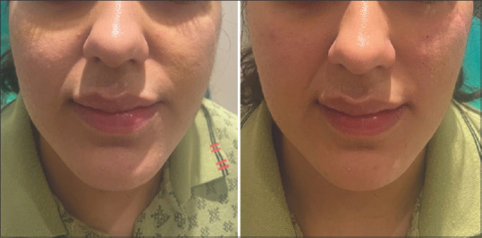
Volume restoration in a case of post-traumatic facial asymmetry with hyaluronic acid filler
Autologous fat,[2] hyaluronic acid fillers,[3] calcium hydroxylapatite,[4] and polyacrylamide[5] or a combination of fillers[6] have been used to correct residual atrophy in burnt-out disease [Figure 2]. These fillers address volumetric correction and some such as autologous fat and poly-L-lactic acid have bio-stimulatory properties. The required volume of the fillers varies with the nature of the defect. With fat, a mild over correction is desirable, whereas with hyaluronic acid, under correction is usually done to allow volumetric expansion by water absorption later. The advantages of hyaluronic acid filler are the ability to correct or dissolve excess volume of the filler and reduce the chances of complications.[7]
Figure 2.
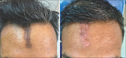
Linear morphea over forehead managed with hyaluronic acid fillers and platelet-rich plasma
Ear lobe ptosis and ear hole piercing correction when surgery is contraindicated or is not desired is another indication that can be treated with dermal fillers. Hyaluronic acid fillers are preferred for this indication [Figure 3].[8]
Figure 3.
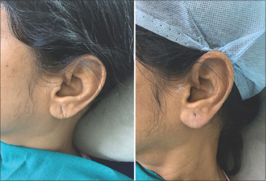
Ear lobe piercing corrected with hyaluronic acid filler
Complications in therapeutic indications, as compared to aesthetic uses, are amplified as the volumes used are usually higher. Nevertheless, standard precautions and safe injection practices need to be followed and over-correction is avoided.[9]
Botulinum toxin: Since its introduction to clinical and later aesthetic medicine, botulinum toxin is one of the most often used aesthetic procedures worldwide.[10]
Its non-aesthetic use in dermatology is ever-expanding[11] although the U.S. Food and Drug Administration clearance in aesthetics is only for dynamic wrinkles of the glabellar complex and crow’s feet.
It inhibits acetylcholine release at the presynaptic vessel along with the release of substance P and Calcitonin Gene-Related Peptide (CGRP). The dilutions are standard for aesthetic indications, using 2.5 mL of saline in a 100 IU vial.[12] Some indications such as palmar and axillary hyperhidrosis, however, use higher dilutions [Figure 4].[13] Reduced sweating and improvement in maceration reduced the disease severity in Hailey–Hailey disease[14] and linear IgA disease.[15] Novel uses have been described in psoriasis, atopic dermatitis, Raynaud’s phenomenon, flushing, acne, rosacea, epidermolysis bullosa, scar prevention and their treatment, pruritus, post-herpetic neuralgia, and other neuropathic pain disorders.[14,16-19]
Figure 4.
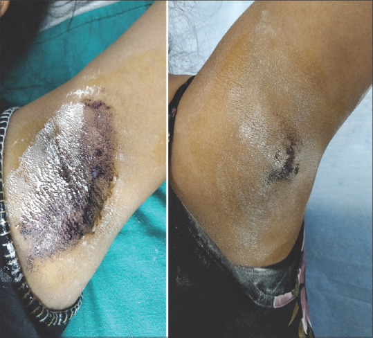
Starch-iodine test in a case of axillary hyperhidrosis: pretreatment (left) and posttreatment (right) with botulinum toxin (20 mg/mL) after 1 week, revealing improvement with a few areas (persistent positive starch-iodine test) needing additional treatment.
Regenerative medicine products: Regenerative medicine is an emerging new interdisciplinary field and its products are being used for acne scars, androgenetic alopecia, and facial rejuvenation amongst others in aesthetic dermatology.[20] Soluble molecules/growth factors utilized in platelet-rich plasma, platelet-rich fibrin, and autologous fat for their accelerated tissue healing apart and improvements in skin surface features are now being used for non-healing wounds [Figure 5], post burns, and post-traumatic scars.[21-23] Their use in morphea and scleroderma for their regenerative potential has also been described.[24,25]
Figure 5.
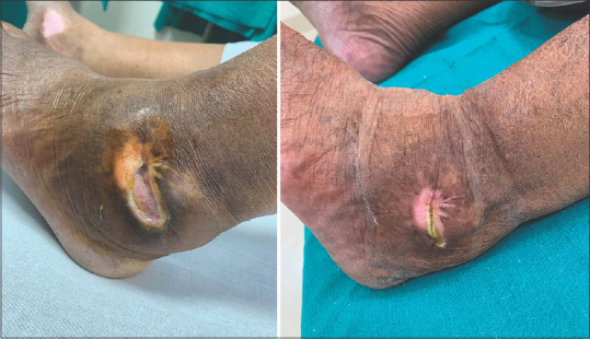
Non-healing ulcer treated with platelet-rich plasma fortnightly sessions for 3 months and saline dressings
Lipolytic injections: Phosphatidylcholine (PDC) and deoxycholate (DC) injections are used for injection lipolysis as an FDA-cleared indication of small pockets of fat, less than 500 mL in volume. PDC and DC form liposomes and micelles, from the larger fat globules, which are easily cleared from the body.[26] It is this mechanism of action that is extrapolated to treat lipomas. PDC also acts as an emulsifier and stimulator of lipase enzyme. Their use has been extrapolated off-label to treat superficial subcutaneous lipomas and also to shrink them before surgery.[27,28] It causes fat necrosis. Shrinkage in the size from 37% to complete resolution has been reported. However, recurrence after treatment has been reported.[29] No standard protocol is devised, and the volume of injection depends on the size of the lipoma.[30,31] In the authors’ experience, smaller lipomas (smaller than 5 cm in diameter) respond well, whereas larger lipomas have at best a moderate reduction in size [Figure 6].
Figure 6.
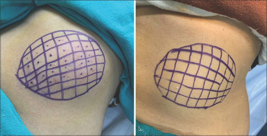
Large lipoma over the abdominal wall showing a moderate response to four sessions of monthly intralesional deoxycholic acid (10 mg/mL) injections
Dermal threads
Absorbable dermal threads of polydioxanone, poly-L-lactic acid, polyglycolic acid, polycaprolactone, and poly L-lactide-co-ϵ-caprolactone induce collagen remodeling and have been used for rejuvenation, volumization, lifting, and augmentation of skin.[32,33] Neo-angiogenesis, fibroblast stimulation with collagen remodeling, results in subsequent skin tightening, which continuously improve over a period of time.[34] They have been used in facial paralysis by targeting the affected side to improve it by volumization and countering the tissue descent and correcting tissue laxity, thus preventing drop of oral commissure.[35-37] Use of barbs to grasp tissue has been used to specifically target the ptotic/sagging skin.[32,35,38] Acne scars improvement is induced by inducing tissue remodeling.[39,40] Hair mass index was observed to improve in androgenic alopecia with monofilament thread injections as monotherapy or in combination with minoxidil.[41,42]
Lasers and light sources
Hair removal and follicular disorders: Hair removal using Ruby, Alexandrite, Diode, Neodymium: Yttrium Aluminum Garnet (Nd: YAG) lasers and Intense Pulse Light (IPL) sources can also be used for their therapeutic applications to treat post graft hypertrichosis,[43] faun tail nevi, and the rarer anterior cervical hypertrichosis [Figure 7].[44] Chronic inflammatory disorders of the follicle such as pilonidal sinus,[45,46] folliculitis decalvans, dissecting cellulitis, hidradenitis suppurativa, pseudofolliculitis barbae[47,48] and keratosis pilaris are now increasingly being treated with laser-assisted hair reduction to obviate the need for prolonged medications and surgery and to improve the quality of life.
Figure 7.
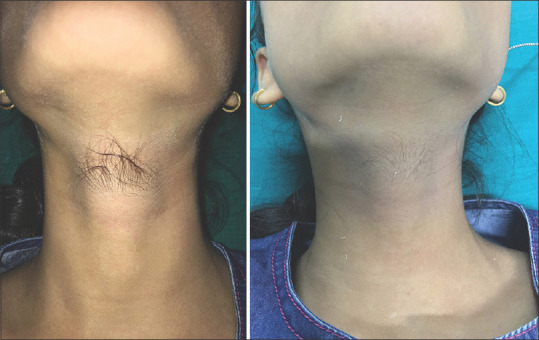
Anterior cervical hypertrichosis pretreatment (left) and post-treatment (right) after five monthly intense pulse light sessions (755–1200 nm)
Diode, long-pulse Nd: YAG laser and IPL are better suited for darker terminal hair in the skin of color and regions with heavily melanized skin. Nevertheless, unlike aesthetic laser, hair removal pain associated is higher in treating these indications. Topical anesthesia is useful, and authors recommend using it as a routine.
For pilonidal sinus, it is a necessary addon to surgery and has proven effective in preventing recurrences. Six or more sessions 4–6 weeks apart are needed for long-lasting results. The first session may be administered up to a week before surgery and followed up thereafter with additional sessions. More than 80% reduction in recurrences has been reported.[45,46]
Hidradenitis suppurativa needs higher doses with double or triple stacking to treat the inflamed hair follicles and scarring, which may have already been present. In a meta-analysis on the use of non-ablative lasers, long-pulse Nd: YAG, IPL, and Alexandrite proved useful. Higher fluences and management of Hurley stage I/II for up to 10 sessions administered 4–6 weeks apart produced the best results.[49,50] Unlike the safer, conventional use of IPL in the super pulse mode (750–1200 nm) here, a 400–1200 nm filter is used.
Vascular disorders: Lasers such as Pulse Dye Laser targeting oxyhemoglobin with absorption peaks at 542 nm and 577 nm are used for the management of vascular malformations either by themselves or by administering photodynamic therapy.[51] Increasingly, other lasers and light sources such as eodymium-doped, yttrium–aluminum–garnet (Nd-YAG) laser, 532 nm Potassium-Titanyl-Phosphate (KTP), 595 nm PDL, dual-wavelength long-pulsed 775 nm alexandrite/neodymium: yttrium-aluminum (Nd: YAG) 1064 nm, and IPL are being used for spider nevi, telangiectasia, and cherry angiomas. Among these PDL, Nd: YAG, and IPL are usually preferred. Telangiectasias with a diameter less than a 30 gauge needle is amenable to laser therapy.[52] Larger spot sizes of Nd: YAG owing to its longer wavelength help treat deeper reticular vessels. Superficial spider nevi and fine telangiectasias respond to IPL with a 500–1200 nm filter. However, erythema, blistering purpura, and erosions at high fluences restrict widespread IPL use [Figure 8].
Figure 8.
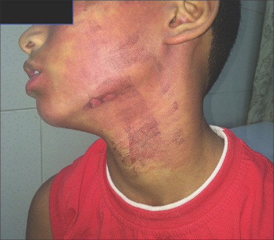
Port wine stain in a 9-year-old boy: bruising and superficial erosions 5 days after a session of intense pulse light (550–1200 nm)
Inflammatory acne and its vascular component respond to PDL 595 nm and 1319 nm, IPL 400–1200 nm, by upregulating TGFB2, an anti-inflammatory cytokine.[53,54] PDL 595 nm has no depilatory effects and can hence be used safely in males too.
Hypertrophic scars respond better than keloids to PDL and long-pulse Nd: YAG. Mechanisms of action postulated are capillary destruction resulting in hypoxemia, a decrease in cytokine or growth factor levels with a resultant reduction in local collagen production.[55] Combination treatments with intralesional triamcinolone and 5-fluorouracil improve the results.[56] Superficial vessels can be targeted by Nd: YAG and deeper with PDL. Repeated sessions are needed until there is no erythema/induration. Contact mode Nd: YAG has a better response owing to its deeper penetration as compared to non-contact mode Nd: YAG.[55]
Erythematotelangiectatic rosacea responds well to PDL, Nd: YAG, and IPL [Figure 9]. A combination with retinoids or follow-up with topical brimonidine 1% improves results.[57,58] Papulopustular lesions showed a good response to treatment by IPL. Epidermal cooling helps to reduce the side effects of treating superficial vessels with higher fluences and including a margin of 1–2 mm of normal skin helps improve results. IPL is also useful for other conditions causing superficial erythema, e.g., Poikiloderma of Civatte and post steroid abuse red skin.[57-62]
Figure 9.
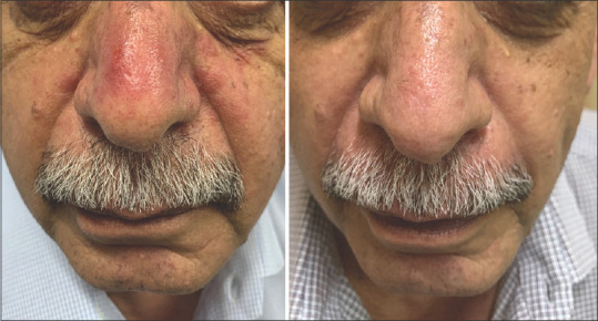
Rosacea in a 64-year-old male patient: Pretreatment (left) and posttreatment (right) after six sessions of intense pulse light (550–1200 nm)
Tissue ablative/non-ablative resurfacing and scar revision: Skin resurfacing and scar revision are common aesthetic procedures used for facial rejuvenation and acne scar treatment. Initial treatment of scars with ablative lasers was described in the early 1980s when ablative continuous wave CO2 lasers were used.[63] With the introduction of fractional photothermolysis, the side effect profile improved and downtime reduced considerably. Ablative fractional resurfacing (AFR) lasers include the fractionated CO2 (10,600 nm), Erbium: Yttrium Aluminum Garnet (Er: YAG 2990 nm) and Yttrium Scandium Gallium Garnet (YSGG 2790 nm) lasers that have a high affinity for water.
Burn scar pliability, texture, and sclerosis improve with PDL (585–595 nm) if the scar is less than 1.2 mm and if instituted after 3 to 6 months of burn injury.[64]
The use of fractional CO2 alone or in combination with platelet-rich plasma, micro fragmented adipose tissue, or stromal vascular fraction administered as monthly sessions improve post-traumatic scars.[63–67] Burns and post-traumatic scars need higher fluences of up to 70 mJ/cm2 as compared to acne scars.[68-70] Ablative CO2 laser is also used to debulk phymatous rosacea.[71]
An ideal approach would be to treat erythema and texture with PDL or IPL or wait for it to settle as mentioned above and then use the ablative lasers for deeper scar management.
Non-ablative management of scar using 1,550 nm non-ablative fractional Erbium laser, which has a penetration depth of 2 mm had shown a good response as reported by Waibel et al.[72] They reported an excellent response in up to 60% of cases. Other non-ablative lasers described for acne scar management such as 1540 nm Erbium glass laser, long-pulsed neodymium-doped yttrium aluminum garnet laser 1320 nm or 1064 nm need to be explored for post-burn and traumatic scars.[73] Although they are safer in Fitzpatrick skin types IV–VI with a lower downtime, they are limited by their depth of penetration.
Combination with regenerative products may enhance results [Figure 10].[74]
Figure 10.
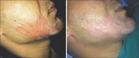
Post-traumatic scar on the face in a 30-year-old male patient: pretreatment (left) and post-treatment (right) after six monthly sessions of fractional CO2 laser (10,600 nm) with platelet-rich plasma
Ablative lasers are used for therapeutic removal of benign and early malignant skin growths such as actinic keratosis, angiofibroma, Bowen’s patches, comedones, colloid milium, milia, superficial basal cell carcinoma, xanthelasma palpebrarum, dermatosis papulosa nigra, epidermal nevi amongst other skin conditions [Table 1].[75,76]
Table 1.
Applications of medical aesthetics
| Treatment modality | Indication | |
|---|---|---|
| Lasers and light sources | Low-level laser light therapy | Vitiligo |
| Acne | ||
| Wound healing | ||
| Burns | ||
| Psoriasis | ||
| Alopecia areata | ||
| Alopecia | ||
| Recurrent herpes | ||
| Photoprotection | ||
| Hypertrophic scars and keloids | ||
| Aphthous ulcers | ||
| CO2 Laser | Scars atrophic and hypertrophic | |
| Verruca vulgaris | ||
| Seborrheic keratosis | ||
| Xanthelasma palpebrarum | ||
| Acrochordons | ||
| Nevi | ||
| Angiokeratomas | ||
| Contractures | ||
| Calcinosis cutis | ||
| Alopecia | ||
| Laser-assisted drug delivery | ||
| Onychomycosis | ||
| Nd: YAG | Nevi | |
| Onychomycosis | ||
| Spider nevi, telangiectasia, cherry angiomas | ||
| Keloids and hypertrophic scars | ||
| Lichen planus pigmentosus | ||
| Fixed drug eruption | ||
| Intense pulse light | Acne | |
| Rosacea | ||
| Vascular malformations | ||
| Therapeutic hair removal | ||
| Regenerative medicine | Micro fat | Scars |
| Morphea, scleroderma, | ||
| lipodystrophy in HIV | ||
| Biofillers in atrophic scars | ||
| Autologous micrografting | Androgenetic alopecia | |
| Vitiligo | ||
| PRP/PRF | Scars | |
| Androgenetic alopecia | ||
| Alopecia areata | ||
| Non-healing ulcers | ||
| Biofillers | ||
| Lichen sclerosus | ||
| Vitiligo | ||
| Microdermabrasion | Acne | |
| Acne scars | ||
| Post-inflammatory hyperpigmentation | ||
| Transepidermal drug delivery | ||
| Primary cutneous amyloidosis | ||
| Microneedling | Dermaroller | Hypertrophic scars |
| Dermastamp | Burn and posttraumatic scars | |
| Automated microneedling device | Atrophic acne scars | |
| Microneedling radiofrequency | Hyperhidrosis | |
| Hidradenitis suppurativa | ||
| Transdermal drug delivery | ||
| Dermal threads | Facial nerve palsy | |
| Atrophic acne scars | ||
| Alopecia | ||
| Dermal fillers | Facial deformities | |
| Depressed scars | ||
| Lip augmentation for angular cheilitis | ||
| Acquired lipodystrophy | ||
| Parry–Romberg syndrome | ||
| Lupus panniculitis | ||
| AIDS lipodystrophy | ||
| Linear morphea | ||
| Earlobe plumping, earring ptosis | ||
| Ear lobe repair | ||
| Botulinum toxin | Palmoplantar and axillary hyperhidrosis | |
| Auriculotemporal syndrome | ||
| Hailey–Hailey disease | ||
| Chromhidrosis and Bromhidrosis | ||
| Psoriasis | ||
| Hidradenitis suppurativa Pompholyx Eccrine nevus | ||
| Keloids and hypertrophic scars | ||
| Raynaud’s phenomenon | ||
| Facial flushing | ||
| Linear IgA bullous dermatosis, epidermolysis bullosa simplex, Darier disease, pachyonychia congenita | ||
| Notalgia paresthetica Postherpetic neuralgia | ||
| Alopecia areata Androgenetic alopecia | ||
| Chemical peeling | Active acne | |
| Acne scarring | ||
| Superficial scars | ||
| Seborrheic keratoses | ||
| Actinic keratoses | ||
| Warts | ||
| Milia | ||
| Sebaceous hyperplasia | ||
| Dermatosis papulosa nigra | ||
| Electroporation | Transdermal drug delivery | |
| Tattoo | Vitiligo | |
Their use in the management of onychomycosis from the clearance of the affected nail to the treatment of the infected nail is being increasingly described. Fractional CO2 lasers function to destroy the fungal elements with heat and promote transdermal drug delivery on topical antifungals. Long-pulse and Q-switched Nd: YAG target the chromophores such as xanthomegnin found in the fungal cell wall. The authors have reported that their therapeutic response is as good as oral agents. We found that using Nd: YAG is much more painful than a fractional CO2 laser. Repeated passes administered in sessions every 4–6 weeks for 3–6 sessions are effective [Figure 11].[77-81] Non-onychomycotic onychogryphosis also responds to fractional CO2 laser ablation.
Figure 11.
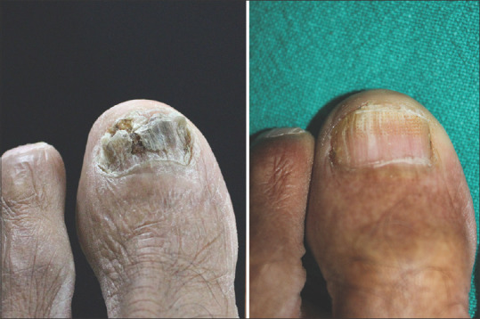
Total dystrophic onychomycosis: Pretreatment (left) and post-treatment (right) after six sessions of monthly fractional CO2 laser (10,600nm, 256 spots/cm2, pulse interval of 0.5 mm, pulse duration of 0.1 ms) and a follow-up period of 3 months
Hyperpigmentation and laser: Tattoo removal has been synonymous with pigment lasers apart from their application in skin and color rejuvenation. Q-switched ruby laser (694 nm), alexandrite (755 nm), 1064 nm, and 532 nm Nd: YAG lasers are the main lasers used. Actinic lentigo, lento simplex, ephelides, siderosis, and hemosiderosis need shorter wavelength lasers with adequate epidermal cooling, and acquired dermal melanocytosis responds better to longer 1064 nm wavelengths. Fixed-drug eruptions [Figure 12], drug-induced argyrosis, and chrysiasis, ochronosis, and lichen planus pigmentosus respond well to 1064 nm Qs Nd: YAG and low-fluence 1064 nm Nd: YAG picosecond laser.[82] Skin toning treatment with 1064 nm Qs Nd: YAG 1.8–4.6 J/cm2 in up to 10 passes and multiple sessions alone or combination with tacrolimus have shown good response.[82-84] Bhari et al.[85] reported reduced tyrosinase activity with the use of 1064 nm Q-switched Nd-YAG laser, with no significant change in erythema or pigmentation index. Larger spot sizes of 8–10 mm with low fluences have been proposed to be safer and effective with the least side effects in darker skin types.[26]
Figure 12.

Lightening of pigmentation in a case of fixed drug eruption after three sessions administered once a month with Q-switched neodymium-doped yttrium aluminum garnet laser 1064 nm
The use of ablative fractional CO2 and Erbium YAG before the use of pigment laser helps enhance their response when targeting thicker lesions.[86]
Low-level laser therapy
The use of red and infrared spectrum wavelengths used as cold lasers at non-thermal irradiance to prevent thermal damage and at the same time cause biological activity has been reported for some time. Their mechanism of action is not fully understood. The wavelengths penetrate deep enough to cause cellular changes. Cytochrome C oxidase acts as a mitochondrial chromophore along with photo acceptors in the plasma membrane, resulting in increased enzymatic activity, ATP production, and mitochondrial activity, thereby enhancing intracellular signaling and resulting in cellular proliferation and tissue repair.
As a therapeutic modality, it has been used in pigmentary conditions, papulosquamous disorders, burns and wounds, and alopecia.[87,88]
Microneedling
Microneedling was initially described in 1995 when Orentreich and Orentreich demonstrated dermal needling for scar treatment. Traditionally used as a collagen induction therapy for facial scars and skin rejuvenation, it now finds itself being useful for post-traumatic, post-varicella, hypertrophic and burn scars, alopecia, drug delivery, and hyperhidrosis, amongst other indications. The delivery device may be a manual dermaroller, dermastamp, or an automated device, with or without combinations with other technologies, such as radiofrequency.[89]
Burn scars have been shown to respond up to 80% with normalization of the dermis within a year of treatment. Camirand and Doucet initially used a tattoo gun with needles in the absence of pigment to treat scars.[90] Like ablative lasers, microneedling radiofrequency using non-insulated needles targets the entire depth of the scar, surface down. They are used in the destruction of apocrine and eccrine glands in focal hyperhidrosis and hidradenitis suppurativa.[49] Microneedles are increasingly being used to deliver macromolecules such as insulin, growth hormones, immunobiological, proteins, peptides, and vaccines apart from cosmeceuticals in aesthetic practice.[91,92]
The addition of bilayer dissolving needles containing triamcinolone and 5-fluorouracil have been described for the management of hypertrophic scars.[93] Solid needles can be used for access to deeper layers with application of topical agents after microneedling as in the treatment of alopecia areata with triamcinolone acetonide.[94]
Chemical peels
Peeling agents exhibit an ability to produce chemical burns at controlled skin depths, induce improvement, modulate the function of the stratum corneum barrier,[95] and reduce sebum production by their antibacterial, anti-inflammatory, keratolytic, and comedolytic effects. Because of these actions, they have been used in acne vulgaris, xerosis and ichthyosis, and superficial scarring.[95] Chen et al.[96] in their systematic review on acne management noted a positive response to chemical peels. Salicylic acid, trichloroacetic acid, mandelic acid, and Jessner’s peel have been used with recommendations to avoid deep peels in darker skin types. Salicylic acid and mandelic acid combinations were more effective in mild to moderate acne than glycolic acid. ‘‘Field-directed’’ therapy with chemical peels is useful in multiple actinic keratoses and seborrheic keratoses. A combination of 5-fluorouracil with Jessner’s solution or with 70% buffered glycolic acid solution, carried out for 8 weeks, offered higher clearance than just peeling alone.[97] Glycolic and trichloroacetic acid have been used for warts and superficial scars.[98,99] Combination peels have been used for verruca plana.[100]
Conclusion
The extrapolation of aesthetic products and procedures into the therapeutic arena is fast growing. Thorough knowledge of the changes in local anatomy and dermatological condition induced is paramount in providing the best clinical outcomes as then the robust science behind the procedure, product, or equipment can be best utilized.[11] Their use in aesthetics is revolutionizing the treatment of many medical conditions, with several therapeutic indications now being treated with conventional aesthetic products and equipment. Medical aesthetics, hence, has the potential to fill a therapeutic gap that conventional dermatological practices are unable to address.
Financial support and sponsorship
Nil.
Conflicts of interest
There are no conflicts of interest.
References
- 1.Arora S, Arora G. Recognizing “medical aesthetics”in dermatology:The need of the hour. Indian J Dermatol Venereol Leprol. 2021;87:1–2. doi: 10.25259/IJDVL_678_20. [DOI] [PubMed] [Google Scholar]
- 2.Hunstad JP, Shifrin DA, Kortesis BG. Successful treatment of Parry-Romberg syndrome with autologous fat grafting:14-Year Follow-up and Review. Ann Plast Surg. 2011;67:423–5. doi: 10.1097/SAP.0b013e31820b3aa8. [DOI] [PubMed] [Google Scholar]
- 3.Jo M, Ahn H, Ju H, Park E, Yoo J, Kim MS, et al. Parry-Romberg syndrome augmented by hyaluronic acid filler. Ann Dermatol. 2018;30:704–7. doi: 10.5021/ad.2018.30.6.704. [DOI] [PMC free article] [PubMed] [Google Scholar]
- 4.Cox SE, Soderberg JM. Idiopathic hemifacial atrophy treated with serial injections of calcium hydroxylapatite. Dermatol Surg. 2010;36:542–5. doi: 10.1111/j.1524-4725.2010.01499.x. [DOI] [PubMed] [Google Scholar]
- 5.Al-Niaimi F, Taylor JA, Lyon CC. Idiopathic hemifacial atrophy treated with permanent polyacrylamide subdermal filler. Dermatol Surg. 2012;38:143–5. doi: 10.1111/j.1524-4725.2011.02241.x. [DOI] [PubMed] [Google Scholar]
- 6.Ha D-L, Oh C-K, Kim M-B. Parry-Romberg syndrome treated with injectable poly-L-lactic acid and hyaluronic acid filler:A case report. J Eur Acad Dermatol Venereol. 2020;34:e275–6. doi: 10.1111/jdv.16258. [DOI] [PubMed] [Google Scholar]
- 7.Arora G. Fillers for aesthetics on the face –newer perspectives. Cosmoderma. 2021;1:6. doi:10.25259/CSDM_6_2021. [Google Scholar]
- 8.Arora G, Arora S. Rejuvenating earlobe esthetics with dermal fillers. J Cosmet Dermatol. 2022;21:2788–92. doi: 10.1111/jocd.14552. [DOI] [PubMed] [Google Scholar]
- 9.Vedamurthy M IADVL Dematosurgery Task Force. Standard guidelines for the use of dermal fillers. Indian J Dermatol Venereol Leprol. 2008;74(Suppl):S23–7. [PubMed] [Google Scholar]
- 10.Small R. Botulinum toxin injection for facial wrinkles. Am Fam Physician. 2014;90:168–75. [PubMed] [Google Scholar]
- 11.Arora G. Botulinum toxin beyond aesthetics in dermatology. Cosmoderma. 2022;2:15. doi:10.25259/CSDM_8_2022. [Google Scholar]
- 12.Arora G, Arora S. Where and how to use botulinum toxin on the face and neck –Indications and techniques. Cosmoderma. 2021;1:17. doi:10.25259/CSDM_16_2021. [Google Scholar]
- 13.Bhidayasiri R, Truong DD. Evidence for effectiveness of botulinum toxin for hyperhidrosis. J Neural Transm Vienna Austria 1996. 2008;115:641–5. doi: 10.1007/s00702-007-0812-7. [DOI] [PubMed] [Google Scholar]
- 14.Farahnik B, Blattner CM, Mortazie MB, Perry BM, Lear W, Elston DM. Interventional treatments for Hailey–Hailey disease. J Am Acad Dermatol. 2017;76:551–8.e3. doi: 10.1016/j.jaad.2016.08.039. [DOI] [PubMed] [Google Scholar]
- 15.Legendre L, Maza A, Almalki A, Bulai-Livideanu C, Paul C, Mazereeuw-Hautier J. Botulinum toxin A:An effective treatment for linear immunoglobulin A bullous dermatosis located in the axillae. Acta Derm Venereol. 2016;96:122–3. doi: 10.2340/00015555-2178. [DOI] [PubMed] [Google Scholar]
- 16.Kim YS, Hong ES, Kim HS. Botulinum toxin in the field of dermatology:Novel indications. Toxins. 2017;9:403. doi: 10.3390/toxins9120403. doi:10.3390/toxins9120403. [DOI] [PMC free article] [PubMed] [Google Scholar]
- 17.Antonucci F, Rossi C, Gianfranceschi L, Rossetto O, Caleo M. Long-distance retrograde effects of botulinum neurotoxin A. J Neurosci Off J Soc Neurosci. 2008;28:3689–96. doi: 10.1523/JNEUROSCI.0375-08.2008. [DOI] [PMC free article] [PubMed] [Google Scholar]
- 18.Xiao L, Mackey S, Hui H, Xong D, Zhang Q, Zhang D. Subcutaneous injection of botulinum toxin a is beneficial in postherpetic neuralgia. Pain Med Malden Mass. 2010;11:1827–33. doi: 10.1111/j.1526-4637.2010.01003.x. [DOI] [PubMed] [Google Scholar]
- 19.Wang TS, Tsai TF. Intralesional therapy for psoriasis. J Dermatol Treat. 2013;24:340–7. doi: 10.3109/09546634.2012.672706. [DOI] [PubMed] [Google Scholar]
- 20.Yepuri V, Venkataram M. Platelet-rich plasma with microneedling in androgenetic alopecia:Study of efficacy of the treatment and the number of sessions required. J Cutan Aesthetic Surg. 2021;14:184–90. doi: 10.4103/JCAS.JCAS_33_20. [DOI] [PMC free article] [PubMed] [Google Scholar]
- 21.Dieckmann C, Renner R, Milkova L, Simon JC. Regenerative medicine in dermatology:Biomaterials, tissue engineering, stem cells, gene transfer and beyond:Skin substitution by tissue engineering. Exp Dermatol. 2010;19:697–706. doi: 10.1111/j.1600-0625.2010.01087.x. [DOI] [PubMed] [Google Scholar]
- 22.Klinger M, Klinger F, Caviggioli F, Maione L, Catania B, Veronesi A, et al. Fat grafting for treatment of facial scars. Clin Plast Surg. 2020;47:131–8. doi: 10.1016/j.cps.2019.09.002. [DOI] [PubMed] [Google Scholar]
- 23.Schulz A, Schiefer JL, Fuchs PC, Kanho CH, Nourah N, Heitzmann W. Does platelet-rich fibrin enhance healing of burn wounds?Our first experiences and main pitfalls. Ann Burns Fire Disasters. 2021;34:42–52. [PMC free article] [PubMed] [Google Scholar]
- 24.Mura S, Fin A, Parodi PC, Denton CP, Howell KJ, Rampino Cordaro E. Autologous fat transfer in the successful treatment of upper limb linear morphoea. Clin Exp Rheumatol. 2018;36(Suppl 113):183. [PubMed] [Google Scholar]
- 25.Strong AL, Rubin JP, Kozlow JH, Cederna PS. Fat grafting for the treatment of scleroderma:Plast Reconstr Surg. 2019;144:1498–507. doi: 10.1097/PRS.0000000000006291. [DOI] [PubMed] [Google Scholar]
- 26.Aurangabadkar S. Optimizing Q-switched lasers for melasma and acquired dermal melanoses. Indian J Dermatol Venereol Leprol. 2019;85:10–7. doi: 10.4103/ijdvl.IJDVL_1086_16. [DOI] [PubMed] [Google Scholar]
- 27.Amber KT, Ovadia S, Camacho I. Injection therapy for the management of superficial subcutaneous lipomas. J Clin Aesthetic Dermatol. 2014;7:46–8. [PMC free article] [PubMed] [Google Scholar]
- 28.Hasengschwandtner F. Injection lipolysis for effective reduction of localized fat in place of minor surgical lipoplasty. Aesthet Surg J. 2006;26:125–30. doi: 10.1016/j.asj.2006.01.008. [DOI] [PubMed] [Google Scholar]
- 29.Pindur L, Sand M, Altmeyer P, Bechara FG. Recurrent growth of lipomas after previous treatment with phosphatidylcholine and deoxycholate. J Cosmet Laser Ther. 2011;13:95–6. doi: 10.3109/14764172.2011.564630. [DOI] [PubMed] [Google Scholar]
- 30.Bechara FG, Sand M, Sand D, Rotterdam S, Stücker M, Altmeyer P, et al. Lipolysis of lipomas in patients with familial multiple lipomatosis:An ultrasonography-controlled trial. J Cutan Med Surg. 2006;10:155–9. doi: 10.2310/7750.2006.00040. [DOI] [PubMed] [Google Scholar]
- 31.Thomas MK, D’Silva JA, Borole AJ. Injection lipolysis:A systematic review of literature and our experience with a combination of phosphatidylcholine and deoxycholate over a period of 14 years in 1269 patients of Indian and South East Asian Origin. J Cutan Aesthetic Surg. 2018;11:222–8. doi: 10.4103/JCAS.JCAS_117_18. [DOI] [PMC free article] [PubMed] [Google Scholar]
- 32.Arora G, Arora S. Neck rejuvenation with thread lift. J Cutan Aesthetic Surg. 2019;12:196–200. doi: 10.4103/JCAS.JCAS_181_18. [DOI] [PMC free article] [PubMed] [Google Scholar]
- 33.Wong V. The science of absorbable poly (L-Lactide-Co-ϵ-Caprolactone) threads for soft tissue repositioning of the face:An evidence-based evaluation of their physical properties and clinical application. Clin Cosmet Investig Dermatol. 2021;14:45–54. doi: 10.2147/CCID.S274160. [DOI] [PMC free article] [PubMed] [Google Scholar]
- 34.Arora G, Arora S. Thread lift in breast ptosis. J Cutan Aesthetic Surg. 2017;10:228. doi: 10.4103/JCAS.JCAS_91_17. [DOI] [PMC free article] [PubMed] [Google Scholar]
- 35.Dua A, Bhardwaj B. A case report on use of cog threads and dermal fillers for facial-lifting in facioscapulohumeral muscular dystrophy. J Cutan Aesthetic Surg. 2019;12:52–5. doi: 10.4103/JCAS.JCAS_128_18. [DOI] [PMC free article] [PubMed] [Google Scholar]
- 36.Costan VV, Popescu E, Sulea D, Stratulat IS. A new indication for barbed threads:Static reanimation of the paralyzed face. J Oral Maxillofac Surg. 2018;76:639–45. doi: 10.1016/j.joms.2017.07.176. [DOI] [PubMed] [Google Scholar]
- 37.Navarrete ML, Palao R, Torrent L, Fuentes JF, González M. Facial asymmetry correction in facial palsy patients with silhouette sutures. Int J Clin Med. 2012;03:55–9. [Google Scholar]
- 38.Choe WJ, Kim HD, Han BH, Kim J. Thread lifting:A minimally invasive surgical technique for long-standing facial paralysis. HNO. 2017;65:910–5. doi: 10.1007/s00106-017-0367-3. [DOI] [PubMed] [Google Scholar]
- 39.Shin JJ, Park TJ, Kim BY, Kim CM, Suh DH, Lee SJ, et al. Comparative effects of various absorbable threads in a rat model. J Cosmet Laser Ther. 2019;21:158–62. doi: 10.1080/14764172.2018.1493511. [DOI] [PubMed] [Google Scholar]
- 40.Donnarumma M, Vastarella M, Ferrillo M, Cantelli M, D’Andrea M, Fabbrocini G. An innovative treatment for acne scars with thread-lift technique:our experience. Ital J Dermatol Venerol. 2021:15640–41. doi: 10.23736/S2784-8671.19.06313-2. [DOI] [PubMed] [Google Scholar]
- 41.Khattab FM, Bessar H. Accelerated hair growth by combining thread monofilament and minoxidil in female androgenetic alopecia. J Cosmet Dermatol. 2020;19:1738–44. doi: 10.1111/jocd.13228. [DOI] [PubMed] [Google Scholar]
- 42.Metwalli M, Khattab FM, Mandour S. Monofilament threads in treatment of female hair loss. J Dermatol Treat. 2021;32:521–5. doi: 10.1080/09546634.2019.1682499. [DOI] [PubMed] [Google Scholar]
- 43.García-Zamora E, Naz-Villalba E, Pampín-Franco A, Vicente-Martín FJ, López-Estebaranz J. Laser therapy for hair removal on grafts and flaps. Dermatol Ther. 2019;32:e12880. doi: 10.1111/dth.12880. doi:10.1111/dth. 12880. [DOI] [PubMed] [Google Scholar]
- 44.Arora S, Arora G, Totlani S, Chandra M. Faun tail nevus:A series of 15 cases and their management with intense pulse light. Med J Armed Forces India. 2019;75:389–94. doi: 10.1016/j.mjafi.2018.06.002. [DOI] [PMC free article] [PubMed] [Google Scholar]
- 45.Ganjoo A. Laser hair reduction for pilonidal sinus-my experience. J Cutan Aesthetic Surg. 2011;4:196. [PMC free article] [PubMed] [Google Scholar]
- 46.Oram Y, Kahraman F, Karincaoğlu Y, Koyuncu E. Evaluation of 60 patients with pilonidal sinus treated with laser epilation after surgery. Dermatol Surg. 2010;36:88–91. doi: 10.1111/j.1524-4725.2009.01387.x. [DOI] [PubMed] [Google Scholar]
- 47.Aleem S, Majid I. Unconventional uses of laser hair removal:A review. J Cutan Aesthetic Surg. 2019;12:8–16. doi: 10.4103/JCAS.JCAS_97_18. [DOI] [PMC free article] [PubMed] [Google Scholar]
- 48.Jfri A, Saxena A, Rouette J, Netchiporouk E, Barolet A, O’Brien E, et al. The efficacy and effectiveness of non-ablative light-based devices in hidradenitis suppurativa:A systematic review and meta-analysis. Front Med. 2020;7:591580. doi: 10.3389/fmed.2020.591580. doi:10.3389/fmed. 2020.591580. [DOI] [PMC free article] [PubMed] [Google Scholar]
- 49.Tchero H, Herlin C, Bekara F, Fluieraru S, Teot L. Hidradenitis suppurativa:A systematic review and meta-analysis of therapeutic interventions. Indian J Dermatol Venereol Leprol. 2019;85:248–57. doi: 10.4103/ijdvl.IJDVL_69_18. [DOI] [PubMed] [Google Scholar]
- 50.Jain A, Jain V. Use of lasers for the management of refractory cases of hidradenitis suppurativa and pilonidal sinus. J Cutan Aesthetic Surg. 2012;5:190–2. doi: 10.4103/0974-2077.101377. [DOI] [PMC free article] [PubMed] [Google Scholar]
- 51.Liu A, Moy RL, Ross EV, Hamzavi I, Ozog DM. Pulsed dye laser and pulsed dye laser–mediated photodynamic therapy in the treatment of dermatologic disorders. Dermatol Surg. 2012;38:351–66. doi: 10.1111/j.1524-4725.2011.02293.x. [DOI] [PubMed] [Google Scholar]
- 52.Nakano LC, Cacione DG, Baptista-Silva JC, Flumignan RL Cochrane Vascular Group. Treatment for telangiectasias and reticular veins. Cochrane Database Syst Rev. 2021. [Last accessed on 2022 Apr 13]. Available from:https://doi.wiley.com/10.1002/14651858. CD012723 . [DOI] [PMC free article] [PubMed]
- 53.Kassir M, Arora G, Galadari H, Kroumpouzos G, Katsambas A, Lotti T, et al. Efficacy of 595- and 1319-nm pulsed dye laser in the treatment of acne vulgaris:A narrative review. J Cosmet Laser Ther. 2020;22:111–4. doi: 10.1080/14764172.2020.1774063. [DOI] [PubMed] [Google Scholar]
- 54.Kumaresan M, Srinivas CR. Efficacy of ipl in treatment of acne vulgaris:Comparison of single- and burst-pulse mode in ipl. Indian J Dermatol. 2010;55:370–2. doi: 10.4103/0019-5154.74550. [DOI] [PMC free article] [PubMed] [Google Scholar]
- 55.Koike S, Akaishi S, Nagashima Y, Dohi T, Hyakusoku H, Ogawa R. Nd:YAG laser treatment for keloids and hypertrophic scars:An analysis of 102 cases. Plast Reconstr Surg Glob Open. 2014;2:e272. doi: 10.1097/GOX.0000000000000231. [DOI] [PMC free article] [PubMed] [Google Scholar]
- 56.Asilian A, Darougheh A, Shariati F. New combination of triamcinolone, 5-fluorouracil, and pulsed-dye laser for treatment of keloid and hypertrophic scars. Dermatol Surg. 2006;32:907–15. doi: 10.1111/j.1524-4725.2006.32195.x. [DOI] [PubMed] [Google Scholar]
- 57.Anzengruber F, Czernielewski J, Conrad C, Feldmeyer L, Yawalkar N, Häusermann P, et al. Swiss S1 guideline for the treatment of rosacea. J Eur Acad Dermatol Venereol. 2017;31:1775–91. doi: 10.1111/jdv.14349. [DOI] [PubMed] [Google Scholar]
- 58.Kumaresan M, Srinivas C. Efficacy of IPL in treatment of acne vulgaris :Comparison of single- and burst-pulse mode in IPL. Indian J Dermatol. 2010;55:370–2. doi: 10.4103/0019-5154.74550. [DOI] [PMC free article] [PubMed] [Google Scholar]
- 59.Maxwell EL, Ellis DA, Manis H. Acne rosacea:Effectiveness of 532 nm laser on the cosmetic appearance of the skin. J Otolaryngol Head Neck Surg. 2010;39:292–6. [PubMed] [Google Scholar]
- 60.Kim SJ, Lee Y, Seo YJ, Lee JH, Im M. Comparative efficacy of radiofrequency and pulsed dye laser in the treatment of rosacea. Dermatol Surg. 2017;43:204–9. doi: 10.1097/DSS.0000000000000968. [DOI] [PubMed] [Google Scholar]
- 61.Luo Y, Luan X, Zhang J, Wu L, Zhou N. Improved telangiectasia and reduced recurrence rate of rosacea after treatment with 540 nm-wavelength intense pulsed light:A prospective randomized controlled trial with a 2-year follow-up. Exp Ther Med. 2020;19:3543–50. doi: 10.3892/etm.2020.8617. [DOI] [PMC free article] [PubMed] [Google Scholar]
- 62.Campolmi P, Bonan P, Cannarozzo G, Bruscino N, Troiano M, Prignano F, et al. Intense pulsed light in the treatment of non-aesthetic facial and neck vascular lesions:Report of 85 cases:IPL in the treatment of vascular lesions. J Eur Acad Dermatol Venereol. 2011;25:68–73. doi: 10.1111/j.1468-3083.2010.03700.x. [DOI] [PubMed] [Google Scholar]
- 63.Ohshiro T, Ohshiro T, Sasaki K. Laser scar management technique. LASER Ther. 2013;22:255–60. doi: 10.5978/islsm.13-OR-20. [DOI] [PMC free article] [PubMed] [Google Scholar]
- 64.Harithy R, Pon K. Scar treatment with lasers:A review and update. Curr Dermatol Rep. 2012;1:69–75. [Google Scholar]
- 65.Bowen RE. A novel approach to ablative fractional treatment of mature thermal burn scars. J Drugs Dermatol JDD. 2010;9:389–92. [PubMed] [Google Scholar]
- 66.Godara S, Arora S, Dabas R, Arora G, Renganathan G, Choudhary R. A comparative study on the efficacy of fractional CO2 laser and fractional CO2 laser with autologous platelet-rich plasma in scars. Indian Dermatol Online J. 2020;11:930–6. doi: 10.4103/idoj.IDOJ_174_20. [DOI] [PMC free article] [PubMed] [Google Scholar]
- 67.Arora G, Arora S. Platelet rich plasma –where do we stand today?A critical narrative review and analysis. Dermatol Ther. 2020;34:e14343. doi: 10.1111/dth.14343. doi:10.1111/dth. 14343. [DOI] [PubMed] [Google Scholar]
- 68.Onur Erol O, Agaoglu G, Jawad MA. Combined non-ablative laser and microfat grafting for burn scar treatment. Aesthet Surg J. 2019;39:NP55–67. doi: 10.1093/asj/sjy291. [DOI] [PubMed] [Google Scholar]
- 69.Anderson RR, Donelan MB, Hivnor C, Greeson E, Ross EV, Shumaker PR, et al. Laser treatment of traumatic scars with an emphasis on ablative fractional laser resurfacing:Consensus report. JAMA Dermatol. 2014;150:187–93. doi: 10.1001/jamadermatol.2013.7761. [DOI] [PubMed] [Google Scholar]
- 70.Gold MH, McGuire M, Mustoe TA, Pusic A, Sachdev M, Waibel J, et al. Updated international clinical recommendations on scar management:Part 2--algorithms for scar prevention and treatment. Dermatol Surg Off Publ Am Soc Dermatol Surg Al. 2014;40:825–31. doi: 10.1111/dsu.0000000000000050. [DOI] [PubMed] [Google Scholar]
- 71.Tanghetti E, Del Rosso JQ, Thiboutot D, Gallo R, Webster G, Eichenfield LF, et al. Consensus recommendations from the American acne &rosacea society on the management of rosacea, part 4:A status report on physical modalities and devices. Cutis. 2014;93:71–6. [PubMed] [Google Scholar]
- 72.Waibel J, Wulkan AJ, Lupo M, Beer K, Anderson RR. Treatment of burn scars with the 1,550 nm nonablative fractional Erbium Laser:Treatment of burn scars. Lasers Surg Med. 2012;44:441–6. doi: 10.1002/lsm.22038. [DOI] [PubMed] [Google Scholar]
- 73.Sachdev M, Hameed S, Mysore V. Nonablative lasers and nonlaser systems in dermatology:Current status. Indian J Dermatol Venereol Leprol. 2011;77:380. doi: 10.4103/0378-6323.79733. [DOI] [PubMed] [Google Scholar]
- 74.Stachura A, Paskal W, Pawlik W, Mazurek MJ, Jaworowski J. The use of adipose-derived stem cells (ADSCs) and stromal vascular fraction (SVF) in skin scar treatment—A systematic review of clinical studies. J Clin Med. 2021;10:3637. doi: 10.3390/jcm10163637. doi:10.3390/jcm10163637. [DOI] [PMC free article] [PubMed] [Google Scholar]
- 75.Omi T, Numano K. The role of the CO2 laser and fractional CO2 laser in dermatology. LASER Ther. 2014;23:49–60. doi: 10.5978/islsm.14-RE-01. [DOI] [PMC free article] [PubMed] [Google Scholar]
- 76.Madan V. Dermatological applications of carbon dioxide laser. J Cutan Aesthetic Surg. 2013;6:175–7. doi: 10.4103/0974-2077.123393. [DOI] [PMC free article] [PubMed] [Google Scholar]
- 77.Arora S, Lal S, Janney M, Ranjan E, Donaparthi N, Dabas R. Fractional CO2 laser in the management of onychomycosis. J Mar Med Soc. 2020;22:50–3. [Google Scholar]
- 78.Kandpal R, Arora S, Arora D. A study of Q-switched Nd:YAG laser versus itraconazole in management of onychomycosis. J Cutan Aesthet Surg. 2021;14:87–110. doi: 10.4103/JCAS.JCAS_29_20. [DOI] [PMC free article] [PubMed] [Google Scholar]
- 79.Ranjan E, Arora S, Sharma N. Fractional CO2 laser with topical 1% terbinafine cream versus oral itraconazole in the management of onychomycosis:A randomized controlled trial. Indian J Dermatol Venereol Leprol. 2022:1–7. doi: 10.25259/IJDVL_98_2021. doi:10.25259/IJDVL_98_2021. [DOI] [PubMed] [Google Scholar]
- 80.Arora S, Ranjan E. Urea occlusion prior to single session fractional CO 2 laser as a treatment in onychomycosis. Indian J Dermatol Venereol Leprol. 2020;86:331–3. doi: 10.4103/ijdvl.IJDVL_742_19. [DOI] [PubMed] [Google Scholar]
- 81.Bhatta AK, Keyal U, Wang X, Gellén E. A review of the mechanism of action of lasers and photodynamic therapy for onychomycosis. Lasers Med Sci. 2017;32:469–74. doi: 10.1007/s10103-016-2110-9. [DOI] [PubMed] [Google Scholar]
- 82.Wong TH. Picosecond laser treatment for Asian skin pigments:A review. J Cosmet Med. 2019;3:55–63. [Google Scholar]
- 83.Shah SD, Aurangabadkar S, Nikam B. An open-label non-randomized prospective pilot study of the efficacy of Q-switched Nd-YAG laser in management of facial lichen planus pigmentosus. J Cosmet Laser Ther. 2019;21:108–15. doi: 10.1080/14764172.2018.1469770. [DOI] [PubMed] [Google Scholar]
- 84.Wu C, Lin F. A successful combination therapy of tacrolimus, hydroxychloroquine and picosecond laser for lichen planus pigmentosus. Australas J Dermatol. 2019;60:e336–7. doi: 10.1111/ajd.13060. [DOI] [PubMed] [Google Scholar]
- 85.Bhari N, Sharma VK, Singh S, Parihar A, Arava S. Effect of Q-switched Nd-YAG laser on the clinical, pigmentary, and immunological markers in patients with lichen planus pigmentosus:A pilot study. Dermatol Ther. 2020;33:e13208. doi: 10.1111/dth.13208. doi:10.1111/dth. 13208. [DOI] [PubMed] [Google Scholar]
- 86.Passeron T, Genedy R, Salah L, Fusade T, Kositratna G, Laubach H-J, et al. Laser treatment of hyperpigmented lesions:Position statement of the European Society of Laser in Dermatology. J Eur Acad Dermatol Venereol. 2019;33:987–1005. doi: 10.1111/jdv.15497. [DOI] [PubMed] [Google Scholar]
- 87.Avci P, Gupta A, Sadasivam M, Vecchio D, Pam Z, Pam N, et al. Low-level laser (light) therapy (LLLT) in skin:Stimulating, healing, restoring. Semin Cutan Med Surg. 2013;32:41–52. [PMC free article] [PubMed] [Google Scholar]
- 88.Pillai JK, Mysore V. Role of low-level light therapy (LLLT) in androgenetic alopecia. J Cutan Aesthetic Surg. 2021;14:385–91. doi: 10.4103/JCAS.JCAS_218_20. [DOI] [PMC free article] [PubMed] [Google Scholar]
- 89.Singh A, Yadav S. Microneedling:Advances and widening horizons. Indian Dermatol Online J. 2016;7:244–54. doi: 10.4103/2229-5178.185468. [DOI] [PMC free article] [PubMed] [Google Scholar]
- 90.Camirand A, Doucet J. Needle dermabrasion. Aesthetic Plast Surg. 1997;21:48–51. doi: 10.1007/s002669900081. [DOI] [PubMed] [Google Scholar]
- 91.Moore LE, Vucen S, Moore AC. Trends in drug- and vaccine-based dissolvable microneedle materials and methods of fabrication. Eur J Pharm Biopharm. 2022;173:54–72. doi: 10.1016/j.ejpb.2022.02.013. [DOI] [PubMed] [Google Scholar]
- 92.Bariya SH, Gohel MC, Mehta TA, Sharma OP. Microneedles:An emerging transdermal drug delivery system. J Pharm Pharmacol. 2011;64:11–29. doi: 10.1111/j.2042-7158.2011.01369.x. [DOI] [PubMed] [Google Scholar]
- 93.Yang B, Dong Y, Shen Y, Hou A, Quan G, Pan X, et al. Bilayer dissolving microneedle array containing 5-fluorouracil and triamcinolone with biphasic release profile for hypertrophic scar therapy. Bioact Mater. 2021;6:2400–11. doi: 10.1016/j.bioactmat.2021.01.014. [DOI] [PMC free article] [PubMed] [Google Scholar]
- 94.Mysore V, Chandrashekar B, Yepuri V. Alopecia areata-successful outcome with microneedling and triamcinolone acetonide. J Cutan Aesthetic Surg. 2014;7:63. doi: 10.4103/0974-2077.129989. [DOI] [PMC free article] [PubMed] [Google Scholar]
- 95.Berardesca E, Distante F, Vignoli GP, Oresajo C, Green B. Alpha hydroxyacids modulate stratum corneum barrier function. Br J Dermatol. 1997;137:934–8. [PubMed] [Google Scholar]
- 96.Chen X, Wang S, Yang M, Li L. Chemical peels for acne vulgaris:A systematic review of randomised controlled trials. BMJ Open. 2018;8:e019607. doi: 10.1136/bmjopen-2017-019607. [DOI] [PMC free article] [PubMed] [Google Scholar]
- 97.Dianzani C, Conforti C, Giuffrida R, Corneli P, Meo N, Farinazzo E, et al. Current therapies for actinic keratosis. Int J Dermatol. 2020;59:677–84. doi: 10.1111/ijd.14767. [DOI] [PubMed] [Google Scholar]
- 98.Hatta J, Mochizuki T. Successful treatment of plantar warts with topical glycolic acid. J Dermatol. 2017;44:e134–5. doi: 10.1111/1346-8138.13783. [DOI] [PubMed] [Google Scholar]
- 99.Khunger N IADVL Task Force. Standard guidelines of care for chemical peels. Indian J Dermatol Venereol Leprol. 2008;74(Suppl):S5–12. [PubMed] [Google Scholar]
- 100.Rodríguez-Cerdeira C, Sánchez-Blanco E. Glycolic acid 15% plus salicylic acid 2%:A new therapeutic pearl for facial flat warts. J Clin Aesthetic Dermatol. 2011;4:62–4. [PMC free article] [PubMed] [Google Scholar]


