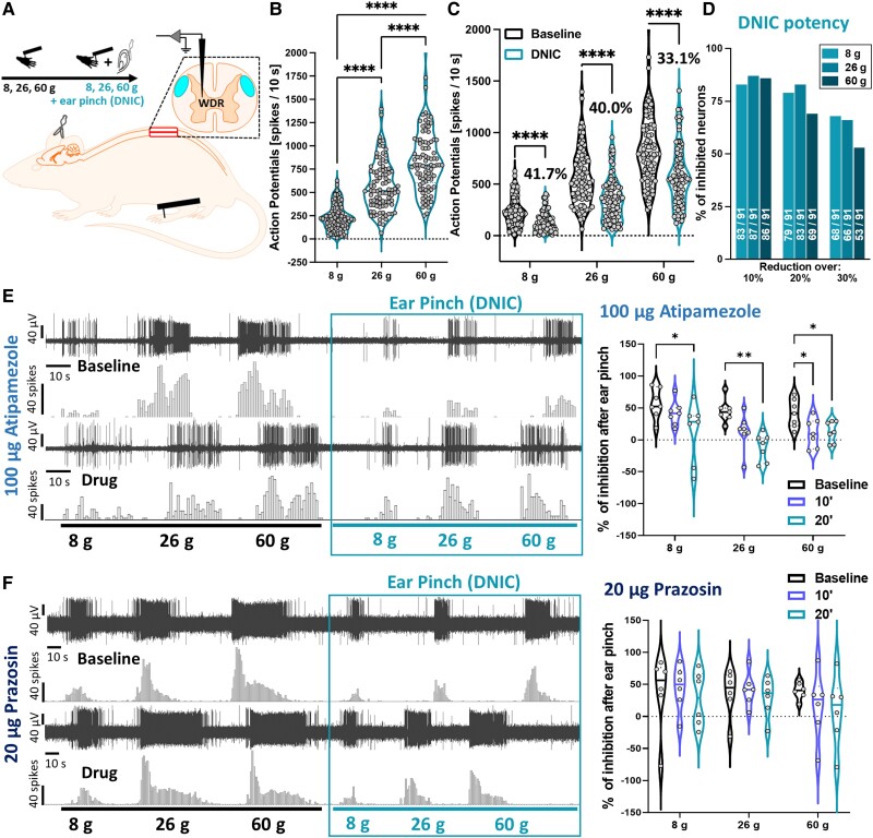
Figure 1.
Spinal α2-adrenoceptors mediate DNIC. (A) Experimental setup. (B) DDH-WDR neurons code stimulus intensity (von Frey-evoked). (C) Application of noxious ear pinch (conditioning stimulus, CS) leads to inhibition of DDH-WDR firing. (D) Percentage of neurons inhibited by CS. Numbers on bars represent units with reduced activity according to a given threshold (out of 91 recorded). (E) Inhibition following CS application (baseline and following α2-adrenoceptor antagonism with spinal atipamezole) with example single unit DDH-WDR neuronal traces. (F) Inhibition following CS application (baseline and following α1-adrenoceptor antagonism with spinal prazosin) with example single unit DDH-WDR neuronal traces. Data represents mean ± SEM. Dots represent individual neuron studied (Baselines: N = 65 rats, n = 91 neurons). For pharmacology one cell was recorded per animal (atipamezole: N/n = 7, prazosin: N/n = 6). Two-way RM-ANOVA with Tukey post hoc: *P < 0.05, **P < 0.01, ****P < 0.0001. See Supplementary Fig. 1.

