Abstract
The relevance of geriatric ankle fractures is continuously increasing.
Treatment of these patients remains challenging and requires adapted diagnostic and therapeutic strategies, as compliance to partial weight bearing is difficult to maintain compared to younger patients.
In addition, in the elderly even low impact injuries may lead to severe soft tissue trauma, influencing timing and operative strategies.
Recently, the direct posterolateral approach and plate fixation techniques, angular stable implants as well as intramedullary nailing of the distal fibula have been found to improve stategical concepts.
This article aims to provide a comprehensive overview of the diagnostic and recent aspects with respect to how this difficult entity of injuries should be approached.
Keywords: ankle, fracture, elderly
Epidemiology
Ankle fractures are very common, with an annual incidence of 74 per 100 000 people and a mean age of 56 years in Germany (1). Interestingly, 60% of the fractures occur in women with an increase of the incidence between the age of 40 and 70 years (1). With the demographic changes, the relevance of ankle fractures particularly in the elderly will increase. However, there are several differences regarding the diagnostics, fracture pattern and the treatment strategies when comparing the elderly population to younger patients.
Pre-existing conditions
Elderly people suffer more frequently from comorbidities, which will affect the incidence and outcome of ankle fractures. Female gender, high BMI, diabetes, polymedication as well as drug abuse and smoking have been determined as independent risk factors for sustaining an ankle fracture (2, 3). In contrast to other fractures, for example proximal femur or vertebral body, a clear causality between osteoporosis and ankle fractures has so far not been proven (4). However, there seems to be a positive correlation between bone mineral density and ankle fractures of the elderly (5, 6). Furthermore, in the geriatric population, changes of the neurovascular status as well as wound healing disorders, skin necrosis and implant failure occur frequently (7). Therefore, the treatment of these fractures remains a challenge, requiring soft tissue-related and treatment strategies.
Trauma mechanism
In the elderly, low-energy trauma is more dominant (8, 9). Still, the fracture pattern seems to be more complex, compared to younger patients, presenting areas with multifragmentary and comminuted pathologies (7, 8). Unstable pronation–abduction injuries (stage III), according to Lauge–Hansen classification, are more common in people older than 60 years (10). These fractures frequently compromise the medial soft tissue envelope, which increases the severity (7).
Diagnostics
The radiographic assessment regularly includes a conventional x-ray of the ankle joint in two planes. As the complex fracture patterns in the elderly may predominate and as x-ray lacks sensitivity in the detection of multifragmentary fibular fractures or bony avulsions of the anterior inferior tibiofibular ligament (11), a preoperative CT scan is therefore recommended.
Furthermore, up to 25% of the posterior malleolus fractures are missed by conventional plain radiographs (11).
Besides the radiologic diagnostics, the evaluation of the soft tissue coverage and pre-existing conditions is not only crucial but affects the timing of as well as the definitive treatment decision. Peripheral artery disease (PAD) is a common and underestimated comorbidity. Therefore, a regular diagnostic algorithm, including the ankle–brachial index, a duplex sonography and – depending on the findings – a CT angiography, is recommended. The standardized diagnostic optimization of the vascular supply in patients with PAD was able to reduce the complication rate in geriatric ankle fractures significantly (12).
Treatment options
The general consideration of treating an ankle fracture in the elderly conservatively or operatively still remains a subject of debate. Fracture dislocations must be reduced and constrained immediately. Here, transfixation with an external fixator becomes increasingly the treatment of choice. It protects the compromised soft tissue, reduces pain and offers stability as well as immediate access to the soft tissue (7). At the moment, these recommendations are not yet state of the art, due to the necessity of an additional operation. However, it opens a window to find the best team, to evaluate preoperative planning as well as to assess the patient’s compliance with the postoperative course.
‘Stable’ ankle fractures may be defined as an isolated fibula fracture with a medial clear space of <4 mm in the mortise view. To detect instability, additional weightbearing x-rays and a GravityView can be performed (7). Stable fractures can be treated conservatively with either a closed contact plaster or a walker for 6–8 weeks.
Besides the fracture pattern, the treatment especially in the geriatric population significantly is affected by the soft tissue status and the pre-existing conditions. The primary goal of an anatomic reconstruction of the articular surface, reduction of the distal fibula and stable retention should carefully be balanced with the status of the patient in general and the soft tissue in particular. A conservative regimen leads to a higher amount of malunions and non-unions of up to 73%, whereas open reduction and internal fixation (ORIF) provides a consolidation rate up to 100% (13, 14). However, the complication rate after surgery in the elderly people is up to 22% (14, 15, 16, 17). Major complications include delayed wound healing, superficial and deep wound infections, malunions and skin necrosis, requiring revision surgery in 11% of patients over 60 years and with an in-hospital mortality rate of 3% (15, 17). The 30-day mortality rate raises up to 5.4% in patients older than 80 years after ORIF (18). Complex bony injuries such as open fractures and more complex bimalleolar and trimalleolar fractures, age, female sex and comorbidities like diabetes, smoking, dementia, osteoporosis and PAD further increase the risk of a peri- or postoperative complications (15, 19, 20).
Regarding the functional outcome, a current systematic review and meta-analysis including eight prospective randomized controlled studies and 1237 patients provides equal results for conservative and surgical treatment in ankle fractures (21). A randomized controlled trial comparing closed contact casting to ORIF in unstable ankle fractures in 593 patients over 60 years describes a similar Olerud and Molander ankle score (OMAS) , quality of life, pain, ankle motion, mobility and patient satisfaction after 6 months. However, 19% of the patients in the casting group were converted to surgery due to loss of reduction (22). Consequently, the decision of either conservative or operative handling remains an individual decision. The treatment aims to achieve a stable union and preserve quality of life, rather than achieving anatomical reduction (23). To achieve this, there are several new techniques for geriatric ankle surgery, which are described.
Posterior malleolus fractures
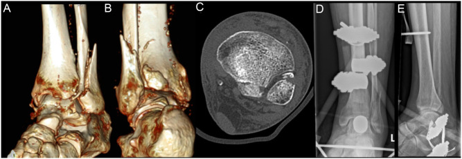
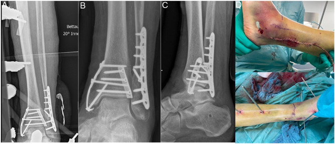
Pronation–abduction injuries regularly lead to a bony avulsion of the posterior syndesmosis – the posterior malleolus (PM). Because of a consecutive instability of the ankle joint, reduction and fixation of the PM is recommended in order to restore the stability of the mortise for PM fractures involving the tibial incisura (24, 25). A historical – size based – indication for operative management of the PM fragment, for example 25% of the articular surface, is now replaced by a morphology-adapted approach. Here the biomechanical aspect of the unstable syndesmosis is the key (25, 26). A direct posterolateral approach clearly increases stability as well as the quality of reduction, compared to an indirect anterior–posterior screw placement (27). Additionally, the stabilization of the PM reduces the risk of complications and enhances the functional outcome compared to an AP lag screw (28, 29). In addition, the need of implantation of a transsyndesmotic fixation is far lower after ORIF (30). Further benefits of the direct posterolateral approach are the improved soft tissue coverage of the implant and the option of a posterior plate osteosynthesis of the distal fibula with a sufficient peroneal tendon soft tissue coverage (7). The possibility of implanting a lag screw through the plate across the fracture and the placement of longer, bicortical screws in the distal fragment of the distal fibula fracture (dorsal antiglide plate) increase the biomechanical stability compared to a standard lateral plate fixation (31) (Figs. 1 and 2).
Figure 1.
Male patient, 86 years suffering an instable trimalleolar ankle fracture after a fall at home. (A, B) 3D CT images. (C) Axial CT image. (D, E) X-rays after transfixation with external fixator.
Figure 2.
ORIF with three-hole one-third tubular plate for PM fixation, ORIF fibula with 2.7 mm lag screw + 3.5 mm LCP, ORIF Mall. medialis with hook plate LCP. (A) Retention of external fixator for soft tissue consolidation after ORIF. (B, C) X-rays after removal of external fixator 1 week after ORIF. (C) Soft tissue condition during ORIF.
Angular-stable locking plates
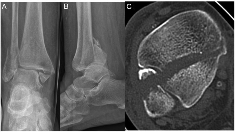
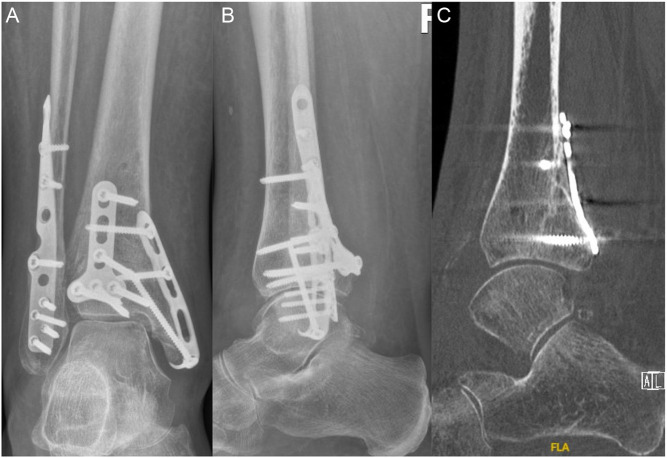
Locking plates were designed predominantly for the elderly population, as they enhance the stability and reduce wound healing problems (32, 33, 34). However, a meta-analysis of biomechanical studies showed no superiority of lateral locking plates compared to conventional lateral plates but an equal stability also in weak bone, indicating a beneficial effect in osteoporotic bone (35). A posterior polyaxial locking plate showed biomechanically no difference to a non-locking posterior plate (36) (Figs. 3, 4 and 5).
Figure 3.
Female patient, 86 years, suffering from diabetes and who had a fall at home. Highly unstable trimalleolar ankle fracture. (A, B) Conventional AP and lateral x-ray with ankle dislocation. (C) Axial CT image with large posterior malleolus fracture (Bartonicek IV).
Figure 4.
(A, B) X-ray 3 months after ORIF of PM with 3.5 mm T-Plate, ORIF fibula with anatomical 3.0/3.5 mm posterolateral plate, ORIF malleolus medialis with 2.7 mm hook plate LCP. (C) Sagittal CT image showing consolidation of PM fracture.
Figure 5.
Soft tissue 3 months after ORIF of complex trimalleolar ankle fracture (see Figs. 3 and 4). Full weightbearing.
Intramedullary fibula nail
The minimal invasive implantation of intramedullary nails (INs) for unstable distal fibula fractures minimizes the soft tissue damage during the fibula osteosynthesis. However, these new implants might lack the reliability of anatomic reconstruction. Previous studies revealed not only fewer complications of INs compared to standard plate osteosynthesis but also an equal functional outcome (37, 38). Intramedullary nailing bears the advantage of immediate full weightbearing and provides the same biomechanical stability compared to a lag screw in combination with a locking plate (39, 40). A systematic review with 627 patients treated with a locked IN stated a consolidation rate of 98% with an infection rate of 1% and skin necrosis in 0.6% (41). A recent meta-analysis consisting of four randomized controlled trials with 359 patients comparing IN to ORIF demonstrated fewer wound-related complications and a better functional short-term 3-month-follow-up for the IN group but no significant differences regarding the overall complications, midterm functional outcome and quality of reduction (42).
Primary hindfoot arthrodesis
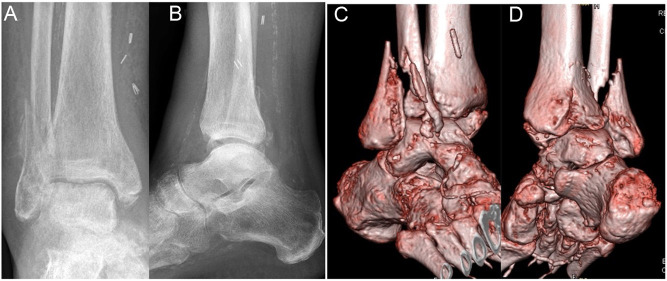
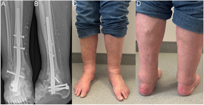
In elderly patients suffering an unstable ankle fracture with severe soft tissue impairment or relevant comorbidities such as diabetes mellitus, an open reduction and internal fixation might be an unsafe option. These patients are often incapable of partial weightbearing and therefore require a treatment option allowing full weightbearing. A primary arthrodesis of the hindfoot including the ankle and subtalar joint (tibiotalocalcaneal arthrodesis (TTC)) might be a safe option for geriatric patients with a consolidation rate of up to 95% and the advantage of allowing immediate full weightbearing (43) and a minimal invasive technique with a hindfoot nail. Furthermore, an atypical, closed reduction and insertion of the nail without an open removal of the cartilage is recommended by some authors, as soft tissue-related complications are minimized by this technique (44). We personally do not have any experience with this modified technique. Recently, a prospective randomized controlled study including 87 patients demonstrated an equal functional outcome of the TTC arthrodesis compared to ORIF, whereas the revision rate seems to be significantly lower (TTC 3% vs ORIF 14%) (45). A systematic review and meta-analysis by Lu et al. revealed high complication rates after TTC arthrodesis with 10% superficial infection, 8% deep infection, 11% implant failure, 11% malunion and 27% all-cause mortality in a high-risk patient cohort with a mean age of 78 years and a diabetes mellitus prevalence rate of 42% (46). These disappointing reports must be seen in relation to the severity of complex bi- or tri-malleolar fractures (Figs 6 and 7 ).
Figure 6.
Male patient, 79 years, who had a fall during stay in cardiology (revision of pacemaker). Complex trimalleolar ankle fracture with multifragmentary fibula fracture, posterior malleolus fracture (Bartonicek II) and multifragmentary malleolus medialis fracture. Comorbidities: coronary heart disease with bypass operation 5 years before. (A, B) Plain radiographs after closed reduction. (C, D) 3D CT scan images.
Figure 7.
Hindfoot arthrodesis with HAN – nail and removal of cartilage in ankle and subtalar joint. (A, B) Plain radiographs 1 year after surgery. (C, D) Clinical images 1 year after surgery with full weightbearing, good soft tissue condition and no pain.
Further options
In cases of a geriatric comminuted distal fibula fracture, some authors propose double plating of the distal fibula in a dorsal and lateral position (47, 48). Compared to an angular stable locking plate, conventional double plating reaches an equal biomechanical stability (49). The incidence of implant irritation seems not to be increased (50).
Tibia-pro-fibula screws, inserted through a fibula plate into the tibia, are another option to strengthen the osteosynthesis in ankle fractures with an increased biomechanical stability compared to locking plates and even lower complication and revision rates compared to intramedullary nailing (51, 52).
Intramedullary (cannulated) partially or fully threaded screws for distal fibula fractures might be an additional option, which allow a minimal invasive implantation with a short skin incision 1–2 cm distal to the fibula tip but bear the disadvantage of a reduced control of reduction (53).
Medial malleolus fractures
Geriatric medial malleolar fractures are often more complex due to a comminuted fracture pattern, decreased bone quality, a high grade of instability and decreased patient compliance. A more rigid and stable fixation compared to the classic fixation with two lag screws might be required. An option to increase the stability is a bicortical placement of the lag screw into the lateral tibial cortex, providing a better biomechanical, radiographic and clinical outcome compared to the monocortical placement (54). However, in cases of a more vertical fracture line or a comminuted fracture, a screw placement can be impossible. Hence, a plate osteosynthesis might be required. A hook plate LCP osteosynthesis not only increased stability but also decreased complication and revision rates compared to screw osteosynthesis in elderly people (55, 56).
Postoperative considerations
The primary goal in the treatment of geriatric ankle fractures remains the achievement of a stable union and the conservation of the quality of life. Regular clinical and radiological follow-ups to detect and treat complications are recommended. An adequate blood sugar concentration in patients with diabetes mellitus (HbA1c <6.5%) increases the radiological and functional outcome and decreases the complication rate in ankle fractures (57, 58).
In cases of unstable ankle fractures, postoperative restricted load for 6–8 weeks might be desirable. However, partial weightbearing can be very challenging or even impossible for elderly people. Casts or walker orthosis may reduce the peak pressure with loading (59), while early physiotherapy is recommended (7). Recently, a systematic review found that early permissive weightbearing might not only be safe but even beneficial to elderly people above 80 years for both operatively and conservatively treated unstable ankle fractures (60).
ICMJE conflict of interest statement
The authors declare that there is no conflict of interest that could be perceived as prejudicing the impartiality of the research reported.
Funding
This work did not receive any specific grant from any funding agency in the public, commercial or not-for-profit sector.
References
- 1.Milstrey A Baumbach SF Pfleiderer A Evers J Boecker W Raschke MJ Polzer H & Ochman S. Trends of incidence and treatment strategies for operatively treated distal fibula fractures from 2005 to 2019: a nationwide register analysis. Archives of Orthopaedic and Trauma Surgery 20221423771–3777. ( 10.1007/s00402-021-04232-0) [DOI] [PMC free article] [PubMed] [Google Scholar]
- 2.Daly PJ Fitzgerald RH & Melton LJ. Llstrup DM. Epidemiology of ankle fractures in Rochester, Minnesota. Acta Orthopaedica Scandinavica 200958539–544. ( 10.3109/17453678709146395) [DOI] [PubMed] [Google Scholar]
- 3.Valtola A Honkanen R Kröger H Tuppurainen M Saarikoski S & Alhava E. Lifestyle and other factors predict ankle fractures in perimenopausal women: a population-based prospective cohort study. Bone 200230238–242. ( 10.1016/s8756-3282(0100649-4) [DOI] [PubMed] [Google Scholar]
- 4.Gunnes M Mellström D & Johnell O. How well can a previous fracture indicate a new fracture? A questionnaire study of 29, 802 postmenopausal women. Acta Orthopaedica Scandinavica 199869508–512. ( 10.3109/17453679808997788) [DOI] [PubMed] [Google Scholar]
- 5.So E Rushing CJ Simon JE Goss DA Prissel MA & Berlet GC. Association between bone mineral density and elderly ankle fractures: a systematic review and meta-analysis. Journal of Foot and Ankle Surgery 2020591049–1057. ( 10.1053/j.jfas.2020.03.012) [DOI] [PubMed] [Google Scholar]
- 6.Biver E Durosier C Chevalley T Herrmann FR Ferrari S & Rizzoli R. Prior ankle fractures in postmenopausal women are associated with low areal bone mineral density and bone microstructure alterations. Osteoporosis International 2015262147–2155. ( 10.1007/s00198-015-3119-9) [DOI] [PubMed] [Google Scholar]
- 7.Ochman S & Raschke MJ. Sprunggelenkfraktur beim älteren Patienten. Unfallchirurg 2021124200–211. ( 10.1007/s00113-021-00953-4) [DOI] [PubMed] [Google Scholar]
- 8.Lee KM Chung CY Kwon SS Won SH Lee SY Chung MK & Park MS. Ankle fractures have features of an osteoporotic fracture. Osteoporosis International 2013242819–2825. ( 10.1007/s00198-013-2394-6) [DOI] [PubMed] [Google Scholar]
- 9.Pichl J & Hoffmann R. Ankle fractures in the elderly. Unfallchirurg 2011114 681–687. ( 10.1007/s00113-011-2023-9) [DOI] [PubMed] [Google Scholar]
- 10.Zwipp H & Amlang M. Frakturversorgung des oberen Sprunggelenks im hohen Lebensalter. Orthopäde 201443332–338. ( 10.1007/s00132-013-2168-z) [DOI] [PubMed] [Google Scholar]
- 11.Jubel A Faymonville C Andermahr J Boxberg S & Schiffer G. Einschränkungen der Aussagekraft des konventionellen Röntgenbilds bei Sprunggelenksfrakturen im Alter. Zeitschrift für Orthopädie und Unfallchirurgie 201615545–51. ( 10.1055/s-0042-113879) [DOI] [PubMed] [Google Scholar]
- 12.Aigner R Lechler P Boese CK Bockmann B Ruchholtz S & Frink M. Standardised pre-operative diagnostics and treatment of peripheral arterial disease reduce wound complications in geriatric ankle fractures. International Orthopaedics 201842395–400. ( 10.1007/s00264-017-3705-x) [DOI] [PubMed] [Google Scholar]
- 13.Anand N & Klenerman L. Ankle fractures in the elderly: MUA versus ORIF. Injury 199324116–120. ( 10.1016/0020-1383(9390202-h) [DOI] [PubMed] [Google Scholar]
- 14.Pagliaro AJ Michelson JD & Mizel MS. Results of operative fixation of unstable ankle fractures in geriatric patients. Foot and Ankle International 200122399–402. ( 10.1177/107110070102200507) [DOI] [PubMed] [Google Scholar]
- 15.Zaghloul A Haddad B Barksfield R & Davis B. Early complications of surgery in operative treatment of ankle fractures in those over 60: a review of 186 cases. Injury 201445780–783. ( 10.1016/j.injury.2013.11.008) [DOI] [PubMed] [Google Scholar]
- 16.Lynde MJ Sautter T Hamilton GA & Schuberth JM. Complications after open reduction and internal fixation of ankle fractures in the elderly. Foot and Ankle Surgery 201218103–107. ( 10.1016/j.fas.2011.03.010) [DOI] [PubMed] [Google Scholar]
- 17.Srinivasan CMS & Moran CG. Internal fixation of ankle fractures in the very elderly. Injury 200132559–563. ( 10.1016/s0020-1383(0100034-1) [DOI] [PubMed] [Google Scholar]
- 18.Shivarathre DG Chandran P & Platt SR. Operative fixation of unstable ankle fractures in patients aged over 80 years. Foot and Ankle International 201132599–602. ( 10.3113/FAI.2011.0599) [DOI] [PubMed] [Google Scholar]
- 19.Day GA Swanson CE & Hulcombe BG. Operative treatment of ankle fractures: A minimum ten-year follow-up. Foot and Ankle International 200122102–106. ( 10.1177/107110070102200204) [DOI] [PubMed] [Google Scholar]
- 20.SooHoo NF Krenek L Eagan MJ Gurbani B Ko CY & Zingmond DS. Complication rates following open reduction and internal fixation of ankle fractures. Journal of Bone and Joint Surgery. American Volume 2009911042–1049. ( 10.2106/JBJS.H.00653) [DOI] [PubMed] [Google Scholar]
- 21.Larsen P Rathleff MS & Elsoe R. Surgical versus conservative treatment for ankle fractures in adults: a systematic review and meta-analysis of the benefits and harms. Foot and Ankle Surgery 201925409–417. ( 10.1016/j.fas.2018.02.009) [DOI] [PubMed] [Google Scholar]
- 22.Willett K, Keene DJ, Mistry D, Nam J, Tutton E, Handley R, Morgan L, Roberts E, Briggs A, Lall R, et al. Close contact casting vs surgery for initial treatment of unstable ankle fractures in older adults: a randomized clinical trial. JAMA 20163161455–1463. ( 10.1001/jama.2016.14719) [DOI] [PubMed] [Google Scholar]
- 23.Klos K Simons P Mückley T Karich B Randt T & Knobe M. Frakturen des oberen Sprunggelenks beim älteren Patienten. Unfallchirurg 2017120979–992. ( 10.1007/s00113-017-0423-1) [DOI] [PubMed] [Google Scholar]
- 24.Bartoníček J Rammelt S Kostlivý K Vaněček V Klika D & Trešl I. Anatomy and classification of the posterior tibial fragment in ankle fractures. Archives of Orthopaedic and Trauma Surgery 2015135505–516. ( 10.1007/s00402-015-2171-4) [DOI] [PubMed] [Google Scholar]
- 25.Bartoníček J Rammelt S & Tuček M. Posterior malleolar fractures changing concepts and recent developments. Foot and Ankle Clinics 201722125–145. ( 10.1016/j.fcl.2016.09.009) [DOI] [PubMed] [Google Scholar]
- 26.Blom RP, Hayat B, Al-Dirini RMA, Sierevelt I, Kerkhoffs GMMJ, Goslings JC, Jaarsma RL, Doornberg JN. & EF3X-trial Study Group. Posterior malleolar ankle fractures. Bone and Joint Journal 2020102–B1229–1241. ( 10.1302/0301-620X.102B9.BJJ-2019-1660.R1) [DOI] [PubMed] [Google Scholar]
- 27.Hartwich K Gomez AL Pyrc J Gut R Rammelt S & Grass R. Biomechanical analysis of stability of posterior antiglide plating in osteoporotic pronation abduction ankle fracture model with posterior tibial fragment. Foot and Ankle International 20173858–65. ( 10.1177/1071100716669359) [DOI] [PubMed] [Google Scholar]
- 28.Choi JY Kim JH Ko HT & Suh JS. Single oblique posterolateral approach for open reduction and internal fixation of posterior malleolar fractures with an associated lateral malleolar fracture. Journal of Foot and Ankle Surgery 201554559–564. ( 10.1053/j.jfas.2014.09.043) [DOI] [PubMed] [Google Scholar]
- 29.Abdelgawad AA Kadous A & Kanlic E. Posterolateral approach for treatment of posterior malleolus fracture of the ankle. Journal of Foot and Ankle Surgery 201150607–611. ( 10.1053/j.jfas.2011.04.022) [DOI] [PubMed] [Google Scholar]
- 30.Baumbach SF Herterich V Damblemont A Hieber F Böcker W & Polzer H. Open reduction and internal fixation of the posterior malleolus fragment frequently restores syndesmotic stability. Injury 201950564–570. ( 10.1016/j.injury.2018.12.025) [DOI] [PubMed] [Google Scholar]
- 31.Minihane KP Lee C Ahn C Zhang LQ & Merk BR. Comparison of lateral locking plate and antiglide plate for fixation of distal fibular fractures in osteoporotic bone&colon: a biomechanical study. Journal of Orthopaedic Trauma 200620562–566. ( 10.1097/01.bot.0000245684.96775.82) [DOI] [PubMed] [Google Scholar]
- 32.Shih CA Jou IM Lee PY Lu CL Su WR Yeh ML & Wu PT. Treating AO/OTA 44B lateral malleolar fracture in patients over 50 years of age: periarticular locking plate versus non-locking plate. Journal of Orthopaedic Surgery and Research 202015 112. ( 10.1186/s13018-020-01622-9) [DOI] [PMC free article] [PubMed] [Google Scholar]
- 33.Zahn RK Frey S Jakubietz RG Jakubietz MG Doht S Schneider P Waschke J & Meffert RH. A contoured locking plate for distal fibular fractures in osteoporotic bone: a biomechanical cadaver study. Injury 201243718–725. ( 10.1016/j.injury.2011.07.009) [DOI] [PubMed] [Google Scholar]
- 34.Switaj PJ Fuchs D Alshouli M Patwardhan AG Voronov LI Muriuki M Havey RM & Kadakia AR. A biomechanical comparison study of a modern fibular nail and distal fibular locking plate in AO/OTA 44C2 ankle fractures. Journal of Orthopaedic Surgery and Research 201611 100. ( 10.1186/s13018-016-0435-5) [DOI] [PMC free article] [PubMed] [Google Scholar]
- 35.Dingemans SA Lodeizen OAP Goslings JC & Schepers T. Reinforced fixation of distal fibula fractures in elderly patients: a meta-analysis of biomechanical studies. Clinical Biomechanics 20163614–20. ( 10.1016/j.clinbiomech.2016.05.006) [DOI] [PubMed] [Google Scholar]
- 36.Hallbauer J Klos K Gräfenstein A Simons P Rausch S Mückley T & Hofmann GO. Does a polyaxial-locking system confer benefits for osteosynthesis of the distal fibula: a cadaver study. Orthopaedics and Traumatology, Surgery and Research 2016102645–649. ( 10.1016/j.otsr.2016.03.014) [DOI] [PubMed] [Google Scholar]
- 37.White TO Bugler KE Appleton P Will E McQueen MM & Court-Brown CM. A prospective randomised controlled trial of the fibular nail versus standard open reduction and internal fixation for fixation of ankle fractures in elderly patients. Bone and Joint Journal 201698-B1248–1252. ( ) [DOI] [PubMed] [Google Scholar]
- 38.Asloum Y Bedin B Roger T Charissoux JL Arnaud JP & Mabit C. Internal fixation of the fibula in ankle fractures. A prospective, randomized and comparative study: plating versus nailing. Orthopaedics and Traumatology, Surgery and Research 2014100S255–S259. ( 10.1016/j.otsr.2014.03.005) [DOI] [PubMed] [Google Scholar]
- 39.Carter TH Wallace R Mackenzie SA Oliver WM Duckworth AD & White TO. The fibular intramedullary nail versus locking plate and lag screw fixation in the management of unstable elderly ankle fractures: a cadaveric biomechanical comparison. Journal of Orthopaedic Trauma 202034e401–e406. ( 10.1097/BOT.0000000000001814) [DOI] [PubMed] [Google Scholar]
- 40.Sain A Garg S Sharma V Meena UK & Bansal H. Osteoporotic distal fibula fractures in the elderly: how to fix them. Cureus 202012 e6552. ( 10.7759/cureus.6552) [DOI] [PMC free article] [PubMed] [Google Scholar]
- 41.Jain S Haughton BA & Brew C. Intramedullary fixation of distal fibular fractures: a systematic review of clinical and functional outcomes. Journal of Orthopaedics and Traumatology 201415245–254. ( 10.1007/s10195-014-0320-0) [DOI] [PMC free article] [PubMed] [Google Scholar]
- 42.Guo W Wu F Chen W Tian K Zhuang R & Pan Y. Can locked fibula nail replace plate fixation for treatment of acute ankle fracture? A systematic review and meta-analysis. Journal of Foot and Ankle Surgery 202362178–185. ( 10.1053/j.jfas.2022.10.003) [DOI] [PubMed] [Google Scholar]
- 43.Fadhel WB Taieb L Villain B Mebtouche N Levante S Bégué T & Aurégan JC. Outcomes after primary ankle arthrodesis in recent fractures of the distal end of the tibia in the elderly: a systematic review. International Orthopaedics 2022461405–1412. ( 10.1007/s00264-022-05317-0) [DOI] [PubMed] [Google Scholar]
- 44.Dimitroulias A. Ankle tibiotalocalcaneal nailing in elderly ankle fractures as an alternative to open reduction internal fixation: technique and literature review. Ota International 20225 e183. ( 10.1097/OI9.0000000000000183) [DOI] [PMC free article] [PubMed] [Google Scholar]
- 45.Georgiannos D Lampridis V & Bisbinas I. Fragility fractures of the ankle in the elderly: open reduction and internal fixation versus tibio-talo-calcaneal nailing: short-term results of a prospective randomized-controlled study. Injury 201748519–524. ( 10.1016/j.injury.2016.11.017) [DOI] [PubMed] [Google Scholar]
- 46.Lu V Tennyson M Zhou A Patel R Fortune MD Thahir A & Krkovic M. Retrograde tibiotalocalcaneal nailing for the treatment of acute ankle fractures in the elderly: a systematic review and meta-analysis. EFORT Open Reviews 20227628–643. ( 10.1530/EOR-22-0017) [DOI] [PMC free article] [PubMed] [Google Scholar]
- 47.Vance DD & Vosseller JT. Double plating of distal fibula fractures. Foot and Ankle Specialist 201710543–546. ( 10.1177/1938640017692416) [DOI] [PubMed] [Google Scholar]
- 48.McKean J Cuellar DO Hak D & Mauffrey C. Osteoporotic ankle fractures: an approach to operative management. Orthopedics 201336936–940. ( 10.3928/01477447-20131120-07) [DOI] [PubMed] [Google Scholar]
- 49.Randall RM Nagle T Steckler A Billow D & Berkowitz MJ. Dual nonlocked plating as an alternative to locked plating for comminuted distal fibula fractures: a biomechanical comparison study. Journal of Foot and Ankle Surgery 201958916–919. ( 10.1053/j.jfas.2019.01.017) [DOI] [PubMed] [Google Scholar]
- 50.Kwaadu KY Fleming JJ & Lin D. Management of complex fibular fractures: double plating of fibular fractures. Journal of Foot and Ankle Surgery 201554288–294. ( 10.1053/j.jfas.2013.08.002) [DOI] [PubMed] [Google Scholar]
- 51.Eyre-Brook AI Ring J Chadwick C Davies H Davies M & Blundell C. A comparison of fibula pro-tibia fixation versus hindfoot nailing for unstable fractures of the ankle in those older than 60 years. Foot and Ankle Specialist 202119386400211017373. ( 10.1177/19386400211017373) [DOI] [PubMed] [Google Scholar]
- 52.Panchbhavi VK Vallurupalli S & Morris R. Comparison of augmentation methods for internal fixation of osteoporotic ankle fractures. Foot and Ankle International 200930696–703. ( 10.3113/FAI.2009.0696) [DOI] [PubMed] [Google Scholar]
- 53.Smith M Medlock G & Johnstone AJ. Percutaneous screw fixation of unstable ankle fractures in patients with poor soft tissues and significant co-morbidities. Foot and Ankle Surgery 20172316–20. ( 10.1016/j.fas.2015.11.008) [DOI] [PubMed] [Google Scholar]
- 54.Ricci WM Tornetta P & Borrelli J. Lag screw fixation of medial malleolar fractures: A biomechanical, radiographic, and clinical comparison of unicortical partially threaded lag screws and bicortical fully threaded lag screws. Journal of Orthopaedic Trauma 201226602–606. ( 10.1097/BOT.0b013e3182404512) [DOI] [PubMed] [Google Scholar]
- 55.Cho BK Kim JB & Choi SM. Efficacy of hook-type locking plate and partially threaded cancellous lag screw in the treatment of displaced medial malleolar fractures in elderly patients. Archives of Orthopaedic and Trauma Surgery 20221422585–2596. ( 10.1007/s00402-021-03945-6) [DOI] [PubMed] [Google Scholar]
- 56.Vajapey SP & Harrison RK. Hook plate fixation of medial malleolar fractures: A comparative study of clinical outcomes. Journal of Foot and Ankle Surgery 202059969–971. ( 10.1053/j.jfas.2018.12.048) [DOI] [PubMed] [Google Scholar]
- 57.Liu J Ludwig T & Ebraheim NA. HbA1c and diabetic ankle fractures. Orthopaedic Surgery 20135203–208. ( 10.1111/os.12047) [DOI] [PMC free article] [PubMed] [Google Scholar]
- 58.Shibuya N Humphers JM Fluhman BL & Jupiter DC. Factors associated with nonunion, delayed union, and malunion in foot and ankle surgery in diabetic patients. Journal of Foot and Ankle Surgery 201352207–211. ( 10.1053/j.jfas.2012.11.012) [DOI] [PubMed] [Google Scholar]
- 59.Ehrnthaller C Rellensmann K Baumbach SF Wuehr M Schniepp R Saller MM Böcker W & Polzer H. Pedobarographic evaluation of five commonly used orthoses for the lower extremity. Archives of Orthopaedic and Trauma Surgery 20221–8. ( 10.1007/s00402-022-04729-2) [DOI] [PMC free article] [PubMed] [Google Scholar]
- 60.Halsema van MS Boers RAR & Leferink VJM. An overview on the treatment and outcome factors of ankle fractures in elderly men and women aged 80 and over: a systematic review. Archives of Orthopaedic and Trauma Surgery 20221423311–3325. ( 10.1007/s00402-021-04161-y) [DOI] [PMC free article] [PubMed] [Google Scholar]


 This work is licensed under a