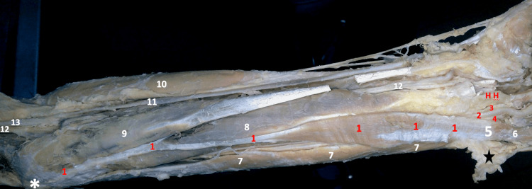
Figure 2. Gross image of the dissected left forearm .
1- Reversed Left Palmaris Longus Muscle (the belly of the reversed muscle reaching the pisiform bone), 2- Ulnar Artery, 3- Deep Branch & 4- Superficial Branch of Ulnar Nerve, 5- Pisiform Bone, 6- Abductor Digiti Minimi, 7- Flexor Carpi Ulnaris, 8- Flexor Digitorum Superficialis Muscle, 9- Flexor Carpi Radialis, 10- Brachioradialis Muscle 11- Radial Artery 12- Median Nerve, 13- Brachial Artery
HH- Hook of Hamate
✱ Medial epicondyle ★ Skin and fascia are reflected

