Abstract
Iatrogenic hallux varus is formed due to an imbalance between the bone, tendon, and ligamentous-articular structures in the first metatarsophalangeal joint (MJ), with the progression of the medial deviation of the big toe. A secondary factor is an imbalance between excessive medial tension and weakness or excessive soft tissue release of lateral capsular-ligamentous structures. The article is devoted to a rare but no less complex postoperative complication of hallux valgus deformity, acquired hallux varus. Based on the literature data on this topic, in the work, the clinical anatomy of the first metatarsophalangeal joint, the pathogenesis of the development of tendon-muscle imbalance in the above joint, and the leading causes of hallux varus are described. Also, the issues of the clinic, X-ray diagnostics, and classification of this type of foot pathology are considered with a description of the appropriate surgical tactics for different types of deformity.
Keywords: Iatrogenic hallux varus, Hallux valgus, Corrective surgery, Complications
1. Context
Iatrogenic hallux varus is formed due to an imbalance between the bone, tendon, and ligamentous-articular structures in the first metatarsophalangeal joint (MJ), with the progression of the medial deviation of the big toe. A secondary factor is an imbalance between excessive medial tension and weakness or excessive soft tissue release of lateral capsular-ligamentous structures ( 1 ).
In varus deformity, the big toe deviates in the medial direction concerning the head of the metatarsal bone ( 2 ). The deformity is often three-plane, with a medial deviation of the big toe; hammer-like deformity of the big toe and supination often occur ( 1 , 3 , 4 ).
The development of hallux varus is often multifactorial and includes an imbalance between bone structures, tendons, and capsular-ligamentous elements of the first metatarsophalangeal joint. Although varus deformity of the big toe is less common than hallux valgus, they are similar in that they have different severities, causes, and treatment methods ( 5 ).
Hallux varus can be congenital or acquired ( 6 ). Deformities develop in systemic diseases such as rheumatoid arthritis, psoriatic arthritis, Charcot-Marie's disease, aseptic necrosis of the metatarsal head, and poliomyelitis. Acquired hallux varusis divided into several subtypes: iatrogenic, traumatic, and post-burn ( 5 , 7 ).
In our article, we will consider the issues of diagnosis and treatment of iatrogenic hallux varus.
Iatrogenic deformity of the big toe often occurs after surgical correction of the hallux valgus. According to various literature sources, the frequency of iatrogenic deformity of the big toe after surgery for hallux valgus is from 2% -17% ( 3 , 5 , 8 ). A large number of sufferers are women, whose average age is 45-50 years ( 9 ).
As a result, of the progression of the varus deformity of the big toe, the patients complain of cosmetic deformity, difficulties in choosing shoes, and pain syndrome ( 4 ).
Therefore, this study was designed to review the literature on the diagnosis and treatment of iatrogenic varus deformity of the big toe after corrective surgery on the hallux valgus.
2. Evidence Acquisition
The search for scientific publications was carried out in search systems: Web of Science, Research Gate, PubMed, and Google Scholar. The criteria for including publications in the literature review are determined - publications with full English text. Since the literature on the treatment of hallux valgus consists of most of a retrospective examination, in our article, we used articles with IV level of evidence.
3. Results
3.1. Anatomy
In a normal foot, the big toe is in the middle position, and the lateral deviation does not exceed 15°. The internal muscles of the foot are fixed at the base of the proximal phalanx and include flexor halluces brevis, extensor halluces brevis, adductor, and abductor hallucis. These short muscles stabilize the big toe and affect the rotation and medial and lateral deviation, especially when imbalanced.
External muscles such as flexor hallucis longus and extensor hallucis longus also affect stability, but they affect the mobility of the metatarsophalangeal joint during flexion and extension. The flexor hallucis brevis has a medial and lateral head that attaches to the proximal phalanx through the sesamoid bones ( 4 ). The plantar surface of the metatarsal head has two, separated by a longitudinal rib, in the pecten of which the medial and lateral sesamoid bones slide. The plantar surface of the head of the metatarsal bone has two grooves, separated by a longitudinal rib, in the pecten of which the medial and lateral sesamoid bones slide (Figure 1) ( 10 ).
Figure 1.
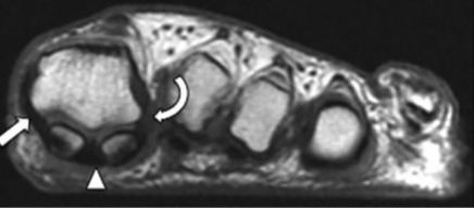
The arrow shows the sesamoid grooves and pectens on the head of the first metatarsal bone
3.2. Pathogenesis
The identified cases of hallux varus are mainly associated with aggressive correction of the hallux valgus. Excessive release of lateral articular structures, excessive tension of the medial articular capsule, and overcorrection of the first intermetatarsal angle are the main reasons for the described cases. The combination of the above factors leads to bone and soft tissue imbalance, which allows normal tendon-muscular efforts to turn towards progressive varus deformity ( 1 , 4 , 5 ).
Tight suturing of the medial capsule may be caused by extensive resection of the excess medial capsule during dissection. An overly aggressive postoperative dressing that holds the metatarsophalangeal joint in the varus position for an extended period can also lead to progressive scarring of the medial joint capsule in a shortened, overly tightened position ( 4 , 8 ).
Another factor in the development of hallux varus is overly extensive resection of the medial eminence of the metatarsal head. It reduces the medial bone support of the metatarsal head and disturbs the proximal phalanx balance. This, in turn, will lead to a varus deflection of the phalanx. Excessive resection of the medial exostosis also destabilizes the tibial sesamoid bone, which subsequently subluxes the medial side and increases varus traction. Since the sagittal groove stabilizes the medial base of the proximal phalanx and helps form the medial border of the tibial sesamoid bone, the sagittal base is recommended groove be preserved during resection of the medial exostosis. Resection of the medial exostosis should begin 2 mm away from the sagittal groove. The direction of the incision should be from dorsolateral to plantomedial. This preserves the plantomedial metatarsal head and provides a fulcrum effect ( 1 , 5 , 8 ).
Overcorrection of 1-2 metatarsal angles can result in hallux varus after distal or proximal metatarsal osteotomy. Furthermore, it is more common after a proximal metatarsal osteotomy if the intermetatarsal angle is zero or negative; the forces of the forming medial varus deformity increase and play a significant role in the formation of hallux varus ( 5 , 8 ).
Maximum lateral movement of the metatarsal head during osteotomy should be avoided. After metatarsal osteotomy and medial capsular suture, the first metatarsophalangeal joint must be maintained in a normal anatomical valgus position to prevent overcorrection of the big toe. The neutral or varus position of the MPJ is the cause of bone imbalance and can lead to phalanx varus deformity ( 4 ).
Although hallux varus occurs as a combination of different surgical interventions, it is mainly described and disassembled after the MacBright procedure with the removal of the fibular sesamoid bone. Fibular sesamidectomy will cause the tibial sesamoid bone to mix inward, while the release of the adductor tendon allows the abductor tendon to act without resistance. The subsequent metatarsophalangeal joint rotates on the varus, and tendons of the big toe's extensor longus, flexor longus, and extensor brevis are displaced inward from the midline and contribute to strengthening the varus setting of the big toe. When the medial head of the flexor brevis is sliding inward along with the tibial sesamoid bone, it can no longer act as an effective flexor and is suppressed by the metatarsophalangeal flexor brevis, which leads to an extension of the big toe in the metatarsophalangeal joint. Extension of the big toe in the metatarsophalangeal joint leads to contraction of the flexor longus and weakening of the extensor longus, which leads to flexion in the interphalangeal joint ( 4 , 5 ).
Akin's osteotomy can also cause varus deformity of the big toe. The normal active axis of the first radius, with a slight varus deviation of the first metatarsal, with a slight hallux valgus, along with the capsular-ligamentous tension, as a rule, creates a straight line of FHL and EHL tendon traction through the first metatarsophalangeal joint. With the deviation of the big toe into the varus with a decrease in the interphalangeal angle (IPA) due to the Akin osteotomy, the tension of the aforementioned muscles can create a varus moment in the first MPJ, which further enhances the varus deformity ( 4 ).
Finally, caution should be exercised in postoperative care, and dressing patients with hallux valgus since aggressive bandaging and taping of the big toe in the varus position can lead to a tightening scar in the medial joint capsule ( 5 , 8 ).
3.3. Clinical Examination
The diagnosis of hallux varus is based on a clinical trial. The medical history should focus on the prescription of previous corrective surgery on hallux valgus, the type of surgery, and the chronology of symptoms. Patients complain more about cosmetic defects and inconvenience in wearing shoes than pain syndrome. During the examination, it is necessary to focus on the MPJ and the interphalangeal joint and their elasticity and rigidity to plan treatment tactics.
Elastic deformations that are passively adjusted should be examined in the sitting and standing positions, as contractures can change depending on the load. At the same time, rigid deformities as a result of prolonged contractures are not reduced. Clinical findings may include:
- medial displacement of the big toe in the first metatarsophalangeal joint;
- supination of the big toe;
- a dorsal extension of the proximal phalanx of the big toe;
- medial tension of the extensor hallucis longus;
- medially combined, painful on palpation of tibial sesamoid bone (tibial sesamoid bone is palpable medially and painful);
- varus change in the position of the big toe;
- hammer-like contracture in the interphalangeal joint and callus on the dorsal surface;
- bursitis in the interphalangeal joint;
- elongated big toe and first radius;
- increased varus deviation of the rest fingers.
Compensatory supination of the posterior part of the foot with overloading of the outer edge of the middle part of the foot with pain syndrome due to a decrease in the support function of the big toe in the gait cycle ( 4 ).
3.4. X-Ray Examination
Dorso-plantar and lateral radiography under load is performed before, one year after, and during the final monitoring. The angle of valgus deviation of the first toe (М1Р1), the angle of varus deviation of the first metatarsal (M1 M2) or the first intermetatarsal angle (IM), the congruence of the first MPJ, and measuring the index of the metatarsal bones are the indicators recommended AOFAS ( 2 ).
Radiography in a straight projection standing under load is valuable for determining the deformation of the MPJ. However, oblique and lateral projection is useful for a better understanding three-plane deformation. The sesamoid position will help determine the position of the sesamoid bone in relation to the head of the first metatarsal bone. The hallux valgus angle is formed between the first metatarsal bone's longitudinal axis and the proximal phalanx's longitudinal axis ( 11 ). Normal values vary from 5 ° to 15 ° ( 12 ).
The above angle will change to 0° or negative in hallux varus cases. Other findings include:
- Overly resected medial eminence;
- Medial subluxation of the tibial sesamoid bone from the sesamoid canal;
- Lack of fibular sesamoid bone;
- The reduced intermetatarsal angle between the longitudinal axis of the first and second metatarsal bones. Normally from 0 ° to 8 °. With hallux varus, it may be 0 ° or negative;
- The first metatarsal bone is longer than the second;
- Varus deformity of the phalanges or incorrectly fused phalanx fracture after a previous complication;
- Cysts or other changes in the metatarsophalangeal and interphalangeal joint;
- Arthrosis, deformation, or hypertrophy of the sesamoid bones ( 1 , 4 ).
Lateral radiography usually reveals:
- Dorsoflexion of the proximal phalanx in MPJ, with or without concomitant plantar flexion in the interphalangeal joint;
- Incorrectly fused fractures of the phalanges in the position of dorsal flexion;
- Elevation of the metatarsal head ( 1 ).
The ideal classification system acts as a reliable and reproducible tool that can be used practically, in terms of a concomitant treatment algorithm, and in terms of providing comparable groups for research. However, such a classification does not exist for iatrogenic hallux varus since each deformity uniquely depends on the degree and nature of iatrogenic damage ( 13 ). In almost all cases, deformity occurs due to corrective surgery on the hallux valgus ( 14 ).
For the first time, the classification of hallux varus was proposed by Hawkins ( 14 ) in 1971. According to him, the acquired hallux varus can be of a static or dynamic type ( 14 ). The static deformity is usually asymptomatic, unilateral, passively corrected, and formed without muscle imbalance. The dynamic deformity is multifaceted, fixed, usually symptomatic, and arises from muscle imbalance in the MPJ ( 4 ). The static deformity occurs due to anatomical factors such as excessive resection of the medial eminence, joint instability after resection arthroplasty, or excessive shortening of the first radius during surgery.
The dynamic deformity is multi-plane and is caused by a muscle imbalance in the first metatarsophalangeal joint. An imbalance occurs after a procedure involving the adductor hallucis muscles with or without a medial capsular suture ( 2 , 8 ).
Akhtar, Malek ( 8 ), 2016 proposed their classification based on the anatomical factors associated with hallux varus. They determine three types of deformity: 1) bone, 2) myoligamentous, and 3) combined. Below is a description of the classification.
Type 1: Bone.
Varus deformity may result from excessive lateral displacement from scarf osteotomy leading to overcorrection of the first intermetatarsal angle (IMA) or excessive resection of the medial eminence of the metatarsal bone behind the sagittal sulcus.
Type 2: Myoligamentous.
Deformation can have internal and external causes. Internal causes usually involve the excessive release of lateral structures (mainly the lateral ligament) and excessive tension in the medial capsule. External causes are inappropriate postoperative bandaging, aggressive remedial gymnastics, joint development, and wearing inappropriate footwear in the postoperative period, which may be a significant trigger in the progression of the deformity.
Type 3: Combined.
Various combinations of bone and ligamentous components where the abductor hallucis plays a vital role in the development of deformity. Strengthening the abductor hallucis without proper resistance of the adductor worsens deformity during functional loading of the foot. Suppose this adds to the loss of integration of the lateral ligament with/or excessive resection of the medial eminence. In that case, the severity of the deformity increases rapidly and often develops very quickly after the onset of functional load on the foot ( 8 ).
3.5. Conservative Treatment
Patients with hallux varus should initially be treated conservatively with bandages, taping, or by fitting appropriate orthopedic shoes. To relieve pain, in the absence of contraindications, anti-inflammatory drugs can be used ( 15 ). For symptomatic deformity, conservative treatment such as custom-made splints and corrective dressing should be used early, with appropriate physical therapy ( 8 ).
When using conservative treatment methods, early recognition of the deformity is essential. Fixed deformations do not develop very quickly. If hallux varus is recognized early after a soft tissue procedure for hallux valgus, weekly bandages and taping of the big toe in a 15 ° valgus position can correct the deformity. Surgical correction is usually required if the deformity is not recognized or not treated early ( 1 , 4 ).
3.6. Surgical Treatment
Surgical treatment options for hallux varus correction include medial soft tissue release, osteotomy of the first metatarsal bone, tendon transposition, arthrodesis of MPJ and IJP, endoprosthetics with implant placement, and resection arthroplasty ( 4 , 7 ).
Choosing a specific type of surgical treatment is the amount of movement in the metatarsophalangeal joint and the reducibility of deformity in the metatarsophalangeal and interphalangeal joints. Ligament reconstruction can be anatomical or palliative with tenodesis. Parallel to soft tissue reconstruction, the metatarsal osteotomy is used in case of excessive decrease or negative values of the angle between the first and second metatarsal bone ( 1 ).
Arthrodesis of the first metatarsophalangeal joint is recommended in inflammatory arthritis, avascular necrosis, osteoarthritis, neuromuscular disorders, and unsuccessful previous hallux varus corrective surgery ( 7 ).
After the main operation, the treatment algorithm for iatrogenic hallux varus depends on the mobility of the interphalangeal and metatarsophalangeal joints and the integration of the joint with soft tissue deformity imbalance. For tendon transposition or tenodesis to be successful, the joint must be movable and reducible. Maintaining joint mobility is preferable but not always possible due to joint arthrosis and/or severe contracture ( 4 ). With arthrosis of the joints, arthrodesis operation is justified ( 1 , 4 , 5 , 7 , 13 ). In rare cases, arthrodesis of both joints is used ( 4 ).
3.7. Soft Tissue Procedures
Medial soft tissue release is an essential initial step for correcting imbalances in overly stretched medial soft tissue elements and weakened lateral capsular and muscle-tendon structures ( 4 , 15 ). Mohan, Dhotare ( 15 ) believes that if the mild compliant deformity is found early, soft tissue release includes medial capsulation, reduction of the sesamoid bone, and 6-week wire fixation in a 10 °-15 ° valgus position.
However, Davies and Blundell ( 13 ) note that, for patients with deformities resulting from excessive stress suture on the medial capsule and ligamentous structures, and in the absence of any contracture in the joint, restoration and suture of the lateral joint capsule are not enough. However, when combined with medial capsule release and abductor tenotomy with postoperative banding, it can be effective in the early detection of deformity. However, the above measures are not very practical for severe deformations.
3.8. Tendon Movements and Renodesis
Tendon transposition has the potential for dynamic correction, while tenodesis provides static correction. The advantage of tendon movement includes maintaining movement and restoring a dynamic balance of forces in the first metatarsophalangeal joint. Contraindications to tendon movements and tenodesis include deformities of the first metatarsal bone and stiffness or arthrosis of the first metatarsophalangeal joint ( 4 ).
The surgical treatment of elastic and passively adjusted hallux varus compensates for the muscle imbalance passing through the first metatarsophalangeal joint ( 13 ).
3.9. Movement of the Abductor Hallucis Tendon
This method was first described by Hawkins ( 14 ) in 1971 with the presentation of the long-term results of surgical treatment of 3 cases of hallux varus caused after the procedure. Mc Bride. O cut off the abductor hallucis tendon from the attachment point at the base of the proximal phalanx and transferred it under the first metatarsal bone and the intermetatarsal ligament in order to pass it through the bony tunnel from the lateral surface of the proximal phalanx and secure it with a suture (Figure 2).
Figure 2.
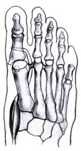
Tenodesis of the abductor hallucis tendon
Even though many researchers have tested this method, there are technical problems associated with achieving a sufficient length of the tendon for movement, and as a result, of the plantar attachment of the transferred tendon, residual supination of the toe may occur ( 13 ).
Leemrijse and Bevernage ( 1 ) described an original technique of "reverse" movement of the abductor hallucis tendon as a reconstruction of the lateral ligament of the first metatarsophalangeal joint. The first step is a wide capsular release into the medial surface of the first metatarsophalangeal joint. Then one-third of the tendon is dissected from the proximal to the distal while maintaining attachment at the base of the first toe. The second approach is performed in the first intermetatarsal space by cleaning fibrous tissues from previous interventions. Two bony tunnels were drilled in the basal phalanx and the first metatarsal bone slightly obliquely. The graft is passed first through a tunnel in the basilar phalanx, pulled through the intermetatarsal space, and passed through the metatarsal tunnel. Then the graft is sutured with non-absorbable sutures in the light valgus 10 °-15 ° transosseous (Figure 3). By this method, five patients, seven feet, were operated on (two patients underwent bilateral procedures). In five cases, patients underwent Mc Bride surgery; scarf osteotomy took place in two cases. According to the author's monitoring over 22.5 months (range 10 to 51 months), the mean on the AOFAS scale improved from 61 to 88.
Figure 3.
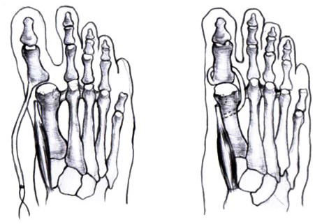
Transposition of the extensor halluces longus
Johnson and Spiegl ( 16 ) developed a more dynamic tendon transposition procedure and, in 1984, presented a new technique that resulted in 15 feet. The extensor hallucis tendon is extended along its length, dissected from its attachment at the base of the main phalanx, then passed under the intermetatarsal ligament, and fixed into the lateral surface of the base of the proximal phalanx through a tunnel drilled in the vertical direction. In addition, the interphalangeal joint undergoes arthrodesis to prevent joint flexion deformity (Figure 4). The authors report excellent results at ten feet and good at four. The result of one operation was unsatisfactory.
Figure 4.
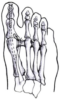
Transposition of the split extensor hallucis longus
After proposing complete movement of the extensor longus tendon, Johnson described a modified technique using split movement. The lateral half of the tendon moves like that described for full EHL, but the other half remains on the proximal phalanx. This makes interphalangeal arthrodesis unnecessary and hypothetically does not affect the ability to extend the hallux. However, when tension is applied distally to the lateral portion of the EHL tendon, it is transferred to the remaining mid-half of the EHL tendon, lengthening it and altering its function ( 4 ). Zielinska, Tubbs ( 17 ) described the results of the three-year monitoring of 12 patients after surgery to move the split extensor longus with soft tissue medial release with elastic hallux varus (Figure 5). Four patients complained of pain during support load (walking barefoot and wearing soft-soled shoes). Another seven patients had a limited range of motion in the first metatarsophalangeal joint. In five patients, plain radiographs showed an average varus position of 14 °. Clinically, varus displacement was evident in only two patients. According to the author, the restriction of movement in the first metatarsophalangeal joints should be associated with the effect of tenodesis on the transferred tendon since the picture of arthrosis was not detected on the X-ray.
Figure 5.
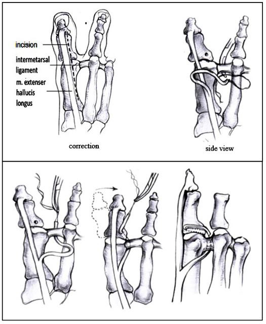
Modified transposition of the split extensor hallucis longus
Lau and Myerson ( 18 ) suggested modifying Johnson's technique due to concerns that the middle half of EHL would be limited after applying stress to the side half. Instead of distal dissection, the lateral half of EHL is dissected proximally, passes under the intermetatarsal ligament, and attaches to the first metatarsal bone as tenodesis. The advantage of proximal release is that pulling the tenodesis does not alter the mechanics of the remaining EHL tendon, providing a more reliable correction (Figure 6).
Figure 6.
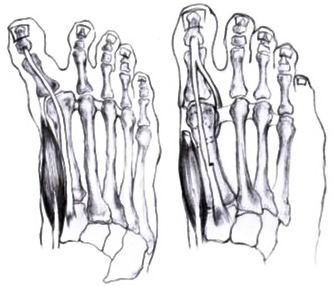
Tenodesis of the extensor hallucis brevis
Dayton, Dujela ( 19 ) suggests using extensor brevis tendon tenodesis as an alternative to extensor longus tendon since EHL may heal from previous surgery. In this procedure, the tendon is mobilized to the muscle-tendon junction and transected, passed plantarly under the intermetatarsal ligament, and attached through the bony tunnel from the lateral surface to the medial one on the body of the first metatarsal bone (Figure 7). The results of the six corrections showed an excellent correction result, preserved with an average monitoring period of 28 months and an improvement in the AOFAS score from 61 to 85.17.
Figure 7.
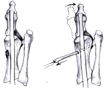
Tenodesis of the tendon of the first intermetatarsal muscle
Valtin. V (1991) proposes the technique of transposition of the tendon of the first interosseous muscle to the base of the proximal phalanx. Dissection of the distal attachment from the base of the main phalanx of the second toe and moving it through the bony tunnel to the base of the proximal phalanx helps generate a thrust vector in the hallux valgus. The technical complexity of refixation due to the small size of the tendon and the unknown long-term consequences of the second finger being deprived of the interosseous muscle are alarming ( 4 ).
3.10. Ligament Plastics Using Synthetic Materials
The lateral ligamentous structures can also be reconstructed as an alternative to autologous tendon movements or tenodesis.
Frontera, Silver ( 20 ) reported on the successful restoration of the lateral ligament (with 1.5 mm Ligapro suture) using the original technique and the obligatory combination of medial release in five patients. Based on the results of 4 years of monitoring, the authors achieved excellent results in all cases.
Two researchers independently published the experience of using mini-suture button constructions for the surgical treatment of varus deformity of the big toe and primary soft tissue imbalance ( 21 , 22 ). After medial release, holes are drilled in the proximal phalanx and metatarsal bone. A ligature is passed through the holes, and the knot is tightened on button support along the medial surface of the metatarsal bone with deformity correction (Figure 8). Given that these were sporadic case reports with favorable outcomes using this reconstructive technique, there is insufficient evidence to recommend the use of such techniques ( 13 ). The disadvantages include the cost of such artificial or allograft reconstructions, poorly defined long-term results, and the potential risk of infectious transmission with allograft ( 23 ).
Figure 8.
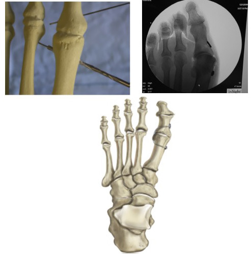
Illustration of the Mini Tight Rope system for hallux varus correction (Submitted by Arthrex, Inc, Naples, FL, USA)
3.11. Bone Procedures
If iatrogenic hallux valgus results from excessive resection of the medial eminence of the metatarsal head, overcorrection of the first intermetatarsal angle, or an incorrectly fused fracture of the proximal phalanx after Acin osteotomy, then work on the bones is necessary ( 1 , 4 , 8 ).
The consequences of excessive resection of the medial surface of the head of the first metatarsal bone are corrected with bone grafting ( 1 , 4 , 8 , 13 ).
Overcorrection of the metatarsal bone with a negative intermetatarsal angle occurs due to proximal or distal osteotomy and soft tissue release. In the case of an incorrectly fused osteotomy fracture of the metatarsal bone, the osteotomy is applicable to correct the deformity of the metatarsal bone, sometimes in combination with tendon movement ( 4 , 13 ).
3.12. Bone Grafting
Suppose the deformity is reducible and secondary after excessive resection of the medial eminence of the head of the first metatarsal bone. In that case, correcting it using bone grafting can be recommended. It restores support for the medial sesamoid bone, prevents medial subluxation, and provides bone support for the main phalanx of the big toe. Stabilizing the metatarsophalangeal joint will help restore muscle balance between the internal and external muscles of the big toe ( 1 , 4 , 8 , 13 ). Rochwerger and colleagues have published the results of bone grafting in restoring lost bone mass after resection of the medial eminence. The results of 7 cases with mean monitoring of 8.6 years showed no relapse, good range of motion in the metatarsophalangeal joint, and 6 satisfactory results, with one bad case with 20° hallux valgus ( 4 ).
3.13. Distal Chevron Osteotomy of the Metatarsal Bone
Monteagudo and Martínez-de-Albornoz ( 9 ) and colleagues presented the distal chevron metatarsal osteotomy results with medial displacement of the distal fragment in 19 patients (19 feet). As a result of treatment, the mean angle of valgus deviation improved from -11.6° to 4.7°, the mean first intermetatarsal angle improved from -0.3° to 2.3°, and the distal metatarsal articular angle decreased from 9.5° to 2.3°. The mean AOFAS score improved from 77 to 95. Two patients had a relapse of hallux varus. One had no symptomatic hallux varus, but the other had clinical manifestations.
3.14. Reverse Scarf and Open-Angle Osteotomy of the Proximal Phalanx
Migliorini, Eschweiler ( 24 ) proposed a correction method for patients who developed Hallux Varus after combined rotational scarf osteotomy and Akin osteotomy after Hallux Valgus resection. They describe medial access to the joint with a gradual release of soft tissues and then a scarf metatarsal osteotomy with an "open wedge" osteotomy of the proximal phalanx. With average monitoring of 38 months, all five patients noted improvement after revision surgery, the mean hallux valgus angle improved from -11° to 10°, and the mean intermetatarsal angle improved from 5° to 9°.
3.15. Joint Procedures
Historically, arthrodesis has been proposed as the standard treatment for patients with fixed deformity or degenerative arthrosis of the first metatarsophalangeal joint. It has been shown to reduce pain and maintain a stable medial column for non-reducible, permanent hallux varus deformities ( 4 , 13 ). Arthrodesis is mainly offered for gross deformities, complications of previous surgery, and severe arthrosis ( 7 , 25 ). The effects of static and dynamic forces on the first metatarsophalangeal joint are usually unpredictable after failed hallux valgus surgery. The authors prefer arthrodesis to stabilize the joint and reduce static and dynamic factors in the joint ( 13 ).
Arthrodesis of the first metatarsophalangeal joint is a reliable option after complications of surgical treatment of hallux valgus. The procedure can be used after many complications of primary surgery on hallux valgus, including recurrence of hallux valgus, hallux varus, hammer-like deformities, degenerative arthrosis of the first metatarsophalangeal joint, with combined deformities of the lesser toes. Arthrodesis of the first metatarsophalangeal joint is used for complications of distal and proximal osteotomy, arthrodesis of the metatarsal joint, Mc Bride procedure, medial exostosectomy, and resection arthroplasty ( 26 ).
The only difficulty with arthrodesis of the first radius is the simultaneous degeneration, contraction, or stiffness of the intermetatarsal joint of the big toe. Arthrodesis of the first metatarsophalangeal and interphalangeal joints can be performed provided there is a degree of shortening of the first radius to reduce the lever arm on the big toe. Intuitively, it is preferable to preserve the interphalangeal joint so that arthrodesis of the first metatarsophalangeal joint can be combined with arthrolysis of the interphalangeal joint or osteotomy of the phalanx in order to reorient the range of motion within the interphalangeal joint.
It is also advisable to consider that the cause of rapid degenerative changes in the first metatarsophalangeal joint in hallux varus after the initial intervention may result from deep joint infection. History of secondary wound healing and reuse of postoperative antibiotics should be considered when assessing the patient's condition. Before planning corrective surgery, a deep tissue biopsy should be performed after an appropriate period of antibiotic abstinence. Suppose an infection is detected during the bacteriological examination. In that case, intraoperative antibiotic therapy should be carried out, taking into account the sensitivity, and in certain circumstances, it may be necessary for a phased arthrodesis ( 13 ).
Iatrogenic deformity of the big toe often occurs after surgical correction of the hallux valgus. According to various literature sources, the frequency of iatrogenic deformity of the big toe after surgery for hallux valgus is from 2% -17%. A large number of sufferers are women, rather than men, whose average age is 45-50 years.
Properly treating iatrogenic hallux valgus requires careful clinical and radiographic examination to identify the cause. Each cause must be corrected in order to achieve stable results over time. In elastic, reversible deformities, tendon movement is possible. If the intermetatarsal angle is less than 6 °, it must be corrected. Arthrodesis is the most reliable method for treating rigidly fixed deformity with arthrosis of the metatarsophalangeal joint.
4. Conclusions
In conclusion, we want to say that hallux valgus is a difficultly tolerated foot deformity. The hallux valgus treatment literature largely consists of a low-evidence retrospective examination of treatment outcomes. Therefore, comparing different methods is rather difficult, especially considering the different contributing factors of deformation. Appropriate treatment requires careful clinical and radiological evaluation to determine the cause of the deformity. When planning treatment tactics, all elements of deformity should be taken into account. To achieve stable results over a long period, each component of this multifactorial deformity must be corrected, and each case must be considered individually. In elastic, reversible deformities, tendon movement is possible. If the intermetatarsal angle is less than 6°, it must be corrected. Arthrodesis is the most reliable method for treating rigidly fixed deformity with arthrosis of the metatarsophalangeal joint.
Authors' Contribution
Study concept and design: B. Z. I.
Acquisition of data: B. Z. I.
Analysis and interpretation of data: M. T. A.
Drafting of the manuscript: O. G. T.
Critical revision of the manuscript for important intellectual content: B. Z. I.
Administrative, technical, and material support: M. T. A.
Conflict of Interest
The authors declare that they have no conflict of interest.
References
- 1.Leemrijse T, Bevernage BD. Surgical treatment of iatrogenic hallux varus. Orthop Traumatol Surg Res. 2020;106(1):S159–S70. doi: 10.1016/j.otsr.2019.05.018. [DOI] [PubMed] [Google Scholar]
- 2.Berezhnoy SY. Iatrogenic Hallux Varus: Causes of Deformity and Possibilities of Percutaneous Surgical Correction (Retrospective Analysis of Case Reports) Traumatol Orthop Russia. 2017;23(4):48–57. [Google Scholar]
- 3.Kobayashi H, Kageyama Y, Shido Y. Gradual correction of traumatic hallux varus with metatarsal hemicallotasis. J Foot Ankle Surg. 2016;55(2):283–7. doi: 10.1053/j.jfas.2014.07.018. [DOI] [PubMed] [Google Scholar]
- 4.Crawford MD, Patel J, Giza E. Iatrogenic hallux varus treatment algorithm. Foot Ankle Clin. 2014;19(3):371–84. doi: 10.1016/j.fcl.2014.06.004. [DOI] [PubMed] [Google Scholar]
- 5.Jastifer J, Coughlin M. Hallux varus: review and surgical treatment. Med Chir Pied. 2014;30(2):41–8. [Google Scholar]
- 6.Samelis PV, Kolovos P, Nikolaou S, Samelis VP, Markeas NG. Primary Congenital Hallux Varus: A Step-Cut Surgical Approach. Cureus. 2022;14(8) doi: 10.7759/cureus.28075. [DOI] [PMC free article] [PubMed] [Google Scholar]
- 7.Lui TH. Arthroscopic first metatarsophalangeal arthrodesis for repair of fixed hallux varus deformity. J Foot Ankle Surg. 2015;54(6):1127–31. doi: 10.1053/j.jfas.2015.06.027. [DOI] [PubMed] [Google Scholar]
- 8.Akhtar S, Malek S, Hariharan K. Hallux varus following scarf osteotomy. Foot. 2016;29:1–5. doi: 10.1016/j.foot.2016.09.004. [DOI] [PubMed] [Google Scholar]
- 9.Monteagudo M, Martínez-de-Albornoz P. Management of complications after hallux valgus reconstruction. Foot Ankle Clin. 2020;25(1):151–67. doi: 10.1016/j.fcl.2019.10.011. [DOI] [PubMed] [Google Scholar]
- 10.Srinivasan R, editor. The hallucal-sesamoid complex: normal anatomy, imaging, and pathology. Seminars in Musculoskeletal Radiology; 2016: Thieme Medical Publishers. [DOI] [PubMed] [Google Scholar]
- 11.Vanore JV, Christensen JC, Kravitz SR, Schuberth JM, Thomas JL, Weil LS, et al. Diagnosis and treatment of first metatarsophalangeal joint disorders. Section 1: Hallux valgus. J Foot Ankle Surg. 2003;42(3):112–23. doi: 10.1016/s1067-2516(03)70014-3. [DOI] [PubMed] [Google Scholar]
- 12.Hecht PJ, Lin TJ. Hallux valgus. Med Clin. 2014;98(2):227–32. doi: 10.1016/j.mcna.2013.10.007. [DOI] [PubMed] [Google Scholar]
- 13.Davies MB, Blundell CM. The treatment of iatrogenic hallux varus. Foot Ankle Clin. 2014;19(2):275–84. doi: 10.1016/j.fcl.2014.02.010. [DOI] [PubMed] [Google Scholar]
- 14.Hawkins F. Acquired hallux varus: cause, prevention and correction. Clin Orthop Relat Res. 1971;76:169–76. doi: 10.1097/00003086-197105000-00024. [DOI] [PubMed] [Google Scholar]
- 15.Mohan R, Dhotare SV, Morgan SS. Hallux varus: A literature review. Foot. 2021;49:101863. doi: 10.1016/j.foot.2021.101863. [DOI] [PubMed] [Google Scholar]
- 16.Johnson KA, Spiegl PV. Extensor hallucis longus transfer for hallux varus deformity. J Bone Joint Surg. 1984;66(5):681–6. [PubMed] [Google Scholar]
- 17.Zielinska N, Tubbs RS, Ruzik K, Olewnik Ł. Classifications of the extensor hallucis longus tendon variations: Updated and comprehensive narrative review. Ann Anat. 2021;238:151762. doi: 10.1016/j.aanat.2021.151762. [DOI] [PubMed] [Google Scholar]
- 18.Lau JT, Myerson MS. Modified split extensor hallucis longus tendon transfer for correction of hallux varus. Foot Ankle Int. 2002;23(12):1138–40. doi: 10.1177/107110070202301212. [DOI] [PubMed] [Google Scholar]
- 19.Dayton PD, Dujela M, Egdorf R. Recurrence and hallux varus. Evidence-Based Bunion Surgery: Springer; 2018. pp. 91–111. [Google Scholar]
- 20.Frontera WR, Silver JK, Rizzo TD. Essentials of physical medicine and rehabilitation e-book: Elsevier Health Sciences; 2018. [Google Scholar]
- 21.Hsu AR, Gross CE, Lin JL. Bilateral hallux varus deformity correction with a suture button construct. Am J Orthop. 2013;42(3):121–4. [PubMed] [Google Scholar]
- 22.Gerbert J, Traynor C, Blue K, Kim K. Use of the Mini TightRope® for correction of hallux varus deformity. J Foot Ankle Surg. 2011;50(2):245–51. doi: 10.1053/j.jfas.2010.12.007. [DOI] [PubMed] [Google Scholar]
- 23.Cho SY, Kim YC, Choi JW. Epidemiology and bone‐related comorbidities of ingrown nail: A nationwide population‐based study. J Dermatol. 2018;45(12):1418–24. doi: 10.1111/1346-8138.14659. [DOI] [PubMed] [Google Scholar]
- 24.Migliorini F, Eschweiler J, Tingart M, Maffulli N. Revision surgeries for failed hallux valgus correction: A systematic review. Surgeon. 2021;19(6):497–506. doi: 10.1016/j.surge.2020.11.010. [DOI] [PubMed] [Google Scholar]
- 25.Geaney LE, Myerson MS. Radiographic results after hallux metatarsophalangeal joint arthrodesis for hallux varus. Foot Ankle Int. 2015;36(4):391–4. doi: 10.1177/1071100714560400. [DOI] [PubMed] [Google Scholar]
- 26.Baumhauer JF, Singh D, Glazebrook M, Blundell C, De Vries G, Le IL, et al. Prospective, randomized, multi-centered clinical trial assessing safety and efficacy of a synthetic cartilage implant versus first metatarsophalangeal arthrodesis in advanced hallux rigidus. Foot Ankle Int. 2016;37(5):457–69. doi: 10.1177/1071100716635560. [DOI] [PubMed] [Google Scholar]


