

Abstract
Patella fractures correspond to 1 % of all fractures. In recent decades, the role of the patella in increasing the lever arm of the quadriceps has been well defined. Surgical treatment is indicated for open fractures, those that compromise the function of the extensor mechanism, those with a joint gap >5 mm and/or joint incongruity >3 mm, a group that corresponds to around 30 % of the total. Anatomical reduction and stabilization with various types of modified tension bands is the most frequently used procedure. Biomechanical studies have shown that stabilization through the use of a tension band replacing the Kirchner wires with cannulated screws presents adequate resistance to fracture displacement and provides greater stability than the classic configuration, maintaining the theoretical principle of converting the forces of anterior tension of the patella generated by the quadriceps in compression at the level of the articular surface. A case of a patient who required reduction and osteosynthesis of an exposed patella fracture, associated with an extensor mechanism lesion, is presented. Clinical and radiographic characteristics of the patient and the resolution of the case are described.
Keywords: Patellar fracture, Open fractures, Knee extensor mechanism, Patellar tendon, Anchor repair
Abbreviations
Introduction
Patella fractures correspond to 1 % of all fractures [1]. Their treatment has been oriented towards bone preservation and vascular supply, and joint restoration and the function of the extensor mechanism. These results have been optimized using the anatomical reduction and fixation technique. However, certain anatomical characteristics of the patella, such as its extensive articular surface and subcutaneous location, the significant biomechanical requirements of the patellofemoral joint, and the need to obtain full and early mobility mean that this continues to be a great challenge [2]. Treatment Surgery is indicated for open fractures, those that compromise the function of the extensor mechanism, those with a joint gap >5 mm and/or joint incongruity >3 mm, a group that corresponds to around 30 % of the total [3]. Anatomical reduction and stabilization with various types of modified tension band is the most frequently used procedure [3].
We present the case of a 59-year-old patient who, after a frontal automobile-locomotive collision (Fig. 1), presented an exposed comminuted fracture of the right patella, associated with an extensor mechanism injury.
Fig. 1.

Post collision locomotive.
Case report
A 59-year-old male patient, diabetic, who is referred from Hospital Claudio Vicuña (San Antonio, Chile), to our center, after a frontal automobile-locomotive collision (Fig. 1), ACLS protocol for polytrauma is performed, with a CT scan of the skull ruling out injuries, X-rays and a CT scan of the right knee, at the level of the physical examination it presents an extensive, anfractuous wound, 20 cm long at the level of the anterior face of the patella, swelling, with bone exposure; ranges not evaluable due to pain, preserved neurovascular examination, regarding the images (AP and Lateral X-ray of the right knee, together with a CT scan of the right knee) revealed a comminuted fracture of the right patella (Fig. 2, Fig. 3).
Fig. 2.

AP and Lateral X-ray of the right knee revealed a comminuted fracture of the right patella.
Fig. 3.

CT scan of the right knee revealed a comminuted fracture of the right patella.
A diagnosis of comminuted fracture of the exposed right patella Gustilo III A + anterior tibial tubercle injury and extensor mechanism is made. Intravenous antibiotic treatment is started with Cefazolin 1 g IV every 8 h and Gentamicin 160 mg IV once a day for 7 days. Reduction and osteosynthesis of the patella with two 3.5 mm cannulated screws (DePuy-Synthes, Warsaw, Indiana, USA) is performed 13 days after admission and cerclage with a 0.8 mm wire loop, two 5.0 mm anchors (Arthrex, Inc. Naples, Florida, USA) are fixed to the distal patella, a Krackow suture is made distally, two 6.5 mm anchors (Arthrex, Inc. Naples, Florida, USA) are positioned under the anterior tuberosity of the tibia, a Krackow suture is made proximal (Fig. 4), a reduction is visualized under fluoroscopic vision (Fig. 5).
Fig. 4.

Tendon repair with Krackow technique.
Fig. 5.
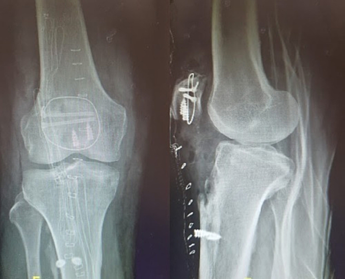
AP and Lateral X-ray of the right knee, showing reduction and osteosynthesis.
The patient has a satisfactory recovery in the immediate postoperative period, with restricted range of motion in extension, then a month later at 0° extension and 30° flexion, which was gradually increased. At six months postoperatively, it is maintained with ranges of 0° and 120°, without associated effusion or sensation of instability (Fig. 6).
Fig. 6.

Postoperative period, with increased range of motion.
Discussion
In recent decades, the role of the patella in increasing the lever arm of the quadriceps has been well defined. Its treatment has been oriented towards bone preservation and vascular supply, and joint restoration and the function of the extensor mechanism. These results have been optimized using the anatomical reduction and fixation technique. However, certain anatomical characteristics of the patella, such as its extensive articular surface and subcutaneous location, the significant biomechanical requirements of the patellofemoral joint, and the need to obtain full and early mobility mean that this continues to be a great challenge [2] Anatomical reduction and Stabilization with various types of modified tension bands is the most frequently used procedure [3]. Biomechanical studies have shown that stabilization using a tension band replacing Kirchner wires with cannulated screws presents adequate resistance to fracture displacement and provides greater stability than the classic configuration, maintaining the theoretical principle of converting the forces of anterior tension of the patella generated by the quadriceps into compression at the level of the articular surface [1]. Comminuted and displaced fractures of the patella require surgical treatment to obtain better clinical and functional results, which represents a complex scenario in which the use of the previously described fixation is insufficient in most cases, which implies the use of more and different osteosynthesis elements [2].
Patellar tendon ruptures are devastating injuries that can cause significant functional impairment attributed to disruption of the knee extensor mechanism. Rupture of the patellar tendon typically occurs in male patients aged <40 years and is caused by sudden quadriceps contraction with the knee in a flexed position, leading to tensile overload of the extensor mechanism [4,5].
The rupture of the extensor mechanism can be at the bone or tendon level [6]; the main cause is the patella fracture, secondly, the rupture of the quadriceps tendon and, finally, the rupture of the patellar tendon. These injuries may be due to direct or indirect trauma [7]. Tendon rupture of the extensor mechanism is rare, representing 3 % of all tendon injuries. It occurs due to trauma related to daily activities, sports or associated with systemic diseases. Quadriceps tendon injury is seen in people >40 years, with a peak between 60 and 70 years, unlike patellar tendon injury which is more common in individuals <40 years [6].
Acute tendon ruptures are typically the result of high eccentric loading, with forces borne by the enthesis, which is primarily made of type II collagen [8]. Bone-tendon failure may be due to abnormal cellular structure at the rupture site [9].
Acute ruptures (within the first 2 weeks) that occur at the junction of the patella with the patellar tendon can be repaired directly with a technique similar to that described for the quadriceps tendon, that is, with sutures extending from the tendon and rest on the patella by performing “Krakow type braided sutures”, anchor sutures have gained popularity with a recent cadaveric study demonstrating a significant decrease in gap formation at significantly higher failure load cycles [10].
Imbergamo et al. performed a systematic review using PRISMA guidelines, searching PubMed, the Cochrane Library, and Embase to identify studies that analyzed the biomechanical properties of SA and TO techniques for repair of a ruptured patellar tendon, resulting that the use of SA fixation results in decreased cyclic gap formation when compared with TO repair, with no significant difference in ultimate load to failure between the groups. These findings may have meaningful clinical implications, coinciding with existing clinical data demonstrating a decreased failure rate for SA repair in comparison with TO repair for patellar tendon rupture [11]. Both patellar and quadriceps tendon ruptures are significant cause of morbidity and can have negative short- and long-term effects on patient lives [12]. Although transosseous fixation of these tendon ruptures has been described as the gold standard, McGowen et al. demonstrated biomechanical superiority in some cadaver studies, with higher load to failure and less gap formation noted [13]. Potential benefits of suture anchor repair include less extensive surgical dissection resulting in less disruption to the blood supply, easier surgical technique, maintained anatomic patella motion and forces, and decreased risk of patella fracture, although cost effectiveness is a concern [14].
Conclusion
Comminuted patellar fracture associated with extensor mechanism injury is an entity that is difficult to manage, associated with significant functional sequelae if it is not adequately evaluated, studied, and managed. The development of post-traumatic osteoarthritis in fractures with joint involvement is a frequent complication that can determine functional alterations. In the context of the recent available literature on the topic, our opinion is that this management was adequate, and it has allowed the patient to present good functional results.
Consent for publication
Written consent to publish this information was obtained from participant.
Declaration of competing interest
All authors declare that they have no conflicts of interest.
References
- 1.Lue T.H., Feng L.W., Jun W.M., Yin L.W. Management of comminuted patellar fracture with non-absorbable suture cerclage and nitinol patellar concentrator. Injury. 2014;45(12):1974–1979. doi: 10.1016/j.injury.2014.10.008. [DOI] [PubMed] [Google Scholar]
- 2.Carredano X., Valderrama J., Valderrama I., Hube M., Bernal N., Mellado C., Espinoza G. Tratamiento de la fractura conminuta de patela: ¿existen diferencias entre realizar o no una eversión? Rev Chil Ortop Traumatol. 2021;62(3):e193–e200. [Google Scholar]
- 3.Melvin J.S., Mehta S. Patellar fractures in adults. J Am Acad Orthop Surg. 2011;19(04):198–207. doi: 10.5435/00124635-201104000-00004. [DOI] [PubMed] [Google Scholar]
- 4.Matava M.J. Patellar tendon ruptures. J Am Acad Orthop Surg. 1996;4(6):287–296. doi: 10.5435/00124635-199611000-00001. [DOI] [PubMed] [Google Scholar]
- 5.Siwek C.W., Rao J.P. Ruptures of the extensor mechanism of the knee joint. J Bone Joint Surg Am. 1981;63(6):932–937. [PubMed] [Google Scholar]
- 6.Alzate Munera M.R., Pereira S., Bidolegui F. Lesiones tendinosas del aparato extensor de la rodilla: Protocolo de tratamiento y rehabilitación. Rev Asoc Argent Ortop Traumatol. 2021;86(3):291–298. [Google Scholar]
- 7.Meyer Z., Ricci W.M. Knee extensor mechanism repairs: standard suture repair and novel augmentation technique. J Orthop Trauma. 2016;30(Suppl. 2):S30–S31. doi: 10.1097/BOT.0000000000000604. [DOI] [PubMed] [Google Scholar]
- 8.Nourissat G., Berenbaum F., Duprez D. Tendon injury: from biology to tendon repair. Nat Rev Rhueumatol. 2015;11:223–233. doi: 10.1038/nrrheum.2015.26. [DOI] [PubMed] [Google Scholar]
- 9.Kannus P., Jozsa L. Hisopathological changes preceding spontaneous rupture of a tendon. A controlled study of 891 patients. J Bone Joint Surg Am. 1991;73:1507–1525. [PubMed] [Google Scholar]
- 10.O’Dowd J.A., Lehoang D., Butler R.K., De Witt D., Mirzayan R. Trans-osseous versus anchor repair of acute patellar tendon ruptures. Orthop J Sports Med. 2018;6(7_suppl4) [Google Scholar]
- 11.Imbergamo C., et al. Failure rates of suture anchor fixation versus transosseous tunnel technique for patellar tendon repair: a systematic review and meta-analysis of biomechanical studies. Orthop J Sports Med. 2022;10(8) doi: 10.1177/23259671221120212. p. 232596712211202. [DOI] [PMC free article] [PubMed] [Google Scholar]
- 12.Greis P.E., Holmstrom M.C., Lahav A. Surgical treatment options for patella tendon rupture. Part 1: acute. Orthopedics. 2005;28(7):672–679. doi: 10.3928/0147-7447-20050701-15. [DOI] [PubMed] [Google Scholar]
- 13.McGowen S., Taylor B., Myers D., Passias B. Suture anchor repair of quadriceps and patellar tendon injuries. J Long-Term Eff Med Implants. 2020;30(1):57–60. doi: 10.1615/JLongTermEffMedImplants.2020034908. [DOI] [PubMed] [Google Scholar]
- 14.Sherman S.L., et al. Biomechanical evaluation of suture anchor versus transosseous tunnel patellar tendon repair techniques. J Knee Surg. 2018;32(08):825–832. doi: 10.1055/s-0038-1669790. [DOI] [PubMed] [Google Scholar]