

Sir,
Sweet syndrome, also called febrile neutrophilic dermatosis, is clinically characterised by asymmetrical distribution of tender, erythematous papules, plaques and nodules usually affecting the face, neck and upper extremities.[1] It can be either idiopathic, malignancy or autoimmune-associated or drug-induced.[1] There are only a few case reports of sweet syndrome localised on the face. We report a case of idiopathic sweet syndrome localised to the left cheek.
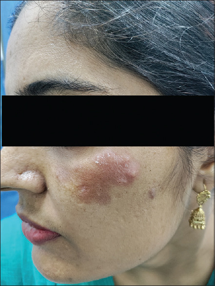
A 36-year-old female presented to us with the complaint of recurrent episodes of red, raised painful lesions on the left cheek. On examination, there was a tender, erythematous plaque of size around 3 × 4 cm2 present on the left cheek in the malar area. The centre of a plaque showed vesiculation [Figure 1]. There was no history suggestive of fever, previous infection or oral medication previous to the appearance of the skin lesion, and the patient was systemically well. A differential diagnosis of cutaneous Jessner's lymphocytic infiltrate, tumid lupus erythematosus, polymorphic light eruption, granuloma faciale, sweets syndrome and Wells syndrome was made [Table 1]. Her erythrocyte sedimentation rate was 45, C- reactive protein was 15, and total leukocyte counts were raised (13000/ml) with a neutrophil percentage of 82%. Due to the recurrent nature of the plaque, the patient agreed for skin biopsy. H and E stain of the section of skin biopsy revealed diffuse infiltration by a mixed inflammatory infiltrate comprised of neutrophils, eosinophils, lymphocytes and occasional plasma cells. Leucocytoclasia and dermal oedema were seen. Vessels were lined by swollen endothelial cells [Figures 2 and 3]. These features were consistent with the diagnosis of sweet syndrome. The workup for malignancy and endocrine disorders did not reveal any abnormality. Hence, a final diagnosis of idiopathic localised facial Sweet syndrome was made. The patient was prescribed oral prednisolone 30 mg once daily after breakfast for one week, to which she responded well [Figure 4].
Figure 1.

Tender erythematous plaque on the left malar area with central vesiculation
Table 1.
Differential diagnosis of the index case.
| Clinical features | Diagnostic and supportive investigations | Histopathology | |
|---|---|---|---|
| Sweet syndrome | Erythematous, tender plaque with pseudovesiculation | Neutrophilia↑CRP@, ↑ESR# | Diffuse neutrophilic infiltrate in the dermis, oedema, fragmentation of the nuclei of neutrophils |
| Wells syndrome | Erythematous, edematous, plaque with superimposed vesicles/bulla | Eosinophilia | Dermal oedema with eosinophilic infiltrate in dermis |
| Jessner’s lymphocytic infiltrate | Erythematous papules or plaques, may be annular, expanding peripherally, in sun-exposed area | UVA and UVB provocative photo testing | Normal epidermis, dense perivascular and periadnexal CD8+T cell lymphocytes and plasmacytoid monocytes infiltrates within the dermis |
| Granuloma faciale | Reddish-brown to violaceous asymptomatic plaques, predominantly on the face showing follicular accentuation and telangiectasia | DIF$: granular deposits of IgG in the perivascular areas or the basement membrane zone may be present | Grenz zone, perivascular infiltration mainly of neutrophils, lymphocytes, plasma cells, eosinophils. dilated follicular ostia and/or follicular plugs |
| Polymorphic light eruption | Pruritic, erythematous papules, plaques and vesicles developing on sun-exposed site | - | Dermal oedema with perivascular, periadnexal lymphocytic infiltrate without vasculitis. (Spongiosis, liquefactive degeneration±) |
| Tumid lupus erythematosus | Annular, erythematous, oedematous, indurated plaque | May be associated with pancytopenia ANA* positive | Lymphocytic interface dermatitis with basal layer degeneration, dermal mucinosis |
@CRP=C- reactive protein, #ESR=Erythrocyte sedimentation rate, $DIF=Direct immunofluorescence, *ANA=Anti-nuclear antibodies
Figure 2.

Histopathologic findings of the section of biopsy specimen (Haematoxylin and eosin stain; 100X
Figure 3.

Dense infiltration of the dermis by a mixed inflammatory infiltrate comprised of neutrophils, eosinophils, lymphocytes and occasional plasma cells. (Haematoxylin and eosin stain; 400X)
Figure 4.
Improvement after administration of an oral steroid
Lesions of sweet syndrome involve predominantly head, neck, upper trunk and upper arms. Neutrophilic dermatosis of the dorsal hands has been classified as a clinical variant of Sweet syndrome.[2] Apart from being localised on hands, it may also occur at sites of post-mastectomy lymphedema,[3] lymphedema of limbs,[4] previously irradiated field,[5] locally at the injection site of azacytidine[6] or pelfilgrastim[7] and at sites of tissue injury.[8] However, there have been very few case reports of Sweet syndrome localised on the face [Table 2].[9,10,11] Although the site of initial appearance of lesions was the oral and perioral area in two cases, the lesions eventually spread to the chest and limbs.[10,11] It has been postulated that the formation of cutaneous lesions occurs as a result of unusual hypersensitivity to infection, drugs, autoimmune diseases and neoplasms. This hypersensitivity is mediated by cytokines, followed by the infiltration of neutrophils.[1] It has also been hypothesised that photosensitivity may play a role in the pathogenesis of disease.[12] Vergara et al.[13] believed that localisation of the lesion at the site of the irradiated field may be explained by the locus minoris resistentae (“a place of less resistance”). Compromised integrity and an impaired barrier function of the irradiated skin may lead to the formation of lesion. In our case, despite repeated enquiries and investigations, we could not find an underlying cause. Hence, the patient may have recurrent idiopathic localised sweet syndrome.
Table 2.
Sweet syndrome localised to face.
| Age/sex | Primary condition | Site of sweets syndrome lesion | Treatment | |
|---|---|---|---|---|
| Brechtel et al. (1994)[9] | 73Y/F | Chronic bronchitis | Right cheek | Oral corticosteroids |
| Van der Meij et al. (1996)[10] | 58/F | Radiation therapy for oral SCC | Oral, perioral→upper chest, back | Spontaneous Resolution |
| Dawe et al. (2003)[11] | 64/M | Radiation therapy for oropharyngeal SCC | Oral, perioral→chest, limbs | Topical corticosteroids |
| Present case | 36/F | Idiopathic | Left cheek | Oral corticosteroids |
Oral corticosteroids are the main modality of treatment, whereas topical corticosteroids may be used for “localized” lesions. Other drugs such as dapsone, potassium iodide, colchicine, cyclosporine, clofazimine and indomethacin may also be used.
It is to be emphasised that a differential diagnosis of Sweet syndrome should be considered even if the lesion is in a localised area with no elicitable underlying cause.
Declaration of patient consent
The authors certify that they have obtained all appropriate patient consent forms. In the form, the patient(s) has/have given his/her/their consent for his/her/their images and other clinical information to be reported in the journal. The patients understand that their names and initials will not be published, and due efforts will be made to conceal their identity, but anonymity cannot be guaranteed.
Financial support and sponsorship
Nil.
Conflicts of interest
There are no conflicts of interest.
References
- 1.Villarreal-Villarreal CD, Ocampo-Candiani J, Villarreal-Martinez A. Sweet syndrome: A review and update. Actas Dermosifiliogr. 2016;107:369–78. doi: 10.1016/j.ad.2015.12.001. [DOI] [PubMed] [Google Scholar]
- 2.Ormerod AD, Hampton PJ. Neutrophilic dermatoses. In: Griffiths C, Barker J, Bleiker T, Chalmers R, Creamer D, editors. Rook's textbook of dermatology. Oxford: Wiley-Blackwell Press; 2016. pp. 49.1–49.18. [Google Scholar]
- 3.García-Río I, Pérez-Gala S, Aragüés M, Fernández-Herrera J, Fraga J, García-Díez A. Sweet's syndrome on the area of postmastectomy lymphoedema. J Eur Acad Dermatol Venereol. 2006;20:401–5. doi: 10.1111/j.1468-3083.2006.01460.x. [DOI] [PubMed] [Google Scholar]
- 4.Chu C-H, Cheng Y-P, Kao H-L, Liang C-W, Chan J-Y, Yu Y. Lymphedema-associated neutrophilic dermatosis: Two cases of localized sweet syndrome on the lymphedematous lower limbs. J Dermatol. 2016;43:1062–6. doi: 10.1111/1346-8138.13379. [DOI] [PubMed] [Google Scholar]
- 5.Lee GY, Do MO, Kim SH, Choi HY, Myung KB, Choi YW. Localized sweet's syndrome in an irradiated field. Ann Dermatol. 2009;21:300–3. doi: 10.5021/ad.2009.21.3.300. [DOI] [PMC free article] [PubMed] [Google Scholar]
- 6.Dev T, Dudani P, Bhari N. Azacytidine induced localized sweets syndrome in myelodysplastic syndrome. Dermatol Ther. 2021;34:e14754. doi: 10.1111/dth.14754. [DOI] [PubMed] [Google Scholar]
- 7.Ichiki T, Sugita K, Goto H, Shindo M, Yamamoto O. Localized pegfilgrastim-induced neutrophilic dermatosis with tissue G-CSF expression: A mimicker of sweet's syndrome. Acta Derm Venereol. 2019;99:685–6. doi: 10.2340/00015555-3170. [DOI] [PubMed] [Google Scholar]
- 8.Gloor AD, Feldmeyer L, Rammlmair A, Schlapbach C, Wallach D, Borradori L. A relapsing localized variant of neutrophilic dermatosis triggered by tissue injury. Br J Dermatol. 2021;184:162–4. doi: 10.1111/bjd.19418. [DOI] [PubMed] [Google Scholar]
- 9.Brechtel B, Haas N, Czarnetzki BM. Localized sweet syndrome. Hautarzt. 1994;45:858–60. doi: 10.1007/s001050050186. [DOI] [PubMed] [Google Scholar]
- 10.van der Meij EH, Epstein JB, Hay J, Ho V, Lerner K. Sweet's syndrome in a patient with oral cancer associated with radiotherapy. Eur J Cancer B Oral Oncol. 1996;32B:133–6. doi: 10.1016/0964-1955(95)00070-4. [DOI] [PubMed] [Google Scholar]
- 11.Dawe SA, Phillips R, Porter W, Francis NA, Bunker CB. Sweet's syndrome as a complication of radiotherapy for squamous carcinoma of the pharynx. Br J Dermatol. 2003;149:884. doi: 10.1046/j.1365-2133.2003.05541.x. [DOI] [PubMed] [Google Scholar]
- 12.Meyer V, Schneider SW, Bonsmann G, Beissert S. Experimentally confirmed induction of sweet's syndrome by phototesting. Acta Derm Venereol. 2011;91:720–1. doi: 10.2340/00015555-1139. [DOI] [PubMed] [Google Scholar]
- 13.Vergara G, Vargas-Machuca I, Pastor MA, Fariña MC, Martín L, Requena L. Localization of sweet's syndrome in radiation-induced locus minoris resistentae. J Am Acad Dermatol. 2003;49:907–9. doi: 10.1016/s0190-9622(03)01832-2. [DOI] [PubMed] [Google Scholar]