

Abstract
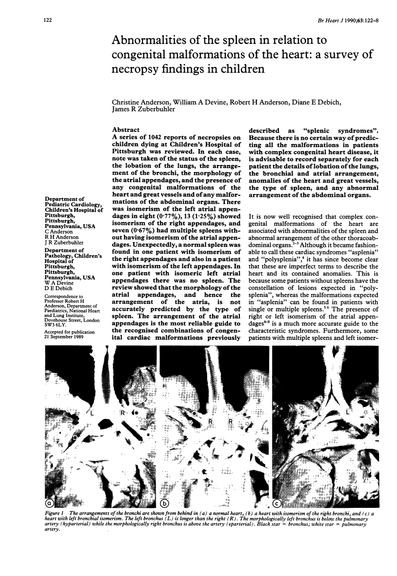
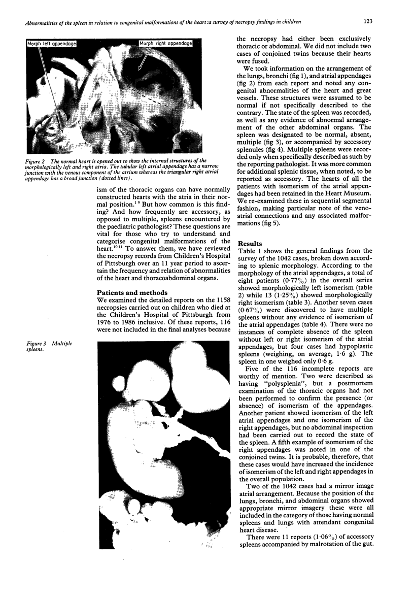
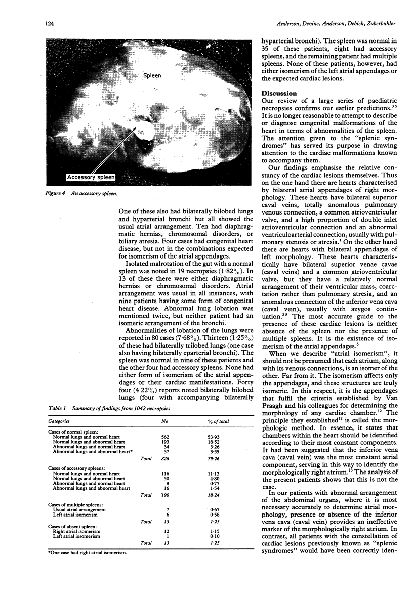
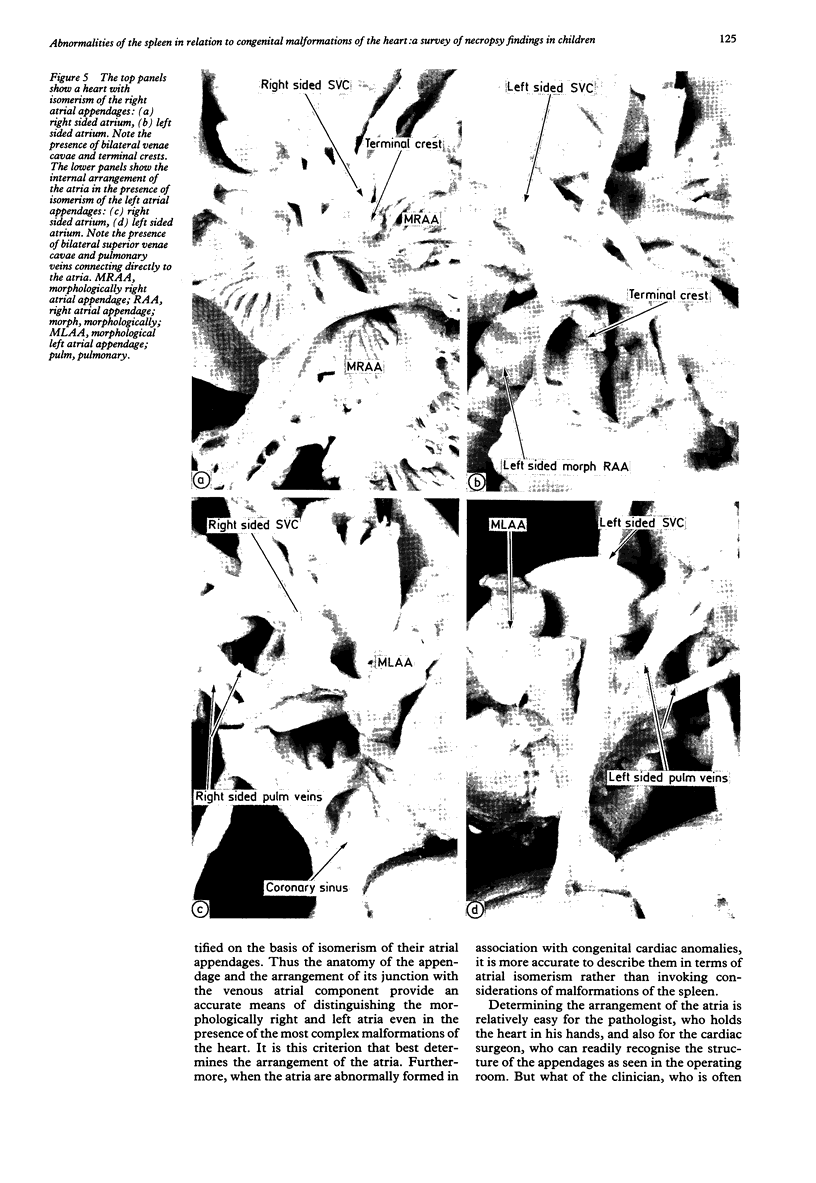
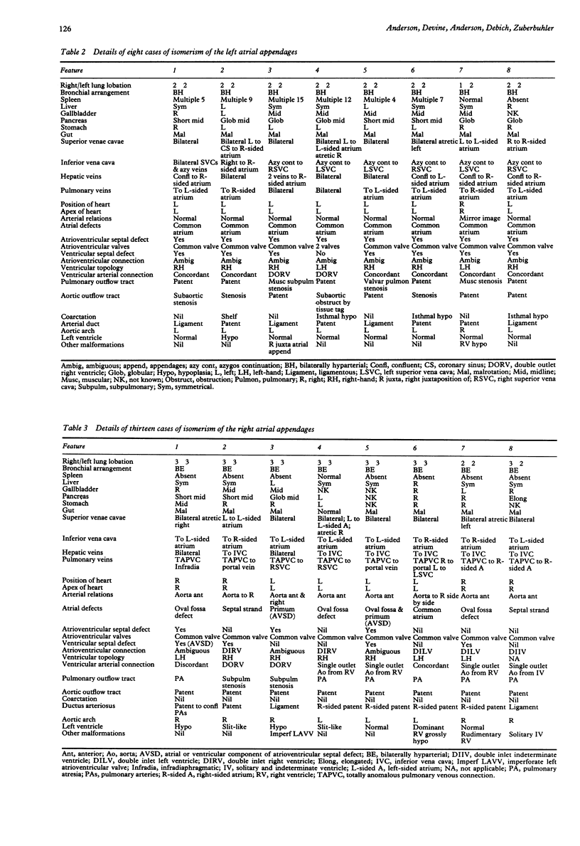
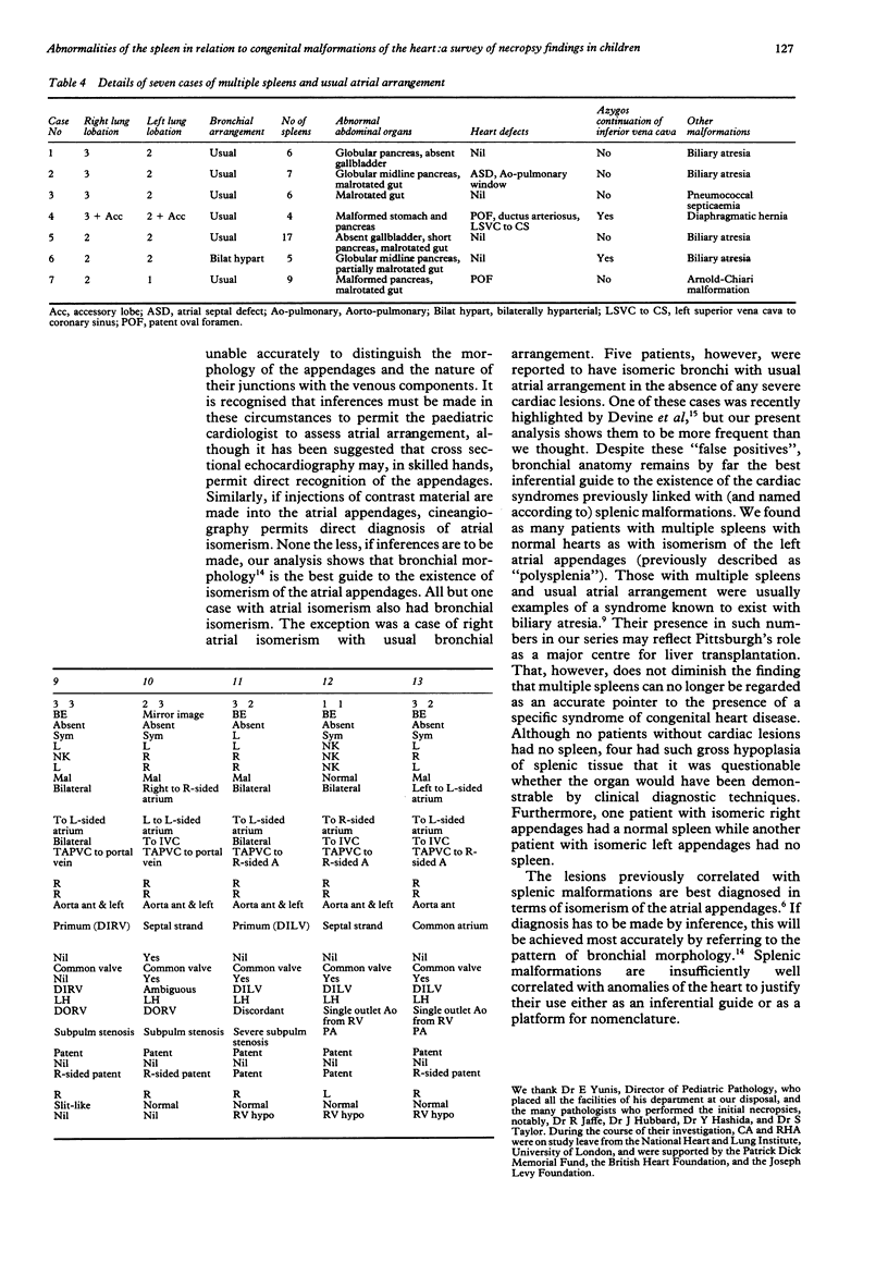
A series of 1042 reports of necropsies on children dying at Children's Hospital of Pittsburgh was reviewed. In each case, note was taken of the status of the spleen, the lobation of the lungs, the arrangement of the bronchi, the morphology of the atrial appendages, and the presence of any congenital malformations of the heart and great vessels and of any malformations of the abdominal organs. There was isomerism of the left atrial appendages in eight (0.77%), 13 (1.25%) showed isomerism of the right appendages, and seven (0.67%) had multiple spleens without having isomerism of the atrial appendages. Unexpectedly, a normal spleen was found in one patient with isomerism of the right appendages and also in a patient with isomerism of the left appendages. In one patient with isomeric left atrial appendages there was no spleen. The review showed that the morphology of the atrial appendages, and hence the arrangement of the atria, is not accurately predicted by the type of spleen. The arrangement of the atrial appendages is the most reliable guide to the recognised combinations of congenital cardiac malformations previously described as "splenic syndromes". Because there is no certain way of predicting all the malformations in patients with complex congenital heart disease, it is advisable to record separately for each patient the details of lobation of the lungs, the bronchial and atrial arrangement, anomalies of the heart and great vessels, the type of spleen, and any abnormal arrangement of the abdominal organs.
Full text
PDF
Images in this article
Selected References
These references are in PubMed. This may not be the complete list of references from this article.
- Anderson R. H., Becker A. E., Freedom R. M., Macartney F. J., Quero-Jimenez M., Shinebourne E. A., Wilkinson J. L., Tynan M. Sequential segmental analysis of congenital heart disease. Pediatr Cardiol. 1984;5(4):281–287. doi: 10.1007/BF02424973. [DOI] [PubMed] [Google Scholar]
- Brandt P. W., Calder A. L. Cardiac connections: the segmental approach to radiologic diagnosis in congenital heart disease. Curr Probl Diagn Radiol. 1977 May-Jun;7(3):1–35. doi: 10.1016/s0363-0188(77)80006-6. [DOI] [PubMed] [Google Scholar]
- Chandra R. S. Biliary atresia and other structural anomalies in the congenital polysplenia syndrome. J Pediatr. 1974 Nov;85(5):649–655. doi: 10.1016/s0022-3476(74)80508-1. [DOI] [PubMed] [Google Scholar]
- Deanfield J. E., Leanage R., Stroobant J., Chrispin A. R., Taylor J. F., Macartney F. J. Use of high kilovoltage filtered beam radiographs for detection of bronchial situs in infants and young children. Br Heart J. 1980 Nov;44(5):577–583. doi: 10.1136/hrt.44.5.577. [DOI] [PMC free article] [PubMed] [Google Scholar]
- Devine W. A., Debich D. E., Taylor S. R. Symmetrical bronchial pattern with normal atrial morphology. Int J Cardiol. 1988 Sep;20(3):395–398. doi: 10.1016/0167-5273(88)90294-x. [DOI] [PubMed] [Google Scholar]
- Gibbons R. J., Hu D. C., Clements I. P., Mankin H. T., Zinsmeister A. R., Brown M. L. Anatomic and functional significance of a hypotensive response during supine exercise radionuclide ventriculography. Am J Cardiol. 1987 Jul 1;60(1):1–4. doi: 10.1016/0002-9149(87)90972-6. [DOI] [PubMed] [Google Scholar]
- Macartney F. J., Zuberbuhler J. R., Anderson R. H. Morphological considerations pertaining to recognition of atrial isomerism. Consequences for sequential chamber localisation. Br Heart J. 1980 Dec;44(6):657–667. doi: 10.1136/hrt.44.6.657. [DOI] [PMC free article] [PubMed] [Google Scholar]
- Peoples W. M., Moller J. H., Edwards J. E. Polysplenia: a review of 146 cases. Pediatr Cardiol. 1983 Apr-Jun;4(2):129–137. doi: 10.1007/BF02076338. [DOI] [PubMed] [Google Scholar]
- Sharma S., Devine W., Anderson R. H., Zuberbuhler J. R. The determination of atrial arrangement by examination of appendage morphology in 1842 heart specimens. Br Heart J. 1988 Sep;60(3):227–231. doi: 10.1136/hrt.60.3.227. [DOI] [PMC free article] [PubMed] [Google Scholar]
- VAN MIEROP L. H., WIGLESWORTH F. W. Isomerism of the cardiac atria in the asplenia syndrome. Lab Invest. 1962 Dec;11:1303–1315. [PubMed] [Google Scholar]
- VANPRAAGH R., VANPRAAGH S., VLAD P., KEITH J. D. ANATOMIC TYPES OF CONGENITAL DEXTROCARDIA: DIAGNOSTIC AND EMBRYOLOGIC IMPLICATIONS. Am J Cardiol. 1964 Apr;13:510–531. doi: 10.1016/0002-9149(64)90159-6. [DOI] [PubMed] [Google Scholar]
- Van Praagh R., David I., Van Praagh S. What is a ventricle? The single-ventricle trap. Pediatr Cardiol. 1982;2(1):79–84. doi: 10.1007/BF02265623. [DOI] [PubMed] [Google Scholar]