

Abstract
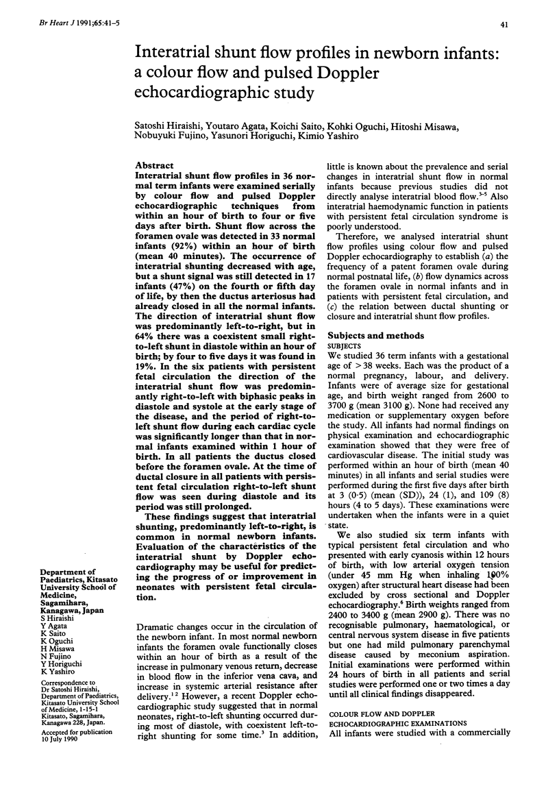
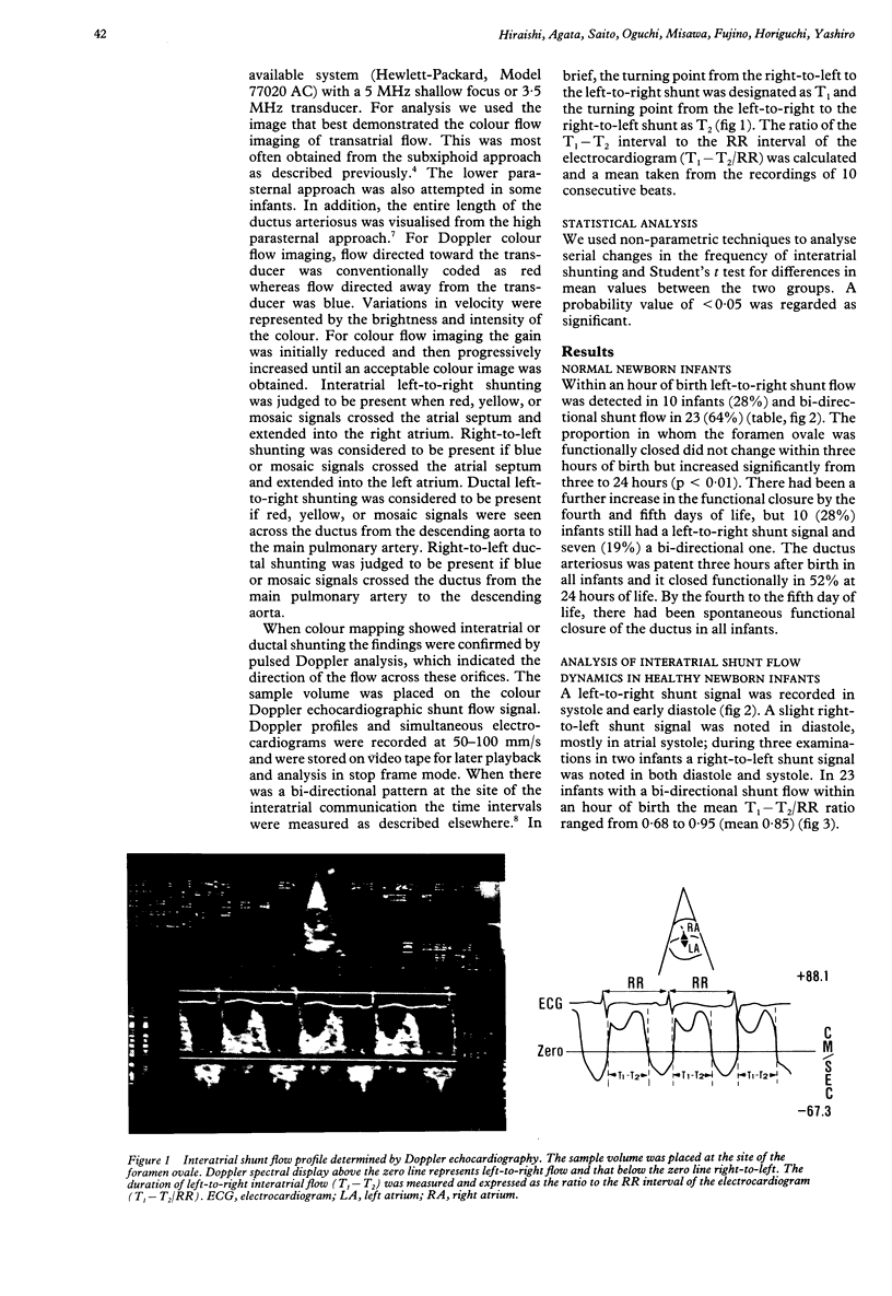
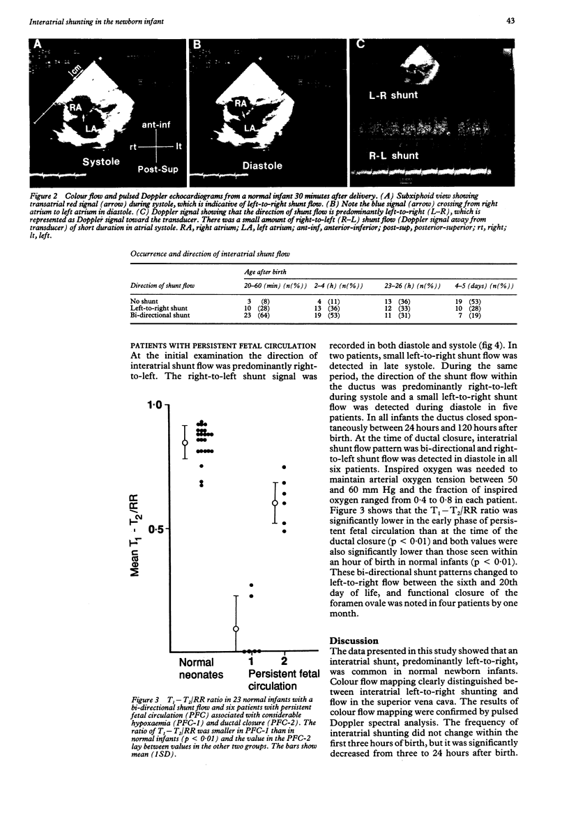
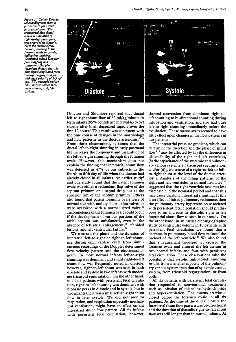
Interatrial shunt flow profiles in 36 normal term infants were examined serially by colour flow and pulsed Doppler echocardiographic techniques from within an hour of birth to four or five days after birth. Shunt flow across the foramen ovale was detected in 33 normal infants (92%) within an hour of birth (mean 40 minutes). The occurrence of interatrial shunting decreased with age, but a shunt signal was still detected in 17 infants (47%) on the fourth or fifth day of life, by then the ductus arteriosus had already closed in all the normal infants. The direction of interatrial shunt flow was predominantly left-to-right, but in 64% there was a coexistent small right-to-left shunt in diastole within an hour of birth; by four to five days it was found in 19%. In the six patients with persistent fetal circulation the direction of the interatrial shunt flow was predominantly right-to-left with biphasic peaks in diastole and systole at the early stage of the disease, and the period of right-to-left shunt flow during each cardiac cycle was significantly longer than that in normal infants examined within 1 hour of birth. In all patients the ductus closed before the foramen ovale. At the time of ductal closure in all patients with persistent fetal circulation right-to-left shunt flow was seen during diastole and its period was still prolonged. These findings suggest that interatrial shunting, predominantly left-to-right, is common in normal newborn infants. Evaluation of the characteristics of the interatrial shunt by Doppler echocardiography may be useful for predicting the progress of or improvement in neonates with persistent fetal circulation.
Full text
PDF
Images in this article
Selected References
These references are in PubMed. This may not be the complete list of references from this article.
- Arcilla R. A., Oh W., Wallgren G., Hanson J. S., Gessner I. H., Lind J. Quantitative studies of the human neonatal circulation. II. Hemodynamic findings in early and late clamping of the umbilical cord. Acta Paediatr Scand. 1967;(Suppl):25+–25+. doi: 10.1111/j.1651-2227.1967.tb05271.x. [DOI] [PubMed] [Google Scholar]
- Bierman F. Z., Williams R. G. Subxiphoid two-dimensional imaging of the interatrial septum in infants and neonates with congenital heart disease. Circulation. 1979 Jul;60(1):80–90. doi: 10.1161/01.cir.60.1.80. [DOI] [PubMed] [Google Scholar]
- Drayton M. R., Skidmore R. Ductus arteriosus blood flow during first 48 hours of life. Arch Dis Child. 1987 Oct;62(10):1030–1034. doi: 10.1136/adc.62.10.1030. [DOI] [PMC free article] [PubMed] [Google Scholar]
- Gessner I., Krovetz L. J., Benson R. W., Prystowsky H., Stenger V., Eitzman D. V. Hemodynamic adaptations in the newborn infant. Pediatrics. 1965 Nov;36(5):752–762. [PubMed] [Google Scholar]
- Hiraishi S., Horiguchi Y., Misawa H., Oguchi K., Kadoi N., Fujino N., Yashiro K. Noninvasive Doppler echocardiographic evaluation of shunt flow dynamics of the ductus arteriosus. Circulation. 1987 Jun;75(6):1146–1153. doi: 10.1161/01.cir.75.6.1146. [DOI] [PubMed] [Google Scholar]
- Hiraishi S., Misawa H., Oguchi K., Kadoi N., Saito K., Fujino N., Hojo M., Horiguchi Y., Yashiro K. Two-dimensional Doppler echocardiographic assessment of closure of the ductus arteriosus in normal newborn infants. J Pediatr. 1987 Nov;111(5):755–760. doi: 10.1016/s0022-3476(87)80263-9. [DOI] [PubMed] [Google Scholar]
- Kupferschmid C., Lang D. The valve of the foramen ovale in interatrial right-to-left shunt: echocardiographic cineangiocardiographic and hemodynamic observations. Am J Cardiol. 1983 May 15;51(9):1489–1494. doi: 10.1016/0002-9149(83)90662-8. [DOI] [PubMed] [Google Scholar]
- Minagoe S., Tei C., Kisanuki A., Arikawa K., Nakazono Y., Yoshimura H., Kashima T., Tanaka H. Noninvasive pulsed Doppler echocardiographic detection of the direction of shunt flow in patients with atrial septal defect: usefulness of the right parasternal approach. Circulation. 1985 Apr;71(4):745–753. doi: 10.1161/01.cir.71.4.745. [DOI] [PubMed] [Google Scholar]
- Musewe N. N., Poppe D., Smallhorn J. F., Hellman J., Whyte H., Smith B., Freedom R. M. Doppler echocardiographic measurement of pulmonary artery pressure from ductal Doppler velocities in the newborn. J Am Coll Cardiol. 1990 Feb;15(2):446–456. doi: 10.1016/s0735-1097(10)80076-2. [DOI] [PubMed] [Google Scholar]
- Pagtakhan R. D., Hartmann A. F., Jr, Goldring D., Kissane J. The valve-incompetent foramen ovale. A report on seven infants with left-to-right atrial shunt. J Pediatr. 1967 Dec;71(6):848–854. doi: 10.1016/s0022-3476(67)80010-6. [DOI] [PubMed] [Google Scholar]
- RUDOLPH A. M., MAYER F. E., NADAS A. S., GROSS R. E. A clinical and hemodynamic study of 23 patients in the first year of life. Pediatrics. 1958 Nov;22(5):892–904. [PubMed] [Google Scholar]
- Riggs T. W., Rodriguez R., Snider A. R., Batton D. Doppler echocardiographic evaluation of right and left ventricular diastolic function in normal neonates. J Am Coll Cardiol. 1989 Mar 1;13(3):700–705. doi: 10.1016/0735-1097(89)90614-1. [DOI] [PubMed] [Google Scholar]
- Satomi G., Nakazawa M., Takao A., Mori K., Touyama K., Konishi T., Tomimatsu H., Nakamura K. Blood flow pattern of the interatrial communication in patients with complete transposition of the great arteries: a pulsed Doppler echocardiographic study. Circulation. 1986 Jan;73(1):95–99. doi: 10.1161/01.cir.73.1.95. [DOI] [PubMed] [Google Scholar]
- Steinfeld L., Almeida O. D., Rothfeld E. L. Asynchronous atrioventricular valve opening as it relates to right to left interatrial shunting in the normal newborn. J Am Coll Cardiol. 1988 Sep;12(3):712–718. doi: 10.1016/s0735-1097(88)80062-7. [DOI] [PubMed] [Google Scholar]