

Abstract
Stenosis of the aortic valve (pressure drop 50 mm Hg) was diagnosed prenatally by Doppler echocardiography in a 33 week old fetus. Measurement of time-velocity integrals through the tricuspid and mitral valves indicated a significantly higher flow in the right heart. The pressure drop across the aortic valve in the 3 hour old infant was 80 mm Hg. The findings in this patient suggest that the usually accepted theory that prenatally the ventricles function in parallel should take into account the chronology of filling and ejection. In this patient the ability of a ventricle to generate a prenatal transvalvar pressure gradient was evidence that the size, compliance, and contractility of the ventricle were sufficient to maintain good function.
Full text
PDF

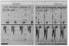
Images in this article

Selected References
These references are in PubMed. This may not be the complete list of references from this article.
- De Geeter B., Marlange J. P., Willard D., Losay J., Bruniaux J., Binet J. P. Rétrécissement aortique valvulaire. Diagnostic prénatal. Une observation. Presse Med. 1985 Jul 13;14(28):1505–1507. [PubMed] [Google Scholar]
- Huhta J. C., Carpenter R. J., Jr, Moise K. J., Jr, Deter R. L., Ott D. A., McNamara D. G. Prenatal diagnosis and postnatal management of critical aortic stenosis. Circulation. 1987 Mar;75(3):573–576. doi: 10.1161/01.cir.75.3.573. [DOI] [PubMed] [Google Scholar]
- Jouk P. S., Rossignol A. M., Denis B., Bost M. L'ostium secundum restrictif: un nouveau syndrome malformatif foetal. Arch Mal Coeur Vaiss. 1987 Apr;80(4):538–542. [PubMed] [Google Scholar]