

Abstract
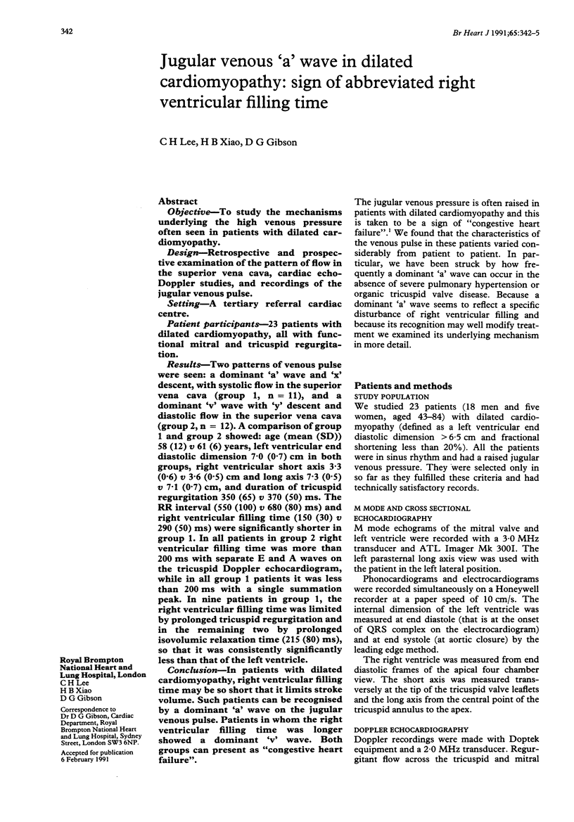
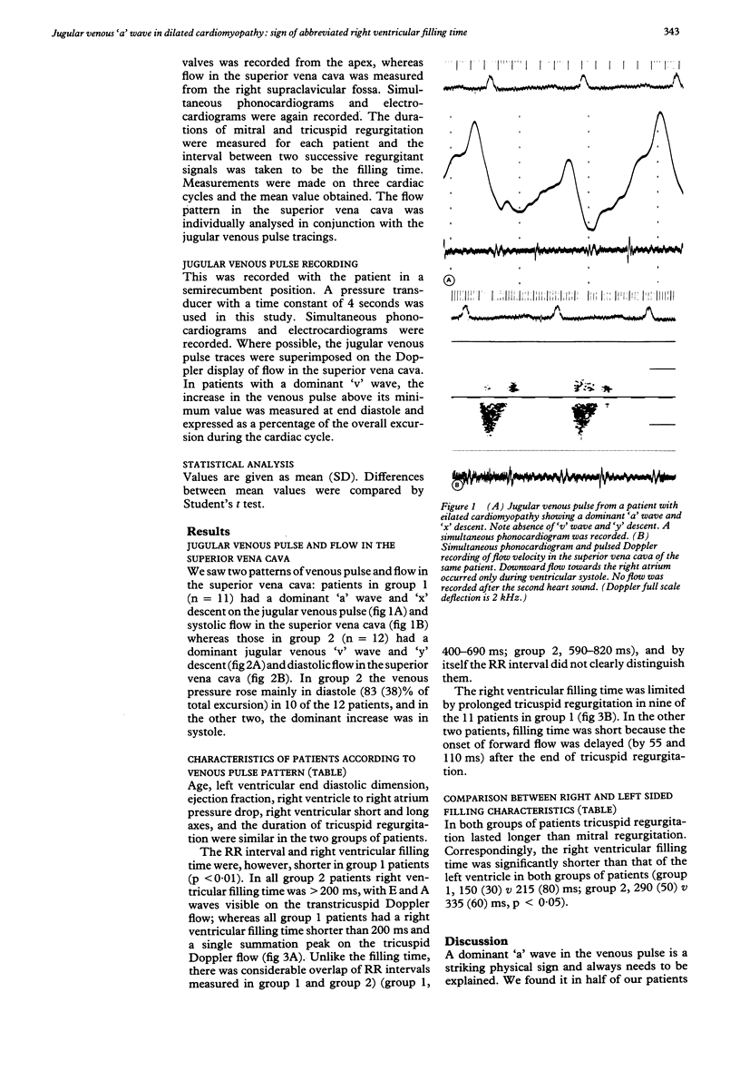
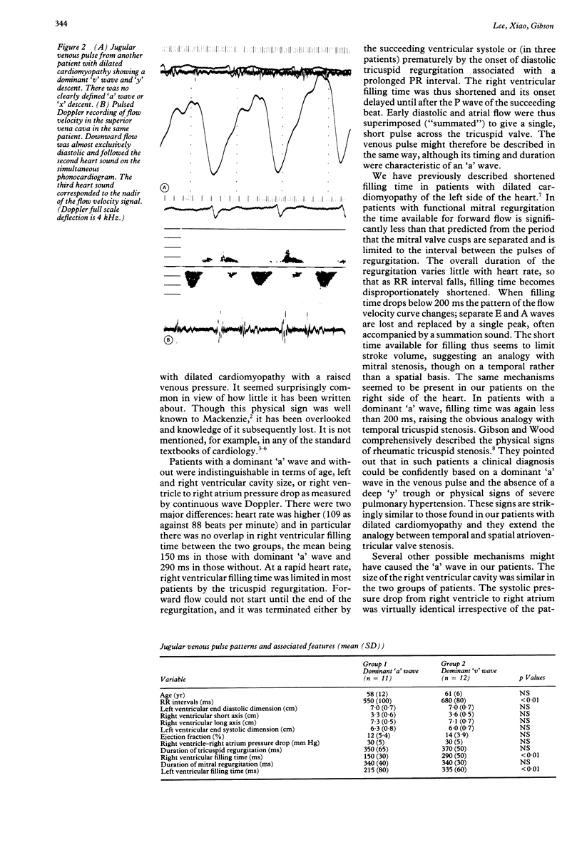
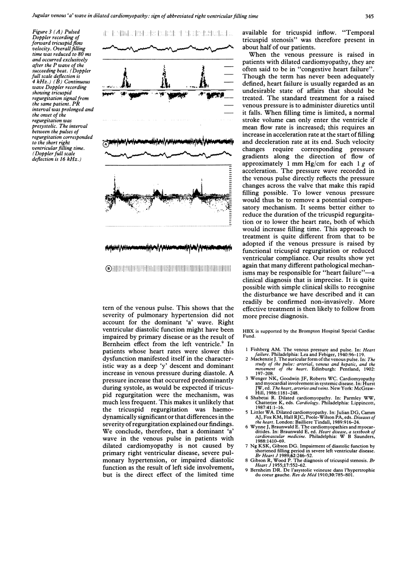
OBJECTIVE--To study the mechanisms underlying the high venous pressure often seen in patients with dilated cardiomyopathy. DESIGN--Retrospective and prospective examination of the pattern of flow in the superior vena cava, cardiac echo-Doppler studies, and recordings of the jugular venous pulse. SETTING--A tertiary referral cardiac centre. PATIENTS PARTICIPANTS--23 patients with dilated cardiomyopathy, all with functional mitral and tricuspid regurgitation. RESULTS--Two patterns of venous pulse were seen: a dominant 'a' wave and 'x' descent, with systolic flow in the superior vena cava (group 1, n = 11), and a dominant 'v' wave with 'y' descent and diastolic flow in the superior vena cava (group 2, n = 12). A comparison of group 1 and group 2 showed: age (mean (SD] 58 (12) v 61 (6) years, left ventricular end diastolic dimension 7.0 (0.7) cm in both groups, right ventricular short axis 3.3 (0.6) v 3.6 (0.5) cm and long axis 7.3 (0.5) v 7.1 (0.7) cm, and duration of tricuspid regurgitation 350 (65) v 370 (50) ms. The RR interval (550 (100) v 680 (80) ms) and right ventricular filling time (150 (30) v 290 (50) ms) were significantly shorter in group 1. In all patients in group 2 right ventricular filling time was more than 200 ms with separate E and A waves on the tricuspid Doppler echocardiogram, while in all group 1 patients it was less than 200 ms with a single summation peak. In nine patients in group 1, the right ventricular filling time was limited by prolonged tricuspid regurgitation and in the remaining two by prolonged isovolumic relaxation time (215 (80) ms), so that it was consistently significantly less than that of the left ventricle. CONCLUSION--In patients with dilated cardiomyopathy, right ventricular filling time may be so short that it limits stroke volume. Such patients can be recognised by a dominant 'a' wave on the jugular venous pulse. Patients in whom the right ventricular filling time was longer showed a dominant 'v' wave. Both groups can present as "congestive heart failure".
Full text
PDF
Selected References
These references are in PubMed. This may not be the complete list of references from this article.
- GIBSON R., WOOD P. The diagnosis of tricuspid stenosis. Br Heart J. 1955 Oct;17(4):552–562. doi: 10.1136/hrt.17.4.552. [DOI] [PMC free article] [PubMed] [Google Scholar]
- Ng K. S., Gibson D. G. Impairment of diastolic function by shortened filling period in severe left ventricular disease. Br Heart J. 1989 Oct;62(4):246–252. doi: 10.1136/hrt.62.4.246. [DOI] [PMC free article] [PubMed] [Google Scholar]