Abstract
Simple Summary
The identification of potential molecular alterations is standard in the diagnostic pathway of non-small-cell lung cancer (NSCLC). The aim of this study is to determine the prevalence of NTRK fusions in NSCLC in a routine diagnostic setting using immunohistochemistry, fluorescence in situ hybridization, and RNA-based next-generation sequencing. A total of 1068 unselected consecutive patients with NSCLC were screened in two scenarios, either with initial IHC followed by RNA-NGS (n = 973) or direct FISH testing (n = 95). In total, 0.2% of all patients were NTRK positive. Both RNA-NGS and FISH are suitable to determine clinically relevant NTRK fusions in a real-world setting. RNA-NGS or FISH NTRK positive results were mutually exclusive with alterations in EGFR/ALK/ROS1/BRAF/RET or KRAS.
Abstract
(1) Background: The main objectives of our study are (i) to determine the prevalence of NTRK (neurotrophic tyrosine kinase) fusions in a routine diagnostic setting in NSCLC (non-small cell lung cancer) and (ii) to investigate the feasibility of screening approaches including immunohistochemistry (IHC) as a first-line test accompanied by fluorescence in situ hybridization (FISH) and RNA-(ribonucleic acid-)based next-generation sequencing (RNA-NGS). (2) Methods: A total of 1068 unselected consecutive patients with NSCLC were screened in two scenarios, either with initial IHC followed by RNA-NGS (n = 973) or direct FISH testing (n = 95). (3) Results: One hundred and thirty-three patients (14.8%) were IHC positive; consecutive RNA-NGS testing revealed two patients (0.2%) with NTRK fusions (NTRK1-EPS15 (epidermal growth factor receptor pathway substrate 15) and NTRK1-SQSTM1 (sequestosome 1)). Positive RNA-NGS was confirmed by FISH, and NTRK-positive patients benefited from targeted treatment. All patients with direct FISH testing were negative. RNA-NGS- or FISH-positive results were mutually exclusive with alterations in EGFR (epidermal growth factor receptor), ALK (anaplastic lymphoma kinase), ROS1 (ROS proto-oncogene 1), BRAF (proto-oncogene B-Raf), RET (rearranged during transfection) or KRAS (kirsten rat sarcoma viral oncogene). Excluding patients with one of these alterations raised the prevalence of NTRK-fusion positivity among panTrk-(tropomyosin receptor kinase-) IHC positive samples to 30.5%. (4) Conclusions: NTRK fusion-positive lung cancers are exceedingly rare and account for less than 1% of patients in unselected all-comer populations. Both RNA-NGS and FISH are suitable to determine clinically relevant NTRK fusions in a real-world setting. We suggest including panTrk-IHC in a diagnostic workflow followed by RNA-NGS. Excluding patients with concurrent molecular alterations to EGFR/ALK/ROS1/BRAF/RET or KRAS might narrow the target population.
Keywords: NSCLC, lung cancer, NTRK, targeted therapy, immunohistochemistry, fluorescence in situ hybridization, FISH, RNA-based next-generation sequencing
1. Introduction
The Trk family of neurotrophic tyrosine kinases, also known as tropomyosin receptor kinases, consists of three proteins, TrkA, TrkB, and TrkC. These proteins are encoded by three genes, NTRK1 on chromosome 1q21-22, NTRK2 on 9q22.1, and NTRK3 on 15q25, respectively [1]. The proteins share structural and functional similarities and are composed of extracellular domains, including IgG-like parts, a transmembrane domain, and a catalytically active intracellular tyrosine kinase domain [1,2,3]. Splice variants that modify protein structure and function and create isoforms have been described for all three genes. Trk proteins are physiologically activated by ligands such as nerve growth factor (NGF), brain-derived neurotrophic factor (BDNF), and neurotrophin-3 (NT-3, also NT-4/5) [4]. As regulators of tissue development during embryogenesis and maintenance of function in adult tissue, Trk kinases are expressed in normal central and peripheral nerve tissue as well as in beta cells of the endocrine pancreas, monocytes, and bone tissue.
Recently, oncogenic translocations involving NTRK1, NTRK2, and NTRK3 genes have been described [5,6,7,8,9,10,11,12,13,14,15,16,17,18,19,20,21,22,23,24,25,26,27,28,29,30,31,32,33,34,35,36,37,38,39,40]; for a compiled overview of the literature of previously published NTRK fusions in NSCLC, including differentiation by histology, see Table 1.
Table 1.
Compiled overview of previously published cases of NTRK-fusion-positive NSCLC.
| NTRK Gene | Fusion Partner | Histology | Described by |
|---|---|---|---|
| NTRK1 | EPS15 | Adenocarcinoma | [14,32], this report |
| SQSTM1 | Adenocarcinoma | [33,34,35], this report | |
| NSCLC | [14,32,35] | ||
| TPM3 | Adenocarcinoma | [14,17,21,22,23,24,32,33,35] | |
| IRF2BP2 | Adenocarcinoma | [14,21,25,32,33,35] | |
| Adenocarcinoma with neuroendocrine features | [33] | ||
| MPRIP | Adenocarcinoma | [20,33] | |
| CD74 | Adenocarcinoma | [20,26,35,36] | |
| TPR | Adenocarcinoma | [32,35] | |
| TGF | Adenocarcinoma | [14] | |
| LMNA | Adenocarcinoma | [35] | |
| PHF20 | Sarcomatoid Carcinoma + Adenocarcinoma | [35] | |
| BCL9 (intergenic region) | Adenocarcinoma | [35] | |
| CLIP1 | Adenocarcinoma | [27] | |
| P2RY8 | Adenocarcinoma | [21] | |
| GRIPAP1 | Adenocarcinoma | [28] | |
| RFWD2 | Large-cell neuroendocrine carcinoma | [29] | |
| F11R | Squamous cell carcinoma | [37] | |
| LIPI | Adenocarcinoma | [38] | |
| NTRK2 | STRN | Adenocarcinoma | [14,23] |
| SQSTM1 | Adenocarcinoma | [17] | |
| TRIM24 | Adenocarcinoma | [16] | |
| NTRK3 | ETV6 | Adenocarcinoma | [17,24,30,32,33,39] |
| Squamous cell carcinoma | [33] | ||
| SQSTM1 | Adenocarcinoma | [14,23,32,36] | |
| Neuroendocrine carcinoma | [33] | ||
| RBPMS | Adenocarcinoma | [14,23] | |
| Intergenic region | Large cell neuroendocrine carcinoma | [31] | |
| EML4 | Adenocarcinoma | [40] |
TPM3, tropomyosin 3; IRF2BP2, interferon regulatory factor 2 binding protein 2; MPRIP, myosin phosphatase rho interacting protein; CD74, cluster of differentiation 74; TPR, translocated promoter region, nuclear basket protein; TGF, transforming growth factor; LMNA, lamin a; PHF20, phd finger protein gene; BCL9, b-cell lymphoma 9; CLIP1, AP-Gly domain containing linker protein 1; P2RY8, P2Y receptor family member 8; GRIPAP1, GRIP1-associated protein 1; RFWD2, also known as COP1 (COP1 E3 ubiquitin ligase); F11R, f11 receptor; LIPI, lipase I; STRN, striatin; TRIM24, tripartite motif containing 24; ETV6, ETS variant transcription factor 6; RBPMS, RNA binding protein; MRNA processing factor; EML4, EMAP like 4.
Biologically, most of these fusions result in a chimeric transcript in which regulatory parts, i.e., extracellular and transmembrane domains, are replaced by parts of another gene that facilitate ligand-independent autonomous dimerization and activation, and thus, the oncogenic transformation of cells. Unsurprisingly, these changes have been found among malignancies of the central nervous system, e.g., adult- and pediatric-type gliomas and glioblastomas [18]. However, a wide variety of non-CNS (central nervous system) tumors turned out to harbor NTRK fusions as well, among them carcinomas of the lung, colorectal cancers, thyroid cancer, and cholangiocellular carcinomas. Exceptionally rare tumors, e.g., secretory carcinomas of the breast and their counterparts in salivary glands, infantile fibrosarcomas, and congenital mesonephric blastomas, are molecularly defined by NTRK fusions. These tumors harbor NTRK fusions in nearly 100% of cases, predominantly the NTRK3-ETV6 variant. Another rare group of tumors, including spitzoid melanomas, papillary thyroid cancer, and inflammatory myofibroblastic tumors, seems to show a moderate frequency of NTRK fusions. However, more prevalent cancers, among them non-small-cell lung cancer, harbor NTRK fusions very infrequently. Data from clinical registers and clinical trials indicate a prevalence of NTRK fusions among lung cancers below 1% [19,22,26,30,31,35,41,42,43]. Population-based data from a clinical all-comer series of Caucasian lung cancer patients are not yet available.
Nevertheless, clinical trials have demonstrated impressive responses of NTRK-fused cancers to two tyrosine kinase inhibitors with anti-Trk activity, Larotrectinib and Entrectinib [44,45]. Larotrectinib and Entrectinib were recently approved by the U.S. Food and Drug Administration (FDA) and the European Medicines Agency (EMA) for the treatment of NTRK-positive tumors irrespective of specific tumor entity [46,47,48,49].
Given the high clinical benefit of Trk inhibitors and the very low prevalence of NTRK fusions among major groups of human cancers, it is extremely important to define a reasonable screening strategy that, on one hand, safely detects NTRK-fusion-positive patients with high sensitivity and specificity and, on the other hand, takes into consideration additional important factors, such as cost-effectiveness, time to diagnosis, and testing feasibility due to tissue characteristics. Basically, several methods are available to detect NTRK fusions: (i) DNA-(desoxyribonucleic acid-) based (hybrid capture) next-generation (NGS) assays, (ii) RNA-based NGS approaches, either target enrichment or hybrid capture methods, (iii) fluorescence in situ hybridization (FISH), and (iv) immunohistochemistry (IHC) [19,50,51]. DNA- and RNA-based sequencing and FISH have been utilized in clinical trials, whereas IHC has been proposed as a pre-screening test [44,45,51]. It has been shown, however, that DNA-based hybrid capture approaches suffer from severe limitations since some specific fusion variants could not be detected by this technology [14]. Therefore, RNA-based NGS is currently considered a gold standard or reference method and is recommended as the primary test approach if available [52]. In contrast, these assays are not always available and extremely demanding in terms of costs, time, and tissue/RNA quality. Therefore, there is an urgent clinical need to develop reasonable screening strategies for major cancer types, such as lung cancer.
In this study, we aimed—as the primary objective—to determine the prevalence of NTRK gene fusions in an unselected series of consecutive lung cancer patients. The secondary objective was to determine the overall feasibility of NTRK screening approaches in a routine diagnostic setting. Furthermore, additional goals were (a) to describe the specificity and potential diagnostic pitfalls of immunohistochemistry as a potential first-level pre-screening approach, (b) to compare the performance and results of IHC, FISH, and RNA-based sequencing as diagnostic techniques, and (c) to describe clinical and histologic phenotypes of NTRK fusion-positive patients.
2. Materials and Methods
2.1. Patients and Samples
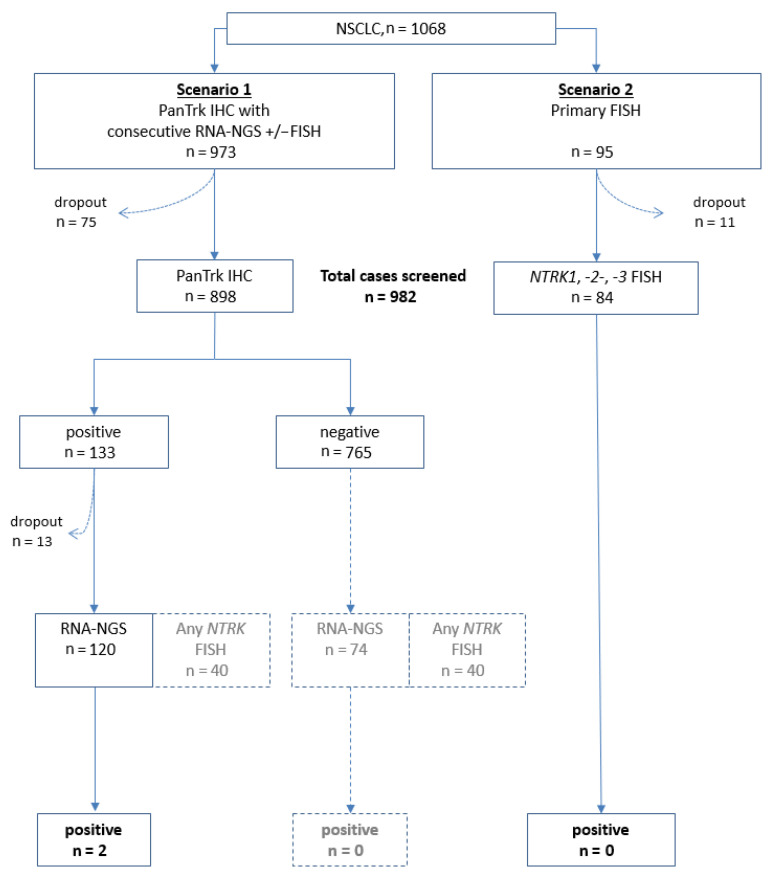
During an 18 month period, 1068 unselected consecutive NSCLC patients were prospectively included in a screening for NTRK fusions as part of our routine diagnostic procedures at the Lung Cancer Center Göttingen (Lungentumorzentrum Universität Göttingen as part of the Göttingen Comprehensive Cancer Center, G-CCC). Patients were included irrespective of tumor stage at the time of diagnosis and independent of tumor histology and molecular subtype. Tumor samples were randomly tested either by IHC and subsequent RNA-based next-generation sequencing with FISH confirmation (Figure 1, Scenario 1) or by direct FISH testing (Figure 1, Scenario 2). After a dropout of 86 cases due to limited material, 982 samples underwent complete prospective screening either with IHC followed by NGS (scenario 1, n = 898) or with FISH (scenario 2, n = 84). A subset of IHC-positive samples of Scenario 1 with sufficient residual tumor tissue was retrospectively cross-tested by FISH (n = 40). A total of 13 IHC-positive cases were not suitable for RNA-NGS analysis due to failure or lack of tumor material or technical errors. In total, 120 IHC-positive cases were examined by RNA-NGS. As part of cross-testing, an additional 74 IHC-negative cases were examined by RNA-NGS. Furthermore, a subset of IHC-negative samples was tested by FISH as part of cross-testing (n = 40; see Figure 1 and Table 2 and Table 3 for details). The molecular diagnostic algorithm for KRAS, EGFR, BRAF and ALK, ROS1, and RET followed the institutional approach as previously described. In brief, mutational analysis was performed by DNA-NGS and the detection of translocations by FISH or RNA assay [53].
Figure 1.
Testing algorithm of this study. A total of 1068 cases were intended to be tested. In total, 982 samples underwent prospective screening either with IHC followed by NGS or with FISH (scenarios 1 and 2). A subset of samples with sufficient residual tumor tissue was retrospectively cross-tested with other methods (grayed-out boxes).
Table 2.
Baseline characteristics of cohort.
| Adenocarcinoma | Squamous Cell Carcinoma | Neuroendocrine Tumor | Sarcomatoid Carcinoma | Others i | Total | ||||||||
|---|---|---|---|---|---|---|---|---|---|---|---|---|---|
| Number (n, %) | 701 | 71.38 ii | 185 | 18.84 | 19 | 1.93 | 16 | 1.63 | 61 | 6.21 | 982 | ||
| Sex (n, %) | Male | 408 | 58.20 | 133 | 71.89 | 11 | 57.89 | 10 | 62.50 | 36 | 59.02 | 598 | 60.90 |
| Female | 286 | 40.80 | 50 | 27.03 | 8 | 42.11 | 5 | 31.25 | 21 | 34.43 | 370 | 37.68 | |
| Unkn. | 7 | 1.00 | 2 | 1.08 | 0 | 0.00 | 1 | 6.25 | 4 | 6.56 | 14 | 1.43 | |
| Age (years) iii | Mean | 66.31 | 67.68 | 65.78 | 66.13 | 69.81 | 66.73 | ||||||
| Median | 66 | 68 | 65.5 | 68 | 71 | 67 | |||||||
| Range | 37–94 | 36–85 | 53–92 | 48–82 | 48–87 | 36–92 | |||||||
| Unkn. (n) | 8 | 3 | 0 | 1 | 4 | 16 | |||||||
| Molecular alterations (n, %) | |||||||||||||
| KRAS mutation | Pos. | 228 | 32.52 | 0 | 0.00 | 1 | 5.26 | 5 | 31.25 | 2 | 3.28 | 236 iv | 24.03 |
| Neg. | 284 | 40.51 | 49 | 26.49 | 6 | 31.58 | 10 | 62.50 | 37 | 60.66 | 386 | 39.31 | |
| Unkn. | 189 | 26.96 | 136 | 73.51 | 12 | 63.16 | 1 | 6.25 | 22 | 36.07 | 360 | 36.66 | |
| EGFR mutation | Pos. | 59 | 8.42 | 0 | 0.00 | 0 | 0.00 | 0 | 0.00 | 1 | 1.64 | 60 v | 6.11 |
| Neg. | 538 | 76.75 | 60 | 32.43 | 9 | 47.37 | 16 | 100 | 48 | 78.69 | 671 | 68.33 | |
| Unkn. | 104 | 14.84 | 125 | 67.57 | 10 | 52.63 | 0 | 0.00 | 12 | 19.67 | 251 | 25.56 | |
| BRAF mutation vi | Pos. | 11 | 1.57 | 0 | 0.00 | 0 | 0.00 | 0 | 0.00 | 0 | 0.00 | 23 | 2.34 |
| Neg. | 513 | 73.18 | 89 | 48.11 | 11 | 57.89 | 16 | 100 | 43 | 70.49 | 660 | 67.21 | |
| Unkn. | 177 | 25.25 | 96 | 51.89 | 8 | 42.11 | 0 | 0.00 | 18 | 29.51 | 299 | 30.45 | |
| ALK fusion | Pos. | 16 | 2.28 | 0 | 0.00 | 0 | 0.00 | 0 | 0.00 | 0 | 0.00 | 16 | 1.63 |
| Neg. | 604 | 86.16 | 56 | 30.27 | 12 | 63.16 | 15 | 93.75 | 50 | 81.97 | 737 | 75.05 | |
| Unkn. | 81 | 11.55 | 129 | 69.73 | 7 | 36.84 | 1 | 6.25 | 11 | 18.03 | 229 | 23.32 | |
| ROS1 fusion | Pos. | 4 | 0.57 | 0 | 0.00 | 0 | 0.00 | 0 | 0.00 | 0 | 0.00 | 4 | 0.41 |
| Neg. | 604 | 86.16 | 51 | 27.57 | 12 | 63.16 | 15 | 93.75 | 50 | 81.97 | 732 | 74.54 | |
| Unkn. | 93 | 13.27 | 134 | 72.43 | 7 | 36.84 | 1 | 6.25 | 11 | 18.03 | 246 | 25.05 | |
| RET fusion | Pos. | 4 | 0.57 | 0 | 0.00 | 0 | 0.00 | 0 | 0.00 | 0 | 0.00 | 4 | 0.41 |
| Neg. | 340 | 48.50 | 39 | 21.08 | 8 | 42.11 | 12 | 75.00 | 30 | 49.18 | 429 | 43.69 | |
| Unkn. | 357 | 50.93 | 146 | 78.92 | 11 | 57.89 | 4 | 25.00 | 31 | 50.82 | 549 | 55.91 | |
| Other vii | Pos. | 96 | 13.69 | 14 | 7.57 | 3 | 15.79 | 8 | 50.00 | 10 | 16.39 | 131 | 13.34 |
| Neg. | 412 | 58.77 | 63 | 34.05 | 7 | 36.84 | 8 | 50.00 | 30 | 49.18 | 520 | 52.95 | |
| Unkn. | 193 | 27.53 | 108 | 58.38 | 9 | 47.37 | 0 | 0.00 | 21 | 34.43 | 331 | 33.71 | |
| PD-L1 (TPS) viii | 0 | 173 | 35.67 | 46 | 30.07 | 9 | 69.23 | 1 | 9.09 | 13 | 35.14 | 242 | 34.62 |
| (n, %) | 1–49 | 153 | 31.55 | 69 | 45.10 | 3 | 23.08 | 1 | 9.09 | 13 | 35.14 | 239 | 34.19 |
| 50–100 | 159 | 32.78 | 38 | 24.84 | 1 | 7.69 | 9 | 81.82 | 11 | 29.73 | 218 | 31.19 | |
| Unkn. | 216 | 30.81 | 32 | 17.30 | 6 | 31.58 | 5 | 31.25 | 24 | 39.34 | 283 | 28.82 | |
i: “Others” include unspecified NSCLC (n = 25), NSCLC, NOS (not otherwise specified) (n = 23), adenosquamous carcinoma (n = 5), large-cell carcinoma (n = 3), clear-cell carcinoma (n = 1), undifferentiated carcinoma (n = 1), adenoid cystic carcinoma (n = 1), carcinosarcoma (n = 1), and giant-cell carcinoma (n = 1); ii: numbers in gray are respective percentages; iii: refers to age at the time of molecular diagnostics; iv: n = 93 with G12C mutation; v: Exon 18: n = 2; Exon 19: n = 31; Exon 20: n = 7; Exon 21: n = 14; Exon 18 + 20: n = 2; Exon 18 + 21: n = 2; Exon 19 + 20: n = 1; Exon 20 + 21: n = 1; vi: all V600E mutations; vii: panel including NRAS (neuroblastoma ras proto-oncogene), AXL (axl receptor tyrosine kinase), CTNNB1 (catenin beta 1), DDR2 (discoidin domain receptor tyrosine kinase 2), ERBB2 (erb-B2 receptor tyrosine kinase 2), FGFR2 (fibroblastic growth factor receptor 2), MAP2K1 (mitogen-activated protein kinase kinase 1), MET (met proto-oncogene), MYB (myb proto-oncogene), NFE2L2 (nuclear factor-erythroid 2-related factor 2), PIK3CA (phosphatidylinositol-4,5-bisphosphate 3-kinase catalytic subunit alpha), and PTEN (phosphatase and tensin homolog); viii: expression of PD-L1 (programmed death ligand 1) described by tumor proportion score (TPS) in %.
Table 3.
Results of NTRK testing.
| Adenocarcinoma | Squamous Cell Carcinoma | Neuroendocrine Tumor | Sarcomatoid Carcinoma | Others | Total | ||||||||
|---|---|---|---|---|---|---|---|---|---|---|---|---|---|
| PanTrk IHC | Pos. | 81 | 11.55 | 31 | 16.76 | 3 | 15.79 | 10 | 62.50 | 8 | 13.11 | 133 | 13.54 |
| (n, %) | Neg. | 547 | 78.03 | 150 | 81.08 | 16 | 84.21 | 5 | 31.25 | 49 | 80.33 | 767 | 78.11 |
| Unkn. | 73 | 10.41 | 4 | 2.16 | 0 | 0.00 | 1 | 6.25 | 4 | 6.56 | 82 | 8.35 | |
| FISH NTRK1 | Pos. | 2 | 0.29 | 0 | 0.00 | 0 | 0.00 | 0 | 0.00 | 0 | 0.00 | 2 | 0.20 |
| (n, %) | Neg. | 113 | 16.12 | 32 | 17.30 | 2 | 10.53 | 5 | 31.25 | 7 | 11.48 | 159 | 16.19 |
| Unkn. | 586 | 83.59 | 153 | 82.70 | 17 | 89.47 | 11 | 68.75 | 54 | 88.52 | 821 | 83.60 | |
| NTRK2 | Pos. | 0 | 0.00 | 0 | 0.00 | 0 | 0.00 | 0 | 0.00 | 0 | 0.00 | 0 | 0.00 |
| Neg. | 90 | 12.84 | 32 | 17.30 | 2 | 10.53 | 4 | 25.00 | 6 | 9.84 | 134 | 13.65 | |
| Unkn. | 611 | 87.16 | 153 | 82.70 | 17 | 89.47 | 12 | 75.00 | 55 | 90.16 | 848 | 86.35 | |
| NTRK3 | Pos. | 0 | 0.00 | 0 | 0.00 | 0 | 0.00 | 0 | 0.00 | 0 | 0.00 | 0 | 0.00 |
| Neg. | 87 | 12.41 | 30 | 16.22 | 2 | 10.53 | 4 | 25.00 | 6 | 9.84 | 129 | 13.14 | |
| Unkn. | 614 | 87.59 | 155 | 83.78 | 17 | 89.47 | 12 | 75.00 | 55 | 90.16 | 853 | 86.86 | |
| RNA-NGS ix (n, %) |
Pos. | 2 | 0.29 | 0 | 0.00 | 0 | 0.00 | 0 | 0.00 | 0 | 0.00 | 2 | 0.20 |
| Neg. | 124 | 17.69 | 43 | 23.24 | 4 | 21.05 | 10 | 62.50 | 11 | 18.03 | 192 | 19.55 | |
| Unkn. | 575 | 82.03 | 142 | 76.76 | 15 | 78.95 | 6 | 37.50 | 50 | 81.97 | 788 | 80.24 | |
ix: Panel including NTRK1, NTRK2, and NTRK3.
Of 982 samples with complete molecular analysis, 523 samples were mediastinal, endobronchial, or transthoracic biopsies of primary tumors, 290 were surgical resections (among them, 189 primary tumors and 101 metastases), 9 were cell blocks of malignant effusions, 143 samples were biopsies from metastases, and 7 samples were from recurrent disease (n = 10 samples from unknown origin). Cytology specimens, i.e., smears or cytospin preparations, were excluded, as were lung metastases of extra-pulmonary primaries and small-cell lung cancers. NSCLC subtypes were established based on the current WHO (world health organization) and IASLC (International Association for the Study of Lung Cancer) classification [54,55] by an experienced lung cancer pathologist (H.-U.S.). Molecular subtyping and PD-L1 testing were carried out as part of routine procedures as previously described [56,57,58,59]. NTRK-fusion-positive patients were enrolled and treated in clinical trials after central confirmation.
2.2. Immunohistochemistry
Slides of 3–4 µm thickness were cut from paraffin blocks and heat-induced epitope retrieval was carried out at pH 9.0 by using the Envision Flex + high pH System at a DAKO Autostainer Link platform (DAKO, Agilent, Santa Clara, CA, USA). Monoclonal rabbit panTrk antibody C17F1 (Cell Signaling Technology) was used (at a 1:25 dilution; 30 min incubation time). DAB served as chromogen. Tissue from cancers with known NTRK fusion status, e.g., secretory breast carcinoma, and brain tissue were used as controls for establishing and validation of staining; brain tissue was used as on-slide controls. Cases were considered “IHC positive” if any staining at any intensity occurred (any staining above background, visible at 200× or 400× magnification, irrespective of sub-cellular location of staining within the tumor, including membranous, cytoplasmic, and nuclear staining) during the screening phase of this project. IHC slides were evaluated by one dedicated pathologist (H.-U.S.).
2.3. RNA-Based Sequencing
Total nucleic acid isolation was performed with the Agencourt FormaPure Kit (Beckman Coulter, Brea, CA, USA), according to the manufacturer’s protocol. RNA was quantified with the Qubit hsRNA Assay and 200 ng were used for subsequent sequencing library preparation with an Anchored Multiplex polymerase chain reaction (AMP; Archer Fusionplex CTL panel). For library sequencing an Illumina MiSeq device (MiSeq Control Software Version 2.6.2.1) was used and data were analyzed with the Archer Analysis bioinformatics platform 4.1. Only fusion variants that were labeled as “strong evidence” were considered NTRK fusion-positive. Evaluation of sequencing data was conducted by a dedicated molecular biologist (K.R.-J.).
2.4. NTRK FISH
Fluorescence in situ hybridization for NTRK1, NTRK2, and NTRK3 translocations was carried out by using the ZytoLight SPEC NTRK1 Dual Color Break Apart Probe, ZytoLight SPEC NTRK2 Dual Color Break Apart Probe, and ZytoLight SPEC NTRK3 Dual Color Break Apart Probe, respectively (all from Zytovision, Bremerhaven, Germany). Hybridization was done as previously described [53,60]. One hundred consecutive tumor cell nuclei were evaluated and the percentage of break apart and/or isolated orange signals was noted. Cases were considered positive if at least 20% of nuclei showed one of these aberrant signal patterns. FISH assays were evaluated by dedicated pathologists with specific experience in ISH evaluation (K.S., H.-U.S.; additional evaluations for retrospective cross-method validations were carried out by Ann.R.).
2.5. Statistics
SPSS software (IBM Corp., IBM SPSS Statistics for Windows, Version 25.0. Armonk, NY, USA) was used for descriptive statistical analysis.
3. Results
3.1. Feasibility of Applied Screening Approaches and Prevalence of NTRK Fusions
A total of 1068 unselected consecutive patients with NSCLC was screened in two scenarios, either with primary initial IHC followed by RNA-NGS (n = 973, Scenario 1) or primary direct FISH testing (n = 95, Scenario 2). Overall, 982 out of 1068 samples could be successfully evaluated (91.9%). The drop-out rate was 11/95 (11.6%) for FISH (Scenario 2), and 75/973 (7.7%) for IHC (Scenario 1). Additionally, 13/133 (9.8%) of IHC-positive samples could not be analyzed by NGS (as part of Scenario 1). The reason for screening failures was the overall amount of available tumor tissue or harsh decalcification. In Scenario 1, 133/898 (14.8%) samples were IHC-positive. A total of 2 out of 120 (1.7%) evaluable IHC-positive samples showed an NTRK fusion using RNA-NGS. In Scenario 2, no NTRK-positive samples were detected. Furthermore, cross-testing (including RNA-NGS of IHC-negative samples and FISH testing of samples from Scenario 1) did not reveal any additional NTRK fusion-positive cases. The overall prevalence of NTRK fusion was 0.2% (2/982). Patients’ characteristics, diagnoses, and molecular findings are displayed in Table 2 and Table 3.
All morphologic subtypes, except adenocarcinomas, were negative for NTRK fusions. The two patients with positive NTRK fusion showed no co-existing driver alteration concerning EGFR, BRAF, ALK, ROS1, RET, or KRAS.
3.2. Characteristics of NTRK-Fusion-Positive Patients
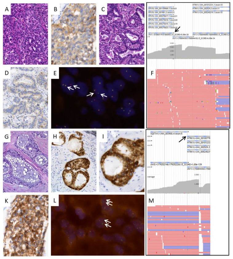
We describe two subtypes of NTRK fusions in NSCLC, one EPS15 (exon 9)—NTRK1 (exon 10) and one SQSTM1 (exon 6)—NTRK1 (exon 10) fusion. Both genes have previously been described as fusion partners for NTRK in NSCLC. EPS15, which encodes the epidermal growth factor receptor substrate 15, has been described as fusing with NTRK1, but with exon 21 of EPS15 as the fusion site [19,32]. Sequestosome-1 (SQSTM1) is known as a translocation partner of NTRK1, -2, and -3 with breakpoints located in exon 5 or 6 of SQSTM1 [33,34,35]. Based on histology, both NTRK-positive cases represent moderately differentiated adenocarcinomas with a predominant acinar morphology. Additional mucinous or solid components occurred (Figure 2). Both cases were negative for KRAS, BRAF, and EGFR mutations, ALK, ROS1, and RET fusions, as well as for MET amplification.
Figure 2.
Morphologic and molecular characteristics of NTRK fusion-positive lung cancer cases. Case 1: EPS15-NTRK1 fusion. (A,C): This adenocarcinoma shows solid and acinar morphology (hematoxylin-eosin staining, specimen from wedge resection). (B,D): Moderate to strong immunohistochemical staining of tumor cell membranes and cytoplasm with panTrk antibody in solid tumor aspect of the primary tumor; weak staining in acinar areas. (E): Fluorescence in situ hybridization (FISH) analysis showing multiple break apart signals (arrows), indicating NTRK1 gene translocation. (F): RNA-based NGS analysis showing multiple reads of an EPS15-NTRK1 fusion gene at the fusion site (arrow), confirming translocation of NTRK1 gene. Case 2: SQSTM1-NTRK1 fusion, (G): Mucinous morphology in a lymph node metastasis (hematoxylin-eosin staining). (H,I): Immunohistochemical staining with panTrk antibody demonstrating strong membranous staining. (K): Immunohistochemical staining in another lymph node metastasis confirming homogeneous strong staining. (L): Fluorescence in situ hybridization (FISH) analysis showing multiple break apart signals (arrows), indicating NTRK1 gene translocation. (M): RNA-based NGS analysis showing multiple reads of an SQSTM1-NTRK1 fusion gene at the fusion site (arrow), confirming translocation of NTRK1 gene. Original magnifications ×100 (A), ×200 (C,D,G,H), ×400 (B,I,K), ×630 (E,I).
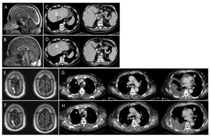
The two patients are male with a comparably long-standing history of their disease and primary diagnosis of lung cancer at younger ages (patient 1 at the age of 51 with oligometastatic disease and patient 2 at the age of 45 with stage IIIA disease). NTRK diagnostic was initiated within progressive and stage IV disease after surgery, radiotherapy, and systemic therapy. Patient 1 is a former smoker with a history of 10 pack years and patient 2 is a current smoker with 45 pack years. Both patients were included in clinical trials with Trk-targeted therapy, patient 1 within 27 months after primary diagnosis of lung cancer and patient 2 15 years and 9 months following the primary diagnosis. Patient 1 received Trk-directed therapy as a second-line treatment for stage IV disease and patient 2 as first line systemic therapy for stage IV disease. Both patients responded with partial remission. After the initiation of Trk-targeted therapy the clinical performance status improved to a normal level in both cases. Patient 1 is still undergoing Trk-targeted therapy at month 45 without recurrent disease. Patient 2 died of an exacerbation of underlying chronic obstructive lung disease, having been treated with Trk-targeted therapy without recurrence for 42 months. Figure 3 illustrates the intra- and extracranial responses in both patients documented by MRI (magnetic resonance imaging) and CT (computerized tomography) scans.
Figure 3.
MRI and CT scans of two patients with NTRK fusion and Trk-directed therapy. Patient 1 (A–D) and patient 2 (E–H) showed responses to Trk-targeted therapy. (A,C,E,G) show MRI and CT scans at baseline. (B,D) show follow-up at month 27 (patient 1) and (F,H) at month 6 (patient 2). Metastatic sites, (A–D): § sellar/suprasellar, & liver parenchyma, (E–H): ↑ right precentral gyrus, * mediastinal, ** hilar, # pericardial effusion and cardiac metastasis.
3.3. Positive Predictive Value and Diagnostic Characteristics of Immunohistochemistry
Our study represents one of the largest series of Trk IHC in NSCLC with roughly 1000 samples. Based on our experience, we noticed that the positive predictive value was low with only 2 out of 133 IHC positive samples where an NTRK fusion could be detected by subsequent sequencing. The NTRK-positive tumors showed weak to moderate cytoplasmic and membranous staining with varying intensities between different tumor manifestations (Figure 2).
We did not notice any major difference in IHC staining patterns between both fusion subtypes. Nuclear staining was not observed in these NTRK1-fused cancers.
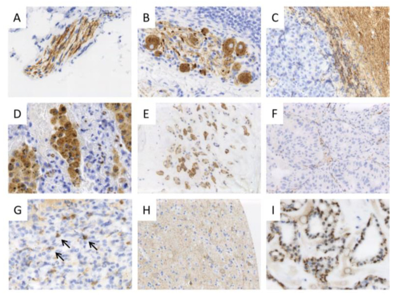
Overall, Trk staining was weak in tumor tissue. We failed to detect a correlation between staining intensity and NTRK fusion since some of the cases with the strongest staining turned out to be fusion negative. Moreover, Trk staining in fusion-positive samples was at least focally remarkably weak. We observed a reproducible and uniform Trk expression in some neoplastic and non-neoplastic tissues, which could be used as controls for IHC, such as secretory breast cancer or brain tissue. Tumor-infiltrating inflammatory cells, structures of peripheral nerval tissue. Or reactively changed skeletal muscle can express Trk at high levels, and therefore represent potential pitfalls for Trk IHC evaluation (Figure 4).
Figure 4.
Pitfalls in evaluating panTrk IHC staining. Trk IHC in non-neoplastic tissue, controls, and NTRK fusion-negative tumors. Trk proteins are physiologically expressed in structures of the peripheral and central nervous system, such as nerval fibers (A), ganglion cells (B), and brain tissue ((C)—right; Trk IHC-negative metastasis of an NSCLC on the left side). Furthermore, macrophages (D), reactively changed muscle fibers (E), and some vessels (F) can demonstrate a Trk expression. (G): Inflammatory cells surrounding a tumor may show positivity (arrows). (H): Brain tissue with weak staining was used as on-slide control. (I): Secretory breast cancer tissue with proven NTRK3-ETV6 fusion can be used as control. Tumor cells show weak cytoplasmic and moderate to strong nuclear staining. Original magnifications ×100 (E,H), ×200 (A–D,F,G,I).
Since panTrk immunohistochemistry does not discriminate between wild-type and fused Trk proteins, it is not surprising that carcinomas can express these proteins as well. The expression levels were the highest among sarcomatoid carcinomas, poorly differentiated NOS carcinomas, and neuroendocrine and squamous cell carcinomas. Staining in these tumors could be both membranous and cytoplasmic; nuclear staining was not observed. Based on our experience within this screening project and with other tumor tissues and previous reports, nuclear Trk staining is probably specific to NTRK3-fused cancers [17]. As mentioned above, we retrospectively examined a number of IHC-negative samples in the context of a cross-method validation by RNA-NGS and/or FISH, but did not observe any IHC-negative/RNA-NGS-positive cases.
3.4. Comparison of Molecular Test Approaches
We further examined NGS-negative cases with positive Trk expression based on IHC with FISH but could not identify additional positive samples. However, RNA-NGS-positive tumors were also clearly positive with NTRK1 FISH. We did not observe any NTRK2- or NTRK3-positive FISH sample. Therefore, we saw a 100% concordance between FISH and RNA-NGS based on the limited number of samples that were analyzed with both methods.
We obtained valid FISH results within two working days, whereas the entire RNA-NGS workflow, including RNA extraction and quantification, library preparation, sequencing, and bioinformatics, took more than 10 working days in our hands.
4. Discussion
Our major goal was to determine the prevalence of NTRK-fused lung cancers under real-world conditions. Based on more than 1000 patients, we detected a frequency of 0.2% in a prospective series of unbiased and unselected lung cancer patients. This prevalence falls within the range of previous reports from clinical trials and more specific NTRK screening programs [19,22,26,30,31,35,41,42,43]. Thus, our study does not only confirm the previously published prevalence but also demonstrates that NTRK-fused NSCLC can be detected by an approach of a two-step screening consisting of IHC as a pre-screening method and subsequent RNA-based NGS of IHC-positive samples. Monitoring the prevalence of NTRK-fusion-positive samples may be used as a validation tool of screening workflows in other institutions as well. Our data indicate that a 0.2% positivity rate in an all-comer population can serve as a reasonable target value.
We demonstrate that NTRK screening is feasible in a routine diagnostic setting as well. The drop-out rate in our proposed workflow of IHC followed by RNA-NGS was 9.0% (as opposed to 11.6% in a FISH-only approach), and we conclude that NTRK testing can be performed on specimens from resections, biopsies, and cell blocks if the tumor tissue fulfills basic requirements for IHC and molecular tests.
In our study, we used IHC as a pre-screening test. Based on our experience with nearly 1000 immunohistochemical samples, we describe a comparably low positive predictive value of IHC: only 1.5% of all IHC-positive NSCLC samples turned out to harbor a NTRK fusion. This is mainly due to the fact that IHC cannot discriminate between the expression of wild-type Trk proteins and chimeric fusion proteins. However, prevalence was higher among adenocarcinomas (2/701 = 0.29%). Among 568 non-squamous NSCLC with available simultaneous testing results of EGFR and KRAS, 297 were wild-type for both genes. In this group, the prevalence for NTRK positivity was 2/297 = 0.67%. Simultaneous testing for EGFR/KRAS/BRAF/ALK/ROS1/RET was available for 339 patients with NSCLC, with negative results for all genes in 175 cases. Among the latter group, the prevalence of NTRK rearrangements was 1.14%. We did not observe any NTRK fusions in tumors other than adenocarcinomas. This is in line with previous reports where the vast majority of NTRK-positive cancers were adenocarcinomas as well. Neuroendocrine carcinomas and squamous carcinomas are infrequently involved [13,15,31,33,34,37].
Based on our experience with Trk IHC, we describe potential diagnostic pitfalls and reasonable control tissues (Figure 4). In our study, we regarded RNA-based NGS as a gold standard and reference method. However, FISH analyses were completely concordant with NGS. Although we could only investigate a relatively small subset of our samples with both methods, we did not find any discrepant cases. Therefore, we conclude that FISH is a valid and reliable method to detect NTRK fusions as well. A major advantage of this technology is the comparably short time to diagnosis, with a disadvantage being the comparably higher number of slides cut from tumor blocks needed to examine all three NTRK genes.
Given the extremely low frequency of NTRK fusions among lung cancer patients on one hand and the significant clinical benefit of Trk inhibitor treatment in NTRK-fusion-positive patients on the other hand, it is extremely important to establish a diagnostic algorithm that is sensitive and specific, tissue-sparing, and cost- and time-efficient and that allows reliable testing in a routine environment.
In this report, we demonstrate that a diagnostic workflow, including IHC and RNA-based sequencing, has the capability to detect NTRK-fusion-positive samples under routine conditions. We identified positive patients at the expected frequency. NTRK fusion positivity could be centrally confirmed as part of a clinical trial, and patients responded to anti-Trk treatment. These clinical data provide further evidence that our workflow was reliable and valid.
It has been demonstrated that RNA-based sequencing should be currently regarded as a reference method or gold standard as it is superior to other methods, including DNA hybrid capture-based sequencing [14]. Therefore, RNA-based testing has been suggested as the method of choice whenever a sequencing device is available [52]. This approach will probably be the most sensitive and specific testing method. However, given the very high prevalence of lung cancer in Western populations, primary RNA-based NGS testing would require enormous financial and technical efforts. Additionally, RNA-based sequencing is known to depend on tissue preservation and RNA quality. Another limitation and potential pitfall of this technology is the need to consider bioinformatically non-pathogenic alterations, which might be detected by these assays. Although RNA-based sequencing can be combined with standard DNA-based mutational analyses to reduce the required time and tissue, these advanced technologies will not be available everywhere. Based on our data, IHC could reduce the number of necessary sequencings. Moreover, as another possible scenario, IHC screening could be carried out locally and only IHC-positive samples could be submitted to a reference laboratory for subsequent sequencing. With this approach, full genomic information on fusion subtypes would be obtained while reducing the number of unnecessary sequencings. However, the reliability of such an algorithm depends extremely on the sensitivity and specificity of immunohistochemistry. This will require rigorous validation and quality management of IHC as well as specific trainings of pathologists to make sure that all positive cases are appropriately recognized. The number of sequencings could be further reduced by considering standard molecular parameters, such as EGFR, KRAS, and BRAF mutations, and ALK, ROS1, and RET rearrangements. Based on our observations and previously published data, these alterations, which reflect specific molecular subtypes of lung cancer, are mutually exclusive with NTRK fusions. Thus, sequencings could be further numerically reduced (without losing true positive cases) if only Trk IHC-positive and EGFR/KRAS/BRAF/ALK/ROS1/RET-negative samples (tested by appropriate methods, including IHC and NGS) undergo RNA-based sequencing. We were able to test 141 EGFR/KRAS/BRAF/ALK/ROS1/RET-negative samples for Trk IHC (Scenario 1) and found only 43 cases positive (30.5%). Certainly, it should be mentioned that in the case of an EGFR-mutant NSCLC with secondary resistance to EGFR TKI therapy, NTRK testing should be always considered as NTRK1 fusions have been described as an escape mechanism against EGFR TKI therapy [35]. As an alternative algorithm, NTRK testing could be performed after histological confirmation of NSCLC and, in the case of adenocarcinoma, immunohistochemical exclusion of ALK or ROS1 alterations. This pathway requires highly sensitive and specific IHC for ALK and ROS1. However, due to the low incidence (together <5%), negative findings for ALK and ROS1 limit the number of cases to be investigated only to a small extent.
Moreover, there are NSCLC subtypes that are very unlikely to harbor NTRK fusions, such as sarcomatoid carcinomas. However, we do not recommend excluding patients from NTRK testing based on tumor morphology since data on correlation with morphologic subtypes of lung cancer are very limited so far and cases of NTRK fusions in non-adenocarcinoma NSCLC have indeed been reported.
Another method for molecular NTRK testing is fluorescence in situ hybridization. Although we have only very limited data from a small subset of our cohort, we did not observe any discordant cases. We could also detect all positive samples with this method and did not observe any false positives. FISH is a comparably fast testing approach, which, however, has two major disadvantages when compared to NGS: (i) it does not provide full genomic information, i.e., fusion partner genes and breakpoints, and (ii) it requires additional tissue slides for all three genes to be tested. The latter could be overcome in the future if multiplex FISH probes covering at least two NTRK genes were available. With a similar amount of material, RNA-NGS has the advantage that further analyses can be carried out on the extracted material, whereas FISH only allows the one examination. Based on the presented data, FISH is also reliable, and therefore it should be included in the proposed workflow as a second choice in case RNA-based NGS is not available, or does not pass quality controls, or exceeds the financial resources.
5. Conclusions
In summary, we provide data on the general feasibility of NTRK screening on a large series of NSCLC samples under real-world conditions. The prevalence of NTRK fusions is 0.2% in unselected lung cancer patients. Our data indicate that a combined approach of Trk IHC pre-screening plus subsequent RNA-based next-generation sequencing is a reliable algorithm to detect patients who would benefit from anti-Trk treatment.
Acknowledgments
The authors are grateful to the expert technical assistance provided by Mercedes Martin-Ortega, Sina Eckstein, Meike Wendt, Judith Wolf-Salgo, Anke Klages, and Nicole Kerl.
Author Contributions
T.R.O., A.R. (Annika Reiffert) and H.-U.S.; Methodology, T.R.O., K.R.-J. and H.-U.S.; Validation, T.R.O., A.R. (Annika Reiffert) and H.-U.S.; Investigation, T.R.O., A.R. (Annika Reiffert), K.R.-J. and H.-U.S.; Data curation, T.R.O., A.R. (Annika Reiffert) and H.-U.S.; Writing—original draft, T.R.O., A.R. (Annika Reiffert) and H.-U.S.; Writing—review & editing, T.R.O., A.R. (Annika Reiffert), K.S., A.R. (Achim Rittmeyer), W.K., S.H., J.S., L.L., T.H., M.H., K.R.-J. and H.-U.S. All authors have read and agreed to the published version of the manuscript.
Institutional Review Board Statement
The study was conducted in accordance with the Declaration of Helsinki, and approved by the Ethics Committee of Georg-August-University of Göttingen (35/9/22, date of approval 23 September 2022).
Informed Consent Statement
Broad consent (University Medical Center Göttingen) was obtained from all subjects involved in the study. Additionally, written informed consent has been obtained from the two patients with evidence of NTRK fusions for scientific publication of their clinical course data as well.
Data Availability Statement
The data presented in this study are available on request from the corresponding author. The data are not publicly available due to patient confidentiality concerns.
Conflicts of Interest
T.R.O. received grants as an advisor or speaker from: AstraZeneca, Boehringer Ingelheim, Daiichi-Sankyo, Roche Pharma, Merck Sharp and Dohme, Novartis Pharma, and Takeda Oncology. T.R.O. received travel support from AstraZeneca and Janssen-Cilag. K.S. received honoraria as advisor or speaker from: Amgen, AstraZeneca, Bayer, Daiichi-Sankyo, Diaceutics, Incyte, Janssen-Cilag, Eli Lilly, Merck Sharp and Dohme, Novartis Pharma, Pfizer, PharmaMar, Roche Pharma, Servier, Stemline, Targos GmbH, and ZytoVision. A.R. received grants as an advisor or speaker from: AbbVie, AstraZeneca, Bristol-Myers Squibb, Boehringer Ingelheim, Eli Lilly, Merck Sharp and Dohme, Novartis Pharma, Pfizer, and Roche Pharma. W.K. received grants as an advisor or speaker from: AstraZeneca, Berlin Chemie, Boehringer Ingelheim, Bristol-Myers Squibb, Eli Lilly, Merck, Merck Sharp and Dohme, Novartis Pharma, Pfizer, Roche Pharma, and Takeda Oncology. M.H. received grants as an advisor or speaker from: AstraZeneca, Merck Sharp and Dohme, and Roche Pharma. K.R.-J. received grants as a speaker from: AstraZeneca, Roche, and ZytoVision. H.-U.S. received a research grant from Novartis Oncology, served as an advisory board member for Abbott Molecular, Abbvie, Bristol-Myers Squibb, Merck Sharp and Dohme, Novartis Oncology, and Pfizer, and received honoraria from Bristol-Myers Squibb, Merck Sharp and Dohme, Pfizer, Roche Pharma, Zytomed, and ZytoVision. The other authors have no conflicts of interest to declare.
Funding Statement
This research was funded by Ignyta, Inc. and Roche Pharma.
Footnotes
Disclaimer/Publisher’s Note: The statements, opinions and data contained in all publications are solely those of the individual author(s) and contributor(s) and not of MDPI and/or the editor(s). MDPI and/or the editor(s) disclaim responsibility for any injury to people or property resulting from any ideas, methods, instructions or products referred to in the content.
References
- 1.Amatu A., Sartore-Bianchi A., Siena S. NTRK gene fusions as novel targets of cancer therapy across multiple tumour types. ESMO Open. 2016;1:e000023. doi: 10.1136/esmoopen-2015-000023. [DOI] [PMC free article] [PubMed] [Google Scholar]
- 2.Luberg K., Wong J., Weickert C.S., Timmusk T. Human TrkB gene: Novel alternative transcripts, protein isoforms and expression pattern in the prefrontal cerebral cortex during postnatal development. J. Neurochem. 2010;113:952–964. doi: 10.1111/j.1471-4159.2010.06662.x. [DOI] [PubMed] [Google Scholar]
- 3.Ichaso N., Rodriguez R.E., Martin-Zanca D., Gonzalez-Sarmiento R. Genomic characterization of the human trkC gene. Oncogene. 1998;17:1871–1875. doi: 10.1038/sj.onc.1202100. [DOI] [PubMed] [Google Scholar]
- 4.Huang E.J., Reichardt L.F. Trk receptors: Roles in neuronal signal transduction. Annu. Rev. Biochem. 2003;72:609–642. doi: 10.1146/annurev.biochem.72.121801.161629. [DOI] [PubMed] [Google Scholar]
- 5.Ardini E., Bosotti R., Borgia A.L., de Ponti C., Somaschini A., Cammarota R., Amboldi N., Raddrizzani L., Milani A., Magnaghi P., et al. The TPM3-NTRK1 rearrangement is a recurring event in colorectal carcinoma and is associated with tumor sensitivity to TRKA kinase inhibition. Mol. Oncol. 2014;8:1495–1507. doi: 10.1016/j.molonc.2014.06.001. [DOI] [PMC free article] [PubMed] [Google Scholar]
- 6.Morosini D., Chmielecki J., Goldberg M., Ross J.S., Stephens P.J., Miller V.A., Davis L.E. Comprehensive genomic profiling of sarcomas from 267 adolescents and young adults to reveal a spectrum of targetable genomic alterations. J. Clin. Oncol. 2015;33:11020. doi: 10.1200/jco.2015.33.15_suppl.11020. [DOI] [Google Scholar]
- 7.Suh J.H., Johnson A., Albacker L., Wang K., Chmielecki J., Frampton G., Gay L., Elvin J.A., Vergilio J.-A., Ali S., et al. Comprehensive Genomic Profiling Facilitates Implementation of the National Comprehensive Cancer Network Guidelines for Lung Cancer Biomarker Testing and Identifies Patients Who May Benefit from Enrollment in Mechanism-Driven Clinical Trials. Oncologist. 2016;21:684–691. doi: 10.1634/theoncologist.2016-0030. [DOI] [PMC free article] [PubMed] [Google Scholar]
- 8.Cocco E., Scaltriti M., Drilon A. NTRK fusion-positive cancers and TRK inhibitor therapy. Nat. Rev. Clin. Oncol. 2018;15:731–747. doi: 10.1038/s41571-018-0113-0. [DOI] [PMC free article] [PubMed] [Google Scholar]
- 9.Prasad M.L., Vyas M., Horne M.J., Virk R.K., Morotti R., Liu Z., Tallini G., Nikiforova M.N., Christison-Lagay E.R., Udelsman R., et al. NTRK fusion oncogenes in pediatric papillary thyroid carcinoma in northeast United States. Cancer. 2016;122:1097–1107. doi: 10.1002/cncr.29887. [DOI] [PubMed] [Google Scholar]
- 10.Ricarte-Filho J.C., Li S., Garcia-Rendueles M.E.R., Montero-Conde C., Voza F., Knauf J.A., Heguy A., Viale A., Bogdanova T., Thomas G.A., et al. Identification of kinase fusion oncogenes in post-Chernobyl radiation-induced thyroid cancers. J. Clin. Investig. 2013;123:4935–4944. doi: 10.1172/JCI69766. [DOI] [PMC free article] [PubMed] [Google Scholar]
- 11.Leeman-Neill R.J., Kelly L.M., Liu P., Brenner A.V., Little M.P., Bogdanova T.I., Evdokimova V.N., Hatch M., Zurnadzy L.Y., Nikiforova M.N., et al. ETV6-NTRK3 is a common chromosomal rearrangement in radiation-associated thyroid cancer. Cancer. 2014;120:799–807. doi: 10.1002/cncr.28484. [DOI] [PMC free article] [PubMed] [Google Scholar]
- 12.Wiesner T., He J., Yelensky R., Esteve-Puig R., Botton T., Yeh I., Lipson D., Otto G., Brennan K., Murali R., et al. Kinase fusions are frequent in Spitz tumors and spitzoid melanomas. Nat. Commun. 2014;5:3116. doi: 10.1038/ncomms4116. [DOI] [PMC free article] [PubMed] [Google Scholar]
- 13.Vaishnavi A., Le A.T., Doebele R.C. TRKing down an old oncogene in a new era of targeted therapy. Cancer Discov. 2015;5:25–34. doi: 10.1158/2159-8290.CD-14-0765. [DOI] [PMC free article] [PubMed] [Google Scholar]
- 14.Solomon J.P., Linkov I., Rosado A., Mullaney K., Rosen E.Y., Frosina D., Jungbluth A.A., Zehir A., Benayed R., Drilon A., et al. NTRK fusion detection across multiple assays and 33,997 cases: Diagnostic implications and pitfalls. Mod. Pathol. 2020;33:38–46. doi: 10.1038/s41379-019-0324-7. [DOI] [PMC free article] [PubMed] [Google Scholar]
- 15.Okamura R., Boichard A., Kato S., Sicklick J.K., Bazhenova L., Kurzrock R. Analysis of NTRK Alterations in Pan-Cancer Adult and Pediatric Malignancies: Implications for NTRK-Targeted Therapeutics. JCO Precis. Oncol. 2018;2018:1–20. doi: 10.1200/PO.18.00183. [DOI] [PMC free article] [PubMed] [Google Scholar]
- 16.Stransky N., Cerami E., Schalm S., Kim J.L., Lengauer C. The landscape of kinase fusions in cancer. Nat. Commun. 2014;5:4846. doi: 10.1038/ncomms5846. [DOI] [PMC free article] [PubMed] [Google Scholar]
- 17.Gatalica Z., Xiu J., Swensen J., Vranic S. Molecular characterization of cancers with NTRK gene fusions. Mod. Pathol. 2019;32:147–153. doi: 10.1038/s41379-018-0118-3. [DOI] [PubMed] [Google Scholar]
- 18.Rubin B.P., Chen C.-J., Morgan T.W., Xiao S., Grier H.E., Kozakewich H.P., Perez-Atayde A.R., Fletcher J.A. Congenital Mesoblastic Nephroma t(12;15) Is Associated with ETV6-NTRK3 Gene Fusion: Cytogenetic and Molecular Relationship to Congenital (Infantile) Fibrosarcoma. Am. J. Pathol. 1998;153:1451–1458. doi: 10.1016/S0002-9440(10)65732-X. [DOI] [PMC free article] [PubMed] [Google Scholar]
- 19.Knezevich S.R., McFadden D.E., Tao W., Lim J.F., Sorensen P.H. A novel ETV6-NTRK3 gene fusion in congenital fibrosarcoma. Nat. Genet. 1998;18:184–187. doi: 10.1038/ng0298-184. [DOI] [PubMed] [Google Scholar]
- 20.Vaishnavi A., Capelletti M., Le A.T., Kako S., Butaney M., Ercan D., Mahale S., Davies K.D., Aisner D.L., Pilling A.B., et al. Oncogenic and drug-sensitive NTRK1 rearrangements in lung cancer. Nat. Med. 2013;19:1469–1472. doi: 10.1038/nm.3352. [DOI] [PMC free article] [PubMed] [Google Scholar]
- 21.Hechtman J.F., Benayed R., Hyman D.M., Drilon A., Zehir A., Frosina D., Arcila M.E., Dogan S., Klimstra D.S., Ladanyi M., et al. Pan-Trk Immunohistochemistry Is an Efficient and Reliable Screen for the Detection of NTRK Fusions. Am. J. Surg. Pathol. 2017;41:1547–1551. doi: 10.1097/PAS.0000000000000911. [DOI] [PMC free article] [PubMed] [Google Scholar]
- 22.Helman E., Nguyen M., Karlovich C.A., Despain D., Choquette A.K., Spira A.I., Yu H.A., Camidge D.R., Harding T.C., Lanman R.B., et al. Cell-Free DNA Next-Generation Sequencing Prediction of Response and Resistance to Third-Generation EGFR Inhibitor. Clin. Lung Cancer. 2018;19:518–530.e7. doi: 10.1016/j.cllc.2018.07.008. [DOI] [PubMed] [Google Scholar]
- 23.Zheng Z., Liebers M., Zhelyazkova B., Cao Y., Panditi D., Lynch K.D., Chen J., Robinson H.E., Shim H.S., Chmielecki J., et al. Anchored multiplex PCR for targeted next-generation sequencing. Nat. Med. 2014;20:1479–1484. doi: 10.1038/nm.3729. [DOI] [PubMed] [Google Scholar]
- 24.Westphalen C.B., Krebs M.G., Le Tourneau C., Sokol E.S., Maund S.L., Wilson T.R., Jin D.X., Newberg J.Y., Fabrizio D., Veronese L., et al. Genomic context of NTRK1/2/3 fusion-positive tumours from a large real-world population. NPJ Precis. Oncol. 2021;5:69. doi: 10.1038/s41698-021-00206-y. [DOI] [PMC free article] [PubMed] [Google Scholar]
- 25.Wang B., Gao Y., Huang Y., Ou Q., Fang T., Tang C., Wu X., Shao Y.W. Durable Clinical Response to Crizotinib in IRF2BP2-NTRK1 Non-small-cell Lung Cancer. Clin. Lung Cancer. 2019;20:e233–e237. doi: 10.1016/j.cllc.2018.12.017. [DOI] [PubMed] [Google Scholar]
- 26.Marchetti A., Di Lorito A., Felicioni L., Buttitta F. An innovative diagnostic strategy for the detection of rare molecular targets to select cancer patients for tumor-agnostic treatments. Oncotarget. 2019;10:6957–6968. doi: 10.18632/oncotarget.27343. [DOI] [PMC free article] [PubMed] [Google Scholar]
- 27.Gautschi O., Bubendorf L., Leyvraz S., Menon R., Diebold J. Challenges in the Diagnosis of NTRK Fusion-Positive Cancers. J. Thorac. Oncol. 2020;15:e108–e110. doi: 10.1016/j.jtho.2019.05.001. [DOI] [PubMed] [Google Scholar]
- 28.Hartmaier R.J., Albacker L.A., Chmielecki J., Bailey M., He J., Goldberg M.E., Ramkissoon S., Suh J., Elvin J.A., Chiacchia S., et al. High-Throughput Genomic Profiling of Adult Solid Tumors Reveals Novel Insights into Cancer Pathogenesis. Cancer Res. 2017;77:2464–2475. doi: 10.1158/0008-5472.CAN-16-2479. [DOI] [PubMed] [Google Scholar]
- 29.Fernandez-Cuesta L., Peifer M., Lu X., Seidel D., Zander T., Leenders F., Ozretić L., Brustugun O.-T., Field J.K., Wright G., et al. Molecular and Cellular Biology, Proceedings of the AACR Annual Meeting 2014, San Diego, CA, USA, 5–9 April 2014. American Association for Cancer Research; Philadelphia, PA, USA: 2014. Abstract 1531: Cross-entity mutation analysis of lung neuroendocrine tumors sheds light into their molecular origin and identifies new therapeutic targets; p. 1531. [Google Scholar]
- 30.von der Thüsen J.H., Dumoulin D.W., Maat A.P.W.M., Wolf J., Sadeghi A.H., Aerts J.G.J.V., Cornelissen R. ETV6-NTRK3 translocation-associated low-grade mucinous bronchial adenocarcinoma: A novel bronchial salivary gland-type non-small cell lung cancer subtype. Lung Cancer. 2021;156:72–75. doi: 10.1016/j.lungcan.2021.04.015. [DOI] [PubMed] [Google Scholar]
- 31.Sigal D.S., Bhangoo M.S., Hermel J.A., Pavlick D.C., Frampton G., Miller V.A., Ross J.S., Ali S.M. Comprehensive genomic profiling identifies novel NTRK fusions in neuroendocrine tumors. Oncotarget. 2018;9:35809–35812. doi: 10.18632/oncotarget.26260. [DOI] [PMC free article] [PubMed] [Google Scholar]
- 32.Drilon A., Kummar S., Moreno V., Patel J., Lassen U., Rosen L., Childs B.H., Nanda S., Cox M.C., Ku N.C., et al. Activity of larotrectinib in TRK fusion lung cancer. Ann. Oncol. 2019;30:ii48–ii49. doi: 10.1093/annonc/mdz063.009. [DOI] [Google Scholar]
- 33.Farago A.F., Taylor M.S., Doebele R.C., Zhu V.W., Kummar S., Spira A.I., Boyle T.A., Haura E.B., Arcila M.E., Benayed R., et al. Clinicopathologic Features of Non-Small-Cell Lung Cancer Harboring an NTRK Gene Fusion. JCO Precis. Oncol. 2018;2018:1–12. doi: 10.1200/PO.18.00037. [DOI] [PMC free article] [PubMed] [Google Scholar]
- 34.Drilon A., Siena S., Ou S.-H.I., Patel M., Ahn M.J., Lee J., Bauer T.M., Farago A.F., Wheler J.J., Liu S.V., et al. Safety and Antitumor Activity of the Multitargeted Pan-TRK, ROS1, and ALK Inhibitor Entrectinib: Combined Results from Two Phase I Trials (ALKA-372-001 and STARTRK-1) Cancer Discov. 2017;7:400–409. doi: 10.1158/2159-8290.CD-16-1237. [DOI] [PMC free article] [PubMed] [Google Scholar]
- 35.Xia H., Xue X., Ding H., Ou Q., Wu X., Nagasaka M., Shao Y.W., Hu X., Ou S.-H.I. Evidence of NTRK1 Fusion as Resistance Mechanism to EGFR TKI in EGFR+ NSCLC: Results from a Large-Scale Survey of NTRK1 Fusions in Chinese Patients with Lung Cancer. Clin. Lung Cancer. 2020;21:247–254. doi: 10.1016/j.cllc.2019.09.004. [DOI] [PubMed] [Google Scholar]
- 36.Bang H., Lee M.-S., Sung M., Choi J., An S., Kim S.-H., Lee S.E., Choi Y.-L. NTRK Fusions in 1113 Solid Tumors in a Single Institution. Diagnostics. 2022;12:1450. doi: 10.3390/diagnostics12061450. [DOI] [PMC free article] [PubMed] [Google Scholar]
- 37.Boulanger M.C., Temel J.S., Mino-Kenudson M., Ritterhouse L.L., Dagogo-Jack I. Primary Resistance to Larotrectinib in a Patient with Squamous NSCLC With Subclonal NTRK1 Fusion: Case Report. JTO Clin. Res. Rep. 2023;4:100501. doi: 10.1016/j.jtocrr.2023.100501. [DOI] [PMC free article] [PubMed] [Google Scholar]
- 38.Xiao Z., Huang X., Xie B., Xie W., Huang M., Lin L. Primary Resistance to Brigatinib in a Patient with Lung Adenocarcinoma Harboring ALK G1202R Mutation and LIPI-NTRK1 Rearrangement. OncoTargets. Ther. 2020;13:4591–4595. doi: 10.2147/OTT.S249652. [DOI] [PMC free article] [PubMed] [Google Scholar]
- 39.Kishikawa S., Hayashi T., Shimizu J., Fuwa B., Nonomura A., Saito T., Yatabe Y., Yao T. Low-grade tracheal adenocarcinoma with ETV6:NTRK3 fusion: Unique morphology akin to subsets of sinonasal low-grade non-intestinal-type adenocarcinoma. Virchows Arch. 2022;481:793–797. doi: 10.1007/s00428-022-03353-0. [DOI] [PubMed] [Google Scholar]
- 40.Lazzari C., Pecciarini L., Doglioni C., Pedica F., Gajate A.M.S., Bulotta A., Gregorc V., Cangi M.G. Case report: EML4:NTRK3 gene fusion in a patient with metastatic lung adenocarcinoma successfully treated with entrectinib. Front. Oncol. 2022;12:1038774. doi: 10.3389/fonc.2022.1038774. [DOI] [PMC free article] [PubMed] [Google Scholar]
- 41.Rosen E.Y., Goldman D.A., Hechtman J.F., Benayed R., Schram A.M., Cocco E., Shifman S., Gong Y., Kundra R., Solomon J.P., et al. TRK Fusions Are Enriched in Cancers with Uncommon Histologies and the Absence of Canonical Driver Mutations. Clin. Cancer Res. 2020;26:1624–1632. doi: 10.1158/1078-0432.CCR-19-3165. [DOI] [PMC free article] [PubMed] [Google Scholar]
- 42.Si X., Pan R., Ma S., Li L., Liang L., Zhang P., Chu Y., Wang H., Wang M., Zhang X., et al. Genomic characteristics of driver genes in Chinese patients with non-small cell lung cancer. Thorac. Cancer. 2021;12:357–363. doi: 10.1111/1759-7714.13757. [DOI] [PMC free article] [PubMed] [Google Scholar]
- 43.Farago A.F., Le L.P., Zheng Z., Muzikansky A., Drilon A., Patel M., Bauer T.M., Liu S.V., Ou S.-H.I., Jackman D., et al. Durable Clinical Response to Entrectinib in NTRK1-Rearranged Non-Small Cell Lung Cancer. J. Thorac. Oncol. 2015;10:1670–1674. doi: 10.1097/01.JTO.0000473485.38553.f0. [DOI] [PMC free article] [PubMed] [Google Scholar]
- 44.Drilon A., Laetsch T.W., Kummar S., DuBois S.G., Lassen U.N., Demetri G.D., Nathenson M., Doebele R.C., Farago A.F., Pappo A.S., et al. Efficacy of Larotrectinib in TRK Fusion-Positive Cancers in Adults and Children. N. Engl. J. Med. 2018;378:731–739. doi: 10.1056/NEJMoa1714448. [DOI] [PMC free article] [PubMed] [Google Scholar]
- 45.Doebele R.C., Drilon A., Paz-Ares L., Siena S., Shaw A.T., Farago A.F., Blakely C.M., Seto T., Cho B.C., Tosi D., et al. Entrectinib in patients with advanced or metastatic NTRK fusion-positive solid tumours: Integrated analysis of three phase 1–2 trials. Lancet Oncol. 2020;21:271–282. doi: 10.1016/S1470-2045(19)30691-6. [DOI] [PMC free article] [PubMed] [Google Scholar]
- 46.FDA Approves Entrectinib for NTRK Solid Tumors and ROS-1 NSCLC. FDA. 20 December 2019. [(accessed on 13 May 2021)]; Available online: https://www.fda.gov/drugs/resources-information-approved-drugs/fda-approves-entrectinib-ntrk-solid-tumors-and-ros-1-nsclc.
- 47.Vitrakvi|European Medicines Agency. [(accessed on 2 October 2022)]. Available online: https://www.ema.europa.eu/en/medicines/human/EPAR/vitrakvi.
- 48.Rozlytrek|European Medicines Agency. [(accessed on 13 May 2021)]. Available online: https://www.ema.europa.eu/en/medicines/human/EPAR/rozlytrek.
- 49.FDA Approves Larotrectinib for Solid Tumors with NTRK Gene Fusions. FDA. 20 December 2019. [(accessed on 9 March 2020)]; Available online: https://www.fda.gov/drugs/fda-approves-larotrectinib-solid-tumors-ntrk-gene-fusions-0.
- 50.Pfarr N., Kirchner M., Lehmann U., Leichsenring J., Merkelbach-Bruse S., Glade J., Hummel M., Stögbauer F., Lehmann A., Trautmann M., et al. Testing NTRK testing: Wet-lab and in silico comparison of RNA-based targeted sequencing assays. Genes Chromosomes Cancer. 2020;59:178–188. doi: 10.1002/gcc.22819. [DOI] [PubMed] [Google Scholar]
- 51.Schildhaus H.-U. Immunohistochemistry-based predictive biomarkers for lung cancer. Pathologe. 2020;41:21–31. doi: 10.1007/s00292-020-00750-7. [DOI] [PubMed] [Google Scholar]
- 52.Marchiò C., Scaltriti M., Ladanyi M., Iafrate A.J., Bibeau F., Dietel M., Hechtman J.F., Troiani T., López-Rios F., Douillard J.-Y., et al. ESMO recommendations on the standard methods to detect NTRK fusions in daily practice and clinical research. Ann. Oncol. 2019;30:1417–1427. doi: 10.1093/annonc/mdz204. [DOI] [PubMed] [Google Scholar]
- 53.Overbeck T.R., Cron D.A., Schmitz K., Rittmeyer A., Körber W., Hugo S., Schnalke J., Lukat L., Hugo T., Hinterthaner M., et al. Top-level MET gene copy number gain defines a subtype of poorly differentiated pulmonary adenocarcinomas with poor prognosis. Transl. Lung Cancer Res. 2020;9:603–616. doi: 10.21037/tlcr-19-339. [DOI] [PMC free article] [PubMed] [Google Scholar]
- 54.Travis W.D., Brambilla E., Nicholson A.G., Yatabe Y., Austin J.H.M., Beasley M.B., Chirieac L.R., Dacic S., Duhig E., Flieder D.B., et al. The 2015 World Health Organization Classification of Lung Tumors: Impact of Genetic, Clinical and Radiologic Advances Since the 2004 Classification. J. Thorac. Oncol. 2015;10:1243–1260. doi: 10.1097/JTO.0000000000000630. [DOI] [PubMed] [Google Scholar]
- 55.Travis W.D., Brambilla E., Noguchi M., Nicholson A.G., Geisinger K.R., Yatabe Y., Beer D.G., Powell C.A., Riely G.J., van Schil P.E., et al. International association for the study of lung cancer/american thoracic society/european respiratory society international multidisciplinary classification of lung adenocarcinoma. J. Thorac. Oncol. 2011;6:244–285. doi: 10.1097/JTO.0b013e318206a221. [DOI] [PMC free article] [PubMed] [Google Scholar]
- 56.Overbeck T.R., Schmitz K., Engelke C., Sahlmann C.-O., Hugo S., Kellner L., Trümper L., Schildhaus H.-U. Partial Response to First-Line Crizotinib in an Elderly Male Patient with ROS1 Translocation-Positive Lung Cancer. Case Rep. Oncol. 2016;9:158–163. doi: 10.1159/000444745. [DOI] [PMC free article] [PubMed] [Google Scholar]
- 57.Schildhaus H.-U., Deml K.-F., Schmitz K., Meiboom M., Binot E., Hauke S., Merkelbach-Bruse S., Büttner R. Chromogenic in situ hybridization is a reliable assay for detection of ALK rearrangements in adenocarcinomas of the lung. Mod. Pathol. 2013;26:1468–1477. doi: 10.1038/modpathol.2013.95. [DOI] [PubMed] [Google Scholar]
- 58.Chen N.-M., Neesse A., Dyck M.L., Steuber B., Koenig A.O., Lubeseder-Martellato C., Winter T., Forster T., Bohnenberger H., Kitz J., et al. Context-Dependent Epigenetic Regulation of Nuclear Factor of Activated T Cells 1 in Pancreatic Plasticity. Gastroenterology. 2017;152:1507–1520.e15. doi: 10.1053/j.gastro.2017.01.043. [DOI] [PubMed] [Google Scholar]
- 59.Schildhaus H.-U. Der prädiktive Wert der PD-L1-Diagnostik. Pathologe. 2018;39:498–519. doi: 10.1007/s00292-018-0507-x. [DOI] [PubMed] [Google Scholar]
- 60.Schildhaus H.-U., Schultheis A.M., Rüschoff J., Binot E., Merkelbach-Bruse S., Fassunke J., Schulte W., Ko Y.-D., Schlesinger A., Bos M., et al. MET amplification status in therapy-naïve adeno- and squamous cell carcinomas of the lung. Clin. Cancer Res. 2015;21:907–915. doi: 10.1158/1078-0432.CCR-14-0450. [DOI] [PubMed] [Google Scholar]
Associated Data
This section collects any data citations, data availability statements, or supplementary materials included in this article.
Data Availability Statement
The data presented in this study are available on request from the corresponding author. The data are not publicly available due to patient confidentiality concerns.


