

Abstract
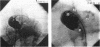
OBJECTIVE--To obtain angiographic views in tetralogy of Fallot that can show whether or not an anomalous coronary artery passes anterior to the right ventricular outflow tract. DESIGN--(a) A 10 year retrospective review of all patients who underwent repair of tetralogy of Fallot up to December 1990; (b) a prospective study of 30 children undergoing routine cardiac catheterisation. PATIENTS AND METHODS--295 cases in whom standard angiographic views had been used were reviewed retrospectively. Thirty non-consecutive children with tetralogy of Fallot were studied prospectively, including one child previously studied in whom diagnosis of an unsuspected anomalous coronary artery was made only at operation. The aortogram was performed with > or = 45 degrees caudocranial and 20 degrees-30 degrees left anterior oblique angles. SETTING--Tertiary referral centre. RESULTS--Ten of the 295 cases reviewed were shown to have a coronary vessel traversing the right ventricular outflow tract. In one case the diagnosis was suspected before operation but it was missed in the others. Even in retrospect we could not be certain of the precise anatomy with the use of standard angiographic views. In the prospective study the caudocranial aortogram showed the aortic valve face on in all the patients. The right ventricular outflow tract lay in a left and anterior (seen as superior) position in relation to the aortic root. Thus any vessel crossing the outflow tract could be identified. Identification of the aortic cusps allowed precise definition of the origin of the coronary arteries. All but four had normal origin and course of the coronary arteries. Four had paired left anterior descending arteries (including the restudied patient), in all cases with a large vessel originating from the right coronary artery passing across the right ventricular outflow tract. CONCLUSIONS--Important anomalies of the coronary arteries in tetralogy of Fallot may remain undiagnosed if standard angiographic projections are used. Aortography with > or = 45 degrees caudocranial and 20 degrees-30 degrees left anterior oblique angles allows precise definition of the anatomy and certainty as to whether any major vessel crosses the right ventricular outflow tract. Interpretation, however, can only be correct if the projection is technically adequate with a view of the aortic valve face on. Furthermore, a normal bifurcation of the left main stem does not exclude a second left anterior descending artery crossing the pulmonary outflow tract.
Full text
PDF

Images in this article
Selected References
These references are in PubMed. This may not be the complete list of references from this article.
- Berry B. E., McGoon D. C. Total correction for tetralogy of Fallot with anomalous coronary artery. Surgery. 1973 Dec;74(6):894–898. [PubMed] [Google Scholar]
- Berry J. M., Jr, Einzig S., Krabill K. A., Bass J. L. Evaluation of coronary artery anatomy in patients with tetralogy of Fallot by two-dimensional echocardiography. Circulation. 1988 Jul;78(1):149–156. doi: 10.1161/01.cir.78.1.149. [DOI] [PubMed] [Google Scholar]
- Dabizzi R. P., Caprioli G., Aiazzi L., Castelli C., Baldrighi G., Parenzan L., Baldrighi V. Distribution and anomalies of coronary arteries in tetralogy of fallot. Circulation. 1980 Jan;61(1):95–102. doi: 10.1161/01.cir.61.1.95. [DOI] [PubMed] [Google Scholar]
- Di Donato R. M., Jonas R. A., Lang P., Rome J. J., Mayer J. E., Jr, Castaneda A. R. Neonatal repair of tetralogy of Fallot with and without pulmonary atresia. J Thorac Cardiovasc Surg. 1991 Jan;101(1):126–137. [PubMed] [Google Scholar]
- Fellows K. E., Freed M. D., Keane J. F., Praagh R., Bernhard W. F., Castaneda A. C. Results of routine preoperative coronary angiography in tetralogy of Fallot. Circulation. 1975 Mar;51(3):561–566. doi: 10.1161/01.cir.51.3.561. [DOI] [PubMed] [Google Scholar]
- Fellows K. E., Smith J., Keane J. F. Preoperative angiocardiography in infants with tetrad of Fallot. Review of 36 cases. Am J Cardiol. 1981 Jun;47(6):1279–1285. doi: 10.1016/0002-9149(81)90259-9. [DOI] [PubMed] [Google Scholar]
- Hawe A., Rastelli G. C., Ritter D. G., DuShane J. W., McGoon D. C. Management of the right ventricular outflow tract in severe tetralogy of Fallot. J Thorac Cardiovasc Surg. 1970 Jul;60(1):131–143. [PubMed] [Google Scholar]
- Jureidini S. B., Appleton R. S., Nouri S. Detection of coronary artery abnormalities in tetralogy of Fallot by two-dimensional echocardiography. J Am Coll Cardiol. 1989 Oct;14(4):960–967. doi: 10.1016/0735-1097(89)90473-7. [DOI] [PubMed] [Google Scholar]
- MENG C. C., ECKNER F. A., LEV M. CORONARY ARTERY DISTRIBUTION IN TETRALOGY OF FALLOT. Arch Surg. 1965 Mar;90:363–366. doi: 10.1001/archsurg.1965.01320090041009. [DOI] [PubMed] [Google Scholar]
- Mandell V. S., Lock J. E., Mayer J. E., Parness I. A., Kulik T. J. The "laid-back" aortogram: an improved angiographic view for demonstration of coronary arteries in transposition of the great arteries. Am J Cardiol. 1990 Jun 1;65(20):1379–1383. doi: 10.1016/0002-9149(90)91331-y. [DOI] [PubMed] [Google Scholar]
- Stein P. D., Sabbah H. N. Orifice-view roentgenography for evaluation of the aortic valve. Am J Roentgenol Radium Ther Nucl Med. 1975 Dec;125(4):847–853. doi: 10.2214/ajr.125.4.847. [DOI] [PubMed] [Google Scholar]
- Touati G. D., Vouhé P. R., Amodeo A., Pouard P., Mauriat P., Leca F., Neveux J. Y. Primary repair of tetralogy of Fallot in infancy. J Thorac Cardiovasc Surg. 1990 Mar;99(3):396–403. [PubMed] [Google Scholar]