

Abstract
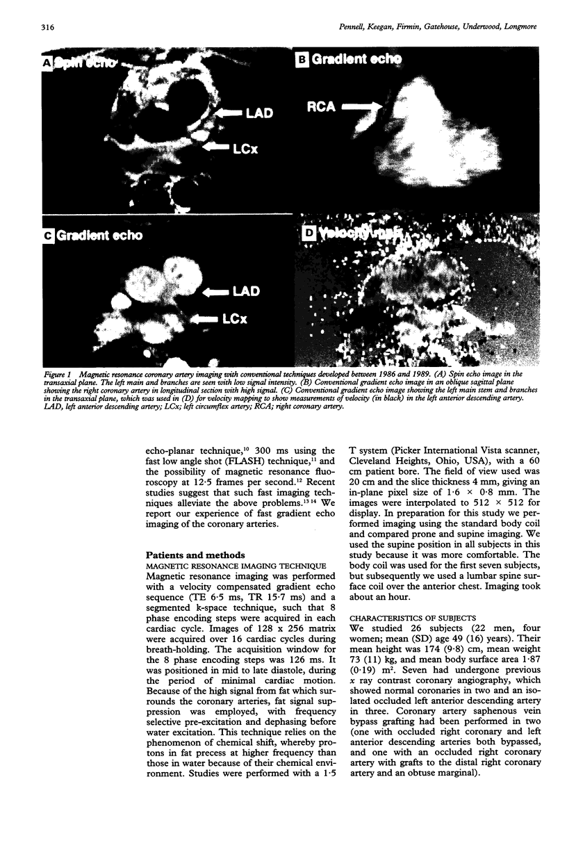
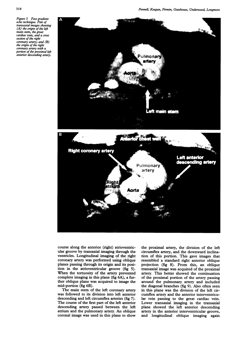
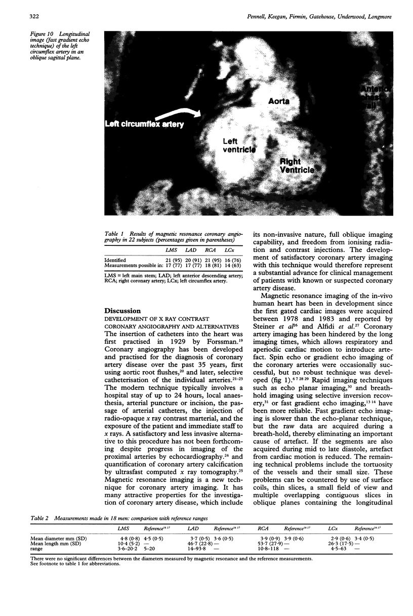
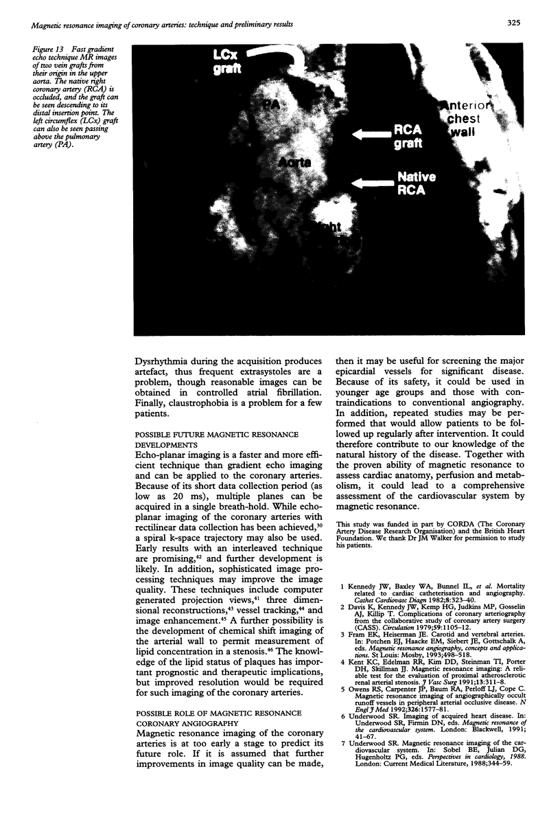
BACKGROUND--Coronary artery imaging is an important investigation for the management of coronary artery disease. The only reliable technique presently available, x ray contrast angiography, is invasive and is associated with a small morbidity and mortality. Alternative non-invasive imaging would be useful, but the small calibre and tortuosity of the coronary vessels, and cardiac and respiratory motion create formidable imaging problems. OBJECTIVE--The development of rapid magnetic resonance imaging of the coronary arteries. PATIENTS--21 healthy controls and five patients with coronary artery disease established by x ray contrast angiography, of whom two had undergone bypass grafting. METHODS--Magnetic resonance imaging was performed with gradient echoes and a segmented k-space technique, such that a complete image was acquired in 16 cardiac cycles during a breathhold. The signal from fat was suppressed and images were acquired in late diastole to reduce artefact from cardiac motion. An imaging strategy was developed for the proximal arteries, including longitudinal imaging from oblique planes defined according to the origins and the continuation of the arteries in the atrioventricular grooves or interventricular sulcus. RESULTS--Of the 26 subjects studied, 22 were imaged successfully. Identification of the artery was possible for the left main stem, left anterior descending, right coronary, and left circumflex arteries respectively in 95%, 91%, 95%, and 76%. The arterial diameter at the origin could be measured in 77%, 77%, 81%, and 63%. The mean (SD) arterial diameter in each case (4.8 (0.8), 3.7 (0.5), 3.9 (0.9), and 2.9 (0.6) mm) was not significantly different from reference values. The mean length of artery visualised was 10.4 (5.2), 46.7 (22.8), 53.7 (27.9), and 26.3 (17.5) mm. In 12 healthy men the total coronary area was 30.9 (9.2) mm2 and the ratio compared with body surface area was 16.4 (4.4) mm2m2 (both p = NS compared with reference values). In seven patients in whom x ray contrast coronary angiography was available, the proximal arterial diameter was 3.9 (1.1) mm measured by magnetic resonance and 3.7 (1.0) mm by x ray contrast angiography (p = NS). The mean difference between the measurements was 0.2 (0.5) mm, and the coefficient of variation was 13.7%. All five occluded coronary arteries were identified, as were all three vein grafts. In two patients insertion of the graft into the native arteries was identified. CONCLUSIONS--Magnetic resonance coronary angiography is feasible. Good results were obtained by a breath-hold, fat suppression technique, gated to late diastole. Arterial occlusions and vein grafts were readily identified. Further studies are required to establish its value in the detection of coronary stenosis and to develop the measurement of coronary flow velocity which could be used to quantify the severity of the stenosis.
Full text
PDF


Images in this article


Selected References
These references are in PubMed. This may not be the complete list of references from this article.
- Alfidi R. J., Haaga J. R., El-Yousef S. J., Bryan P. J., Fletcher B. D., LiPuma J. P., Morrison S. C., Kaufman B., Richey J. B., Hinshaw W. S. Preliminary experimental results in humans and animals with a superconducting, whole-body, nuclear magnetic resonance scanner. Radiology. 1982 Apr;143(1):175–181. doi: 10.1148/radiology.143.1.7063723. [DOI] [PubMed] [Google Scholar]
- Bland J. M., Altman D. G. Statistical methods for assessing agreement between two methods of clinical measurement. Lancet. 1986 Feb 8;1(8476):307–310. [PubMed] [Google Scholar]
- Bryant D. J., Payne J. A., Firmin D. N., Longmore D. B. Measurement of flow with NMR imaging using a gradient pulse and phase difference technique. J Comput Assist Tomogr. 1984 Aug;8(4):588–593. doi: 10.1097/00004728-198408000-00002. [DOI] [PubMed] [Google Scholar]
- DOTTER C. T., FRISCHE L. H. Visualization of the coronary circulation by occlusion aortography: a practical method. Radiology. 1958 Oct;71(4):502–524. doi: 10.1148/71.4.502. [DOI] [PubMed] [Google Scholar]
- Davis K., Kennedy J. W., Kemp H. G., Jr, Judkins M. P., Gosselin A. J., Killip T. Complications of coronary arteriography from the Collaborative Study of Coronary Artery Surgery (CASS). Circulation. 1979 Jun;59(6):1105–1112. doi: 10.1161/01.cir.59.6.1105. [DOI] [PubMed] [Google Scholar]
- Dodge J. T., Jr, Brown B. G., Bolson E. L., Dodge H. T. Lumen diameter of normal human coronary arteries. Influence of age, sex, anatomic variation, and left ventricular hypertrophy or dilation. Circulation. 1992 Jul;86(1):232–246. doi: 10.1161/01.cir.86.1.232. [DOI] [PubMed] [Google Scholar]
- Dumoulin C. L., Souza S. P., Darrow R. D., Adams W. J. A method of coronary MR angiography. J Comput Assist Tomogr. 1991 Jul-Aug;15(4):705–710. doi: 10.1097/00004728-199107000-00036. [DOI] [PubMed] [Google Scholar]
- Edelman R. R., Manning W. J., Burstein D., Paulin S. Coronary arteries: breath-hold MR angiography. Radiology. 1991 Dec;181(3):641–643. doi: 10.1148/radiology.181.3.1947074. [DOI] [PubMed] [Google Scholar]
- Frahm J., Merboldt K. D., Bruhn H., Gyngell M. L., Hänicke W., Chien D. 0.3-second FLASH MRI of the human heart. Magn Reson Med. 1990 Jan;13(1):150–157. doi: 10.1002/mrm.1910130114. [DOI] [PubMed] [Google Scholar]
- Janowitz W. R., Agatston A. S., Viamonte M., Jr Comparison of serial quantitative evaluation of calcified coronary artery plaque by ultrafast computed tomography in persons with and without obstructive coronary artery disease. Am J Cardiol. 1991 Jul 1;68(1):1–6. doi: 10.1016/0002-9149(91)90700-u. [DOI] [PubMed] [Google Scholar]
- Judkins M. P. Selective coronary arteriography. I. A percutaneous transfemoral technic. Radiology. 1967 Nov;89(5):815–824. doi: 10.1148/89.5.815. [DOI] [PubMed] [Google Scholar]
- Kennedy J. W., Baxley W. A., Bunnel I. L., Gensini G. G., Messer J. V., Mudd J. G., Noto T. J., Paulin S., Pichard A. D., Sheldon W. C. Mortality related to cardiac catheterization and angiography. Cathet Cardiovasc Diagn. 1982;8(4):323–340. doi: 10.1002/ccd.1810080402. [DOI] [PubMed] [Google Scholar]
- Kent K. C., Edelman R. R., Kim D., Steinman T. I., Porter D. H., Skillman J. J. Magnetic resonance imaging: a reliable test for the evaluation of proximal atherosclerotic renal arterial stenosis. J Vasc Surg. 1991 Feb;13(2):311–318. [PubMed] [Google Scholar]
- Kleiman N. S., Rodriguez A. R., Raizner A. E. Interobserver variability in grading of coronary arterial narrowings using the American College of Cardiology/American Heart Association grading criteria. Am J Cardiol. 1992 Feb 1;69(4):413–415. doi: 10.1016/0002-9149(92)90245-t. [DOI] [PubMed] [Google Scholar]
- Li D., Paschal C. B., Haacke E. M., Adler L. P. Coronary arteries: three-dimensional MR imaging with fat saturation and magnetization transfer contrast. Radiology. 1993 May;187(2):401–406. doi: 10.1148/radiology.187.2.8475281. [DOI] [PubMed] [Google Scholar]
- MacAlpin R. N., Abbasi A. S., Grollman J. H., Jr, Eber L. Human coronary artery size during life. A cinearteriographic study. Radiology. 1973 Sep;108(3):567–576. doi: 10.1148/108.3.567. [DOI] [PubMed] [Google Scholar]
- Manning W. J., Li W., Boyle N. G., Edelman R. R. Fat-suppressed breath-hold magnetic resonance coronary angiography. Circulation. 1993 Jan;87(1):94–104. doi: 10.1161/01.cir.87.1.94. [DOI] [PubMed] [Google Scholar]
- Manning W. J., Li W., Edelman R. R. A preliminary report comparing magnetic resonance coronary angiography with conventional angiography. N Engl J Med. 1993 Mar 25;328(12):828–832. doi: 10.1056/NEJM199303253281202. [DOI] [PubMed] [Google Scholar]
- Meyer C. H., Hu B. S., Nishimura D. G., Macovski A. Fast spiral coronary artery imaging. Magn Reson Med. 1992 Dec;28(2):202–213. doi: 10.1002/mrm.1910280204. [DOI] [PubMed] [Google Scholar]
- Mohiaddin R. H., Firmin D. N., Underwood S. R., Abdulla A. K., Klipstein R. H., Rees R. S., Longmore D. B. Chemical shift magnetic resonance imaging of human atheroma. Br Heart J. 1989 Aug;62(2):81–89. doi: 10.1136/hrt.62.2.81. [DOI] [PMC free article] [PubMed] [Google Scholar]
- Mohiaddin R. H., Sampson C., Firmin D. N., Longmore D. B. Magnetic resonance morphological, chemical shift and flow imaging in peripheral vascular disease. Eur J Vasc Surg. 1991 Aug;5(4):383–396. doi: 10.1016/s0950-821x(05)80170-7. [DOI] [PubMed] [Google Scholar]
- Mohiaddin R. H., Yang G. Z., Burger P., Firmin D. N., Longmore D. B. Automatic enhancement, animation, and segmentation of flow in peripheral arteries from MR phase-shift velocity mapping. J Comput Assist Tomogr. 1992 Mar-Apr;16(2):176–181. doi: 10.1097/00004728-199203000-00002. [DOI] [PubMed] [Google Scholar]
- Owen R. S., Carpenter J. P., Baum R. A., Perloff L. J., Cope C. Magnetic resonance imaging of angiographically occult runoff vessels in peripheral arterial occlusive disease. N Engl J Med. 1992 Jun 11;326(24):1577–1581. doi: 10.1056/nejm199206113262428. [DOI] [PubMed] [Google Scholar]
- Paulin S., von Schulthess G. K., Fossel E., Krayenbuehl H. P. MR imaging of the aortic root and proximal coronary arteries. AJR Am J Roentgenol. 1987 Apr;148(4):665–670. doi: 10.2214/ajr.148.4.665. [DOI] [PubMed] [Google Scholar]
- Rees S., Firmin D., Mohiaddin R., Underwood R., Longmore D. Application of flow measurements by magnetic resonance velocity mapping to congenital heart disease. Am J Cardiol. 1989 Oct 15;64(14):953–956. doi: 10.1016/0002-9149(89)90854-0. [DOI] [PubMed] [Google Scholar]
- Riederer S. J., Tasciyan T., Farzaneh F., Lee J. N., Wright R. C., Herfkens R. J. MR fluoroscopy: technical feasibility. Magn Reson Med. 1988 Sep;8(1):1–15. doi: 10.1002/mrm.1910080102. [DOI] [PubMed] [Google Scholar]
- SONES F. M., Jr, SHIREY E. K. Cine coronary arteriography. Mod Concepts Cardiovasc Dis. 1962 Jul;31:735–738. [PubMed] [Google Scholar]
- Stehling M. J., Howseman A. M., Ordidge R. J., Chapman B., Turner R., Coxon R., Glover P., Mansfield P., Coupland R. E. Whole-body echo-planar MR imaging at 0.5 T. Radiology. 1989 Jan;170(1 Pt 1):257–263. doi: 10.1148/radiology.170.1.2909106. [DOI] [PubMed] [Google Scholar]
- Stehling M., Howseman A., Chapman B., Coxon R., Glover P., Turner R., Ordidge R. J., Jaroszkiewicz G., Mansfield P., Morris G. K. Real-time NMR imaging of coronary vessels. Lancet. 1987 Oct 24;2(8565):964–965. doi: 10.1016/s0140-6736(87)91441-3. [DOI] [PubMed] [Google Scholar]
- Steiner R. E., Bydder G. M., Selwyn A., Deanfield J., Longmore D. B., Klipsten R. H., Firmin D. Nuclear magnetic resonance imaging of the heart. Current status and future prospects. Br Heart J. 1983 Sep;50(3):202–208. doi: 10.1136/hrt.50.3.202. [DOI] [PMC free article] [PubMed] [Google Scholar]
- Underwood S. R., Firmin D. N., Klipstein R. H., Rees R. S., Longmore D. B. Magnetic resonance velocity mapping: clinical application of a new technique. Br Heart J. 1987 May;57(5):404–412. doi: 10.1136/hrt.57.5.404. [DOI] [PMC free article] [PubMed] [Google Scholar]
- Wang S. J., Hu B. S., Macovski A., Nishimura D. G. Coronary angiography using fast selective inversion recovery. Magn Reson Med. 1991 Apr;18(2):417–423. doi: 10.1002/mrm.1910180216. [DOI] [PubMed] [Google Scholar]
- White R. D., Caputo G. R., Mark A. S., Modin G. W., Higgins C. B. Coronary artery bypass graft patency: noninvasive evaluation with MR imaging. Radiology. 1987 Sep;164(3):681–686. doi: 10.1148/radiology.164.3.3497409. [DOI] [PubMed] [Google Scholar]
- Yoshida K., Yoshikawa J., Hozumi T., Yamaura Y., Akasaka T., Fukaya T., Kato H. Detection of left main coronary artery stenosis by transesophageal color Doppler and two-dimensional echocardiography. Circulation. 1990 Apr;81(4):1271–1276. doi: 10.1161/01.cir.81.4.1271. [DOI] [PubMed] [Google Scholar]
- Zir L. M., Miller S. W., Dinsmore R. E., Gilbert J. P., Harthorne J. W. Interobserver variability in coronary angiography. Circulation. 1976 Apr;53(4):627–632. doi: 10.1161/01.cir.53.4.627. [DOI] [PubMed] [Google Scholar]