Abstract
Diabetes mellitus (DM) is a chronic illness with an increasing global prevalence. More than 537 million cases of diabetes were reported worldwide in 2021, and the number is steadily increasing. The worldwide number of people suffering from DM is projected to reach 783 million in 2045. In 2021 alone, more than USD 966 billion was spent on the management of DM. Reduced physical activity due to urbanization is believed to be the major cause of the increase in the incidence of the disease, as it is associated with higher rates of obesity. Diabetes poses a risk for chronic complications such as nephropathy, angiopathy, neuropathy and retinopathy. Hence, the successful management of blood glucose is the cornerstone of DM therapy. The effective management of the hyperglycemia associated with type 2 diabetes includes physical exercise, diet and therapeutic interventions (insulin, biguanides, second generation sulfonylureas, glucagon-like peptide 1 agonists, dipeptidyl-peptidase 4 inhibitors, thiazolidinediones, amylin mimetics, meglitinides, α-glucosidase inhibitors, sodium-glucose cotransporter-2 inhibitors and bile acid sequestrants). The optimal and timely treatment of DM improves the quality of life and reduces the severe burden of the disease for patients. Genetic testing, examining the roles of different genes involved in the pathogenesis of DM, may also help to achieve optimal DM management in the future by reducing the incidence of DM and by enhancing the use of individualized treatment regimens.
Keywords: type 2 diabetes mellitus, insulin resistance, diabetes complications, hyperglycemia, physical activity, diet, hypoglycemic agents, lifestyle, treatment, remission of diabetes mellitus
1. Introduction
Diabetes mellitus (DM) is a chronic disease characterized by various metabolic abnormalities that lead to high blood glucose levels. At present, the International Diabetes Federation (IDF) reports that more than 537 million individuals (aged between 20 and 70 years) worldwide are diabetic, and it is expected that this figure will increase to 783 million by 2045 [1,2]. In 2021, USD 966 billion was spent on managing DM [2]. Multiple factors contribute to the high prevalence of DM such as urbanization and physical inactivity, which leads to increased rates of obesity. Different drugs are known to predispose individuals to DM after prolonged use, such as glucocorticoids, statins, thiazide diuretics, atypical antipsychotics and nucleoside reverse transcriptase inhibitors [3,4].
DM includes type 1, type 2, gestational diabetes and maturity-onset diabetes of the young (MODY). While the different types of DM share common aspects such as elevated blood glucose levels and dyslipidemia, they differ in etiology, clinical manifestations and management [4,5,6,7]. Type 1 DM (T1DM), previously known as insulin-dependent diabetes mellitus, is characterized by the autoimmune destruction of pancreatic beta cells, which are responsible for the production of insulin. As a result, individuals with this type of disease suffer from the deprivation of insulin. On the other hand, type 2 DM (T2DM), which accounts for about 90% of all cases of DM, is characterized by a partial or complete loss of insulin sensitivity in body cells and tissues, a mechanism called peripheral insulin resistance [8,9]. Gestational diabetes (GD) is a temporary condition that is associated with hyperglycemia during pregnancy [10]. It occurs as a result of perturbations in the levels of several hormones, including estrogen, progesterone, growth hormone, cortisol and human placental lactogen, which cause abnormalities in insulin levels and glucose metabolism [11]. Insulin resistance and central obesity can also lead to the development of GD [12]. Indeed, it has been reported that all the markers (insulin, adiponectin and homeostatic model assessment for insulin resistance) of insulin resistance (IR) increase during the period of GD12. This increased IR significantly promotes the association between the waist–hip ratio (WHR) and waist circumference (WC) and GD.
MODY is the rarest type of DM, comprising approximately 1% of cases, and is characterized by mutations in genes that are involved in glucose metabolism. It can often be confused with T1DM and T2DM, although it can be associated with as many complications as the more common types of DM [13,14,15].
2. Insulin Resistance
Insulin resistance occurs due to lifestyle factors such as obesity, smoking, physical inactivity and alcohol consumption. These lead to a reduction in insulin sensitivity in the liver, adipose tissues and skeletal muscle, which are the major tissues responsible for glucose uptake and metabolism [16].
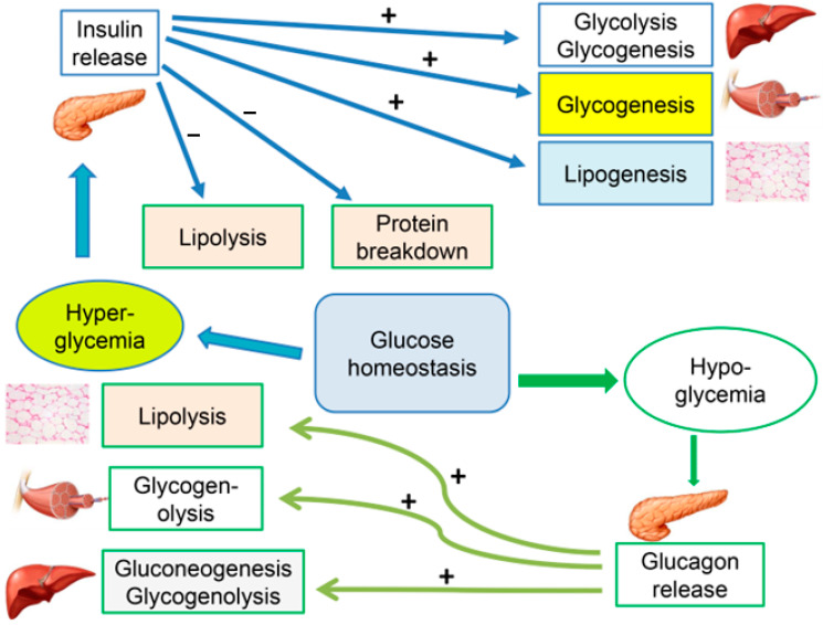
Type 2 DM is a metabolic disorder in which insulin sensitivity is disturbed, affecting its activity in the liver, skeletal muscle and fat cells. Glucose is the main player in insulin metabolism; therefore, maintaining normoglycemia is the main target in the management of T2DM [17]. In order to achieve glucose homeostasis, there are six processes that are tightly regulated both in fed and fasting states. These processes include glycolysis, gluconeogenesis, glycogenolysis, glycogenesis, lipolysis and lipogenesis. In healthy individuals, these processes are finely regulated via the balanced actions of both insulin and glucagon through feedback mechanisms and crosstalk between various organs, including the pancreas, liver, skeletal muscles and adipose tissues (Figure 1) [18].
Figure 1.
Cellular mechanisms for maintaining glucose homeostasis. Note the interplay between glycolysis, gluconeogenesis, glycogenolysis, glycogenesis, lipolysis and lipogenesis, which take place in the liver, muscle and fat tissues. (+) stimulation; (−) inhibition.
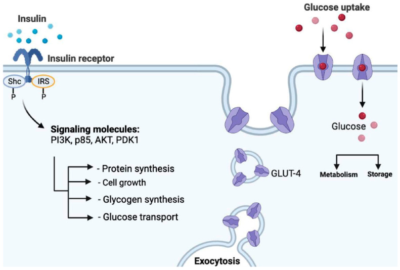
After its secretion, insulin binds to the extracellular domain of its receptor, which will cause a series of phosphorylations of different intracellular proteins. This leads to the migration of insulin-responsive glucose transporter 4 (GLUT4) to the plasma membrane and facilitates glucose uptake into the cell [19,20,21,22,23,24,25] (Figure 2).
Figure 2.
Mechanism of action of insulin: insulin binds with the insulin receptor on the surface of the target cell, leading to the phosphorylation of insulin receptor substrate (IRS) as well as Src homology and collagen protein (Shc). This step is followed by the activation of several signaling molecules (PI3K, p85, AKT and PDK1) which, among others, stimulate the transfer of GLUT4 to the plasma membrane. GLUT4 then assists in the uptake of glucose into the target cell.
Different mechanisms have been proposed to explain the molecular events causing insulin resistance. The first mechanism proposes that a reduction in the IRS-1 and IRS-2 substrates involved in insulin signaling is the main cause for reduced insulin activity [22,23,24]. The reduced phosphorylation of these substrates causes the reduced translocation of GLUT4, which interferes with glucose uptake and leads to hyperglycemia. Another proposed theory suggests the involvement of free fatty acids (FFAs) in reducing the sensitivity of insulin due to their deposition in the liver, pancreas and muscle—this occurs as a result of the decreased capacity of the subcutaneous and visceral adipose tissues to store fatty acids, leading to lipotoxicity [26,27].
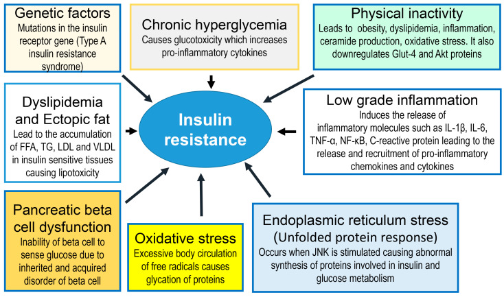
The loss of the effect of insulin results in increased glucose formation in the liver, enhanced glycogenolysis, increased lipolysis, and decreased insulin-mediated glucose uptake by skeletal muscle (Figure 3).
Figure 3.
Tissue pathophysiology of type 2 diabetes mellitus. Insulin resistance is caused by a large variety of factors, including but not limited to genetic factors, chronic hyperglycemia, physical inactivity, dyslipidemia, pancreatic beta cell dysfunction, chronic inflammation and oxidative and endoplasmic reticulum stress. These events lead to hyperglycemia and complications of diabetes. FFA = free fatty acids; TG = triglyceride; LDL = low-density lipoproteins; VLDL = very-low-density lipoproteins.
Moreover, the role of obesity as a risk factor for insulin-resistance-induced diabetes is well-documented. Indeed, adipose tissue serves a role not only in fat storage but also as an endocrine organ [28,29]. Several adipocytokines are secreted from adipocytes such as adiponectin, visfatin, leptin, resistin and tumor-necrosis factor α (TNF-α) [30,31,32,33]. A significant reduction in adiponectin and leptin levels are seen in diabetic individuals; this reduction contributes to the impairment of insulin functions [34]. In contrast, the release of inflammatory cytokines such as TNF-α and resistin is increased in obesity and diabetes, which also contributes to insulin resistance by interfering with the insulin signaling pathway [35,36]. Another molecular event that contributes to insulin sensitivity is the reduced level of the incretins glucagon-like peptide 1 (GLP-1) and gastric inhibitory polypeptide (GIP), which are released in the gastrointestinal tract during nutrient absorption after meals [37,38]. The release of incretins augments insulin secretion in healthy individuals; however, this action diminishes in cases of obesity and T2DM. This could be explained by the reduced secretion of incretins or by their deactivation by dipeptidyl peptidase-4 (DPP-4), an enzyme responsible for incretin breakdown. GLP-1 has additional effects, such as suppressing glucagon secretion, stimulating insulin gene expression and the biosynthesis and restoration of glucose competence in glucose-resistant β-cells, hence the use of GLP-1 agonists as hypoglycemic agents [39,40,41,42].
While insulin resistance is the major cause of T2DM, long-standing DM can also impair the insulin-secreting capacity of the pancreas. The reduction in the ability of pancreatic beta cells to produce insulin is due to insulin resistance, among other factors. Chronic hyperglycemia, which induces insulin resistance, was found to reduce the insulin-secreting capacity of pancreatic beta cells by altering metabolic pathways, causing endoplasmic reticulum stress, altering intracellular Ca2+ levels and altering the activity of K+− ATP channels [43]. Furthermore, diabetic patients were found to have decreased levels of islet amyloid polypeptide (IAPP), which is another pancreatic peptide co-secreted with insulin [44]. This occurs due to the accumulation of IAPP in the pancreas; IAPP then forms insoluble toxic oligomers that deposit in the β-cells, resulting in its dysfunction.
In addition to the above-mentioned mechanisms, other factors have been reported to contribute to the development of insulin resistance. For example, genetic factors involving mutations in the insulin receptor gene may lead to the development of Type A insulin resistance syndrome [45]. Chronic hyperglycemia has also been implicated in the induction of insulin resistance [46,47] because chronic hyperglycemia can cause oxidative stress and initiate glucotoxicity, which is detrimental to beta cell function. Physical inactivity can also contribute to the development of insulin resistance by increasing the risks of obesity, dyslipidemia, inflammation, ceramide production and oxidative stress and downregulating the Glut-4 and Akt proteins [48]. Inflammation, especially the chronic, low-grade type, has been reported to induce the release of inflammatory mediators such as IL-1β, IL-6, TNF-α, NF-κB, C-reactive protein pro-inflammatory chemokines and cytokines [49]. The dysfunction of pancreatic beta cells may also contribute to the pathogenesis of insulin resistance because a defective beta cell is unable to sense glucose concentration [50]. Dyslipidemia and ectopic fat in insulin-sensitive cells can induce insulin resistance via the increased accumulation of free fatty acids (FFAs), triglycerides (TGs), low-density lipoproteins (LDLs) and very-low-density lipoproteins (VLDLs), which, in turn, cause lipotoxicity [51,52]. Oxidative and endoplasmic reticulum stress (unfolded protein response) play key roles in the pathogenesis of insulin resistance [52,53].
It is worth noting that there is a strong interaction between these factors, indicating that the development of insulin resistance is indeed multifactorial in origin.
3. Type 2 DM
Type 2 DM, formerly known as non-insulin-dependent diabetes mellitus (NIDDM), is the most prevalent type of DM [54,55]. The development of this type of DM usually occurs in adulthood, at an age of >40 years, and is characterized by several symptoms such as polyuria, polydipsia, polyphagia and sudden unexplained weight loss. Unlike T1DM, T2DM can be prevented by maintaining a healthy lifestyle that involves regular exercise, smoking cessation, a healthy diet and reversing obesity [8,56,57]. On the other hand, individuals with T2DM are believed to have a higher risk of several complications including stroke, ischemic heart diseases [5], chronic kidney disease [58,59] and increased hospitalization.
Moreover, although it is less commonly seen, poorly controlled T2DM has also been associated with cognitive impairment, symptoms of dementia [60], an increased rate of infections [61] and mild hearing impairment [62]. The time-course of the development of T2DM can take several years, beginning with gradual increments in dysglycemia and moving through prediabetes to overt T2DM [63].
4. Management of DM
4.1. Lifestyle (Diet and Physical Activity)
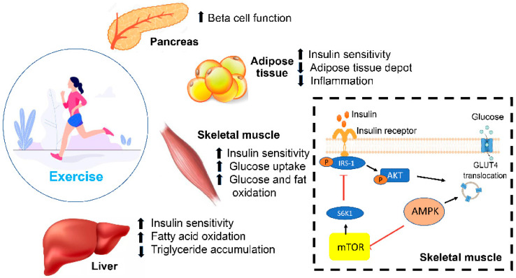
Lifestyle modifications have always been considered the cornerstone of DM management and the first step before advancing to pharmacological intervention. These can be defined as the alteration of eating habits and physical activity on a long-term basis in order to reverse obesity, prevent the occurrence of DM and manage DM and other cardiovascular diseases [64]. The effects of lifestyle modifications in reducing the incidence of DM and/or improving glycemic control are well-documented. Obesity is a well-known risk factor for developing DM. One study showed that both men and women with a BMI of 35 kg/m2 and greater are 20 times more likely to become diabetic compared to those with a BMI of 18.5–24.9 [65]. A study conducted on individuals with pre-diabetes showed a 20% reduction in the incidence of DM after adopting a healthy lifestyle compared to those with an unhealthy diet and sedentary lifestyle [66,67]. Several studies showed that lifestyle modification and/or pharmacological intervention decreases the occurrence of DM [66,67]. A study showed that combining a healthy diet and regular exercise resulted in a 34–69% reduction in DM over a period of 6 years [68]. In addition, a recent study showed that lifestyle modification in addition to the use of metformin led to a 31–58% reduction in DM over 2 years [69]. Other studies also showed the beneficial role of combining oral antidiabetic agents, such as acarbose and rosiglitazone, with lifestyle modifications; however, the reduction in the incidence of DM was not superior to lifestyle modifications alone [70]. In addition to decreasing the conversion from pre-diabetes to diabetes, adhering to a healthy diet and physical activity can also improve the responsiveness to therapy in diabetic individuals. In fact, diet and exercise improved fasting plasma glucose levels in both obese and non-obese individuals [71]. Indeed, it has been reported that physical exercise activates adenosine-monophosphate-activated protein kinase (AMPK), which plays a role in enhancing glucose uptake in the muscle by stimulating GLUT4 translocation. AMPK also regulates mTOR activities by inhibiting the mammalian/mechanistic target of rapamycin (mTOR). In skeletal muscle, mTOR over-activation can generate insulin resistance through the degradation of IRS-1 via S6K1, leading to a reduced glucose uptake [72].
It is worth noting that lifestyle changes (physical activity and diet) can cause the remission of T2DM. Many trials have shown that this is feasible [73,74].
4.2. Diet
One of the most challenging elements in the management of DM is diet and nutrition. While many experts advise diabetic individuals to minimize their intake of carbohydrates and saturated fats, others suggest that eating habits should be individualized, and each diabetic person must be referred to dieticians who are trained to provide diabetes-specific meal plans [75]. There are many diets with proven efficacy in the management of DM, such as the Mediterranean diet, which is high in vegetables, fruits and nuts [76,77], and the Dietary Approaches to Stop Hypertension (DASH) diet [76,78,79]. Although both of these dietary approaches were effective in improving insulin sensitivity and reducing the long-term complications associated with DM, research showed that nutrition distribution for each individual based on current eating patterns, metabolic goals and personal preferences, including financial, traditional and religious factors, is more beneficial in determining the best eating habits [80,81].
Controlling carbohydrate intake has been one of the most important factors for monitoring postprandial hyperglycemia [82,83]. Although many studies showed a 0.2–0.5% reduction in hemoglobin A1C (HbA1c) levels following carbohydrate restriction, the role of a low carbohydrate intake remains unclear, as studies longer than 12 weeks reported no significant improvement in fasting glucose levels or endogenous insulin levels [84].
The literature shows that adjusting daily dietary protein intake provides no evidence in the management of DM in individuals without diabetic kidney disease; however, some studies showed that higher levels of dietary protein may contribute to earlier satiety [78]. In individuals with diabetic kidney disease, careful management of the daily protein intake (0.8 g/kg body weight) is essential in preventing the deterioration of the glomerular filtration rate [85].
The recommended daily intake of fat in diabetic individuals is also controversial. Although the National Academy of Medicine (NAM) encourages a fat intake of 20–35% of the total calorie intake [86], studies showed that the type of fat is more critical than the quantity consumed for achieving metabolic goals and that a minimal consumption of saturated fat should be maintained to meet these outcomes [87,88].
4.3. Physical Activity
Physical activity is another essential lifestyle modification that the community in general and diabetic individuals in particular are advised to adapt to. Several types of training are known, including resistance training, aerobic training and high-intensity interval training. While each type of exercise can produce different beneficial outcomes in improving glucose levels and enhancing insulin activity and weight reduction, studies are inconclusive as to the ideal type of exercise for improving metabolic abnormalities [89] (Figure 4).
Figure 4.
Molecular and physiological effects of physical exercise on glucose metabolism through the pancreas, liver, adipose and skeletal tissues. Physical exercise activates AMPK enhances glucose uptake in the muscle by stimulating GLUT4 translocation.
Resistance exercises, which involve utilizing free weights and body weight exercises, have been shown to cause a threefold reduction in HbA1c in patients with T2DM when compared to inactive patients [90]. Another study showed that an 8-week weight-training protocol in patients with T2DM improved insulin and glucose responses upon oral glucose tolerance testing [91]. In addition, this type of training caused an increase in the skeletal muscle mass, which is believed to be due to enhanced muscle glycogen storage, leading to increased glucose uptake from the bloodstream. These findings support the benefit of implementing this type of training in a diabetes management plan.
Aerobic training is another type of exercise that consists of the continuous movement of large muscles, such as in jogging and walking, for at least 30 min per day for 3–7 days weekly, as per the American Diabetes Association (ADA) guidelines [92]. Aerobic training is a well-established tool in improving HbA1c by improving the lipid metabolism and weight loss [93]. One study showed that in 60 adults with T2DM, 6 months of aerobic training caused a significant reduction in HbA1c and fasting insulin levels [94]. Another study showed that aerobic activity in diabetic patients improved glycemic control, insulin sensitivity and oxidative capacity compared to sedentary individuals [95].
Combining both resistance and aerobic exercise may be the most effective approach to controlling glucose and lipid metabolism in T2DM, as per the current ADA guidelines. Cuff et al. showed that combining both types of exercises led to a significant increase in muscle glucose uptake and insulin sensitivity when compared to aerobic exercises alone [96]. Another distinguished study comparing the effects of both types of exercises alone and their combination in 915 adults showed that individuals utilizing both regimens had a more significant reduction in HbA1c [97].
On the other hand, high-intensity interval training, which consists of four to six repeated intervals of maximal activity interspersed with short periods of rest, recently arose as one of the most practiced exercising modalities [89]. This type of physical activity was found to enhance muscle glycemic control, oxidative capacity and insulin sensitivity in T2DM patients [98,99]. In addition, when comparing the effects of aerobic training to high-intensity interval training, the latter produced a more significant improvement in glucose regulation, insulin resistance and weight reduction [99].
5. Pharmacotherapy
Several groups of injectable and oral hypoglycemic agents have been discovered for the management of DM (Table 1). Each of these groups contains a number of molecules that share a specific mechanism of action but differ in their pharmacokinetic properties, including the duration of action and/or excretion and metabolism [6,54,100]. As previously mentioned, T2DM is characterized by a myriad of pathophysiological processes, including decreased insulin sensitivity, neurotransmitter receptor dysfunction, decreased pancreatic insulin and increased glucagon secretion, increased gluconeogenesis, increased lipolysis, increased renal glucose reabsorption and a reduction in incretin effects [54,101].
Table 1.
Anti-diabetic agents used clinically, their target organs and their mechanisms of action.
| Drugs | Organ Targeted | Mechanism | References |
|---|---|---|---|
| TZD and biguanides | Adipose tissue Skeletal muscle | ↓ Insulin resistance | [102,103,104,105,106,107] |
| TZD and biguanides | Liver | ↓ Gluconeogenesis | [55] |
| SGLT2 inhibitors | Kidney | Glucose elimination in urine | [108] |
| SU and meglitinides | Pancreas | Insulin secretagogues | [109,110] |
| GLP-1R agonists | Pancreas | Improve response to glucose | [111,112,113] |
| Pramlintide | Pancreas | ↓ Glucagon secretion | [114,115,116] |
| Pramlintide | Stomach | Delays gastric emptying | [115] |
| α-glucosidase inhibitors | Small intestine | Slows absorption of starch | [117,118] |
| DPP-4 inhibitors | Plasma | ↓ Incretin breakdown | [119,120] |
TZD = thiazolidinediones; SGLT2 = sodium–glucose transporter-2; SU = sulfonylureas. GLP-1R = glucagon-like peptide-1; DPP-4 = dipeptidyl peptidase 4.
Due to the availability of various hypoglycemic agents, each of these pathways can be targeted to control DM and alleviate the symptoms associated with it, such as hyperglycemia, polyuria and fatigue, as well as the long-term complications. In addition, this cocktail of oral and injectable agents can help clinicians in initiating individualized therapies for diabetic patients considering different elements such as efficacy, side effects, costs, comorbidities, weight gain and glucose levels [2,6,121,122,123,124].
Insulin is the mainstay treatment for T1DM and many individuals with T2DM. Although its use in T2DM is unusual for newly diagnosed patients, there are several instances in which the use of insulin is considered, such as severe hyperglycemia, gestational diabetes, the presence of significant weight loss and ketonuria [125,126,127]. It can be administered intravenously, intramuscularly and subcutaneously; however, subcutaneous administration is the predominant route for long-term administration [43,128]. Different preparations of insulin are classified according to their onset and duration of action (Table 2). This includes short-acting, intermediate-acting and long-acting analogs. The different pharmacokinetic properties of these formulations dictate the dosing frequency and the appropriate time for their administration. Long-acting agents such as glargine and levemir are mostly administered at bedtime to cover basal insulin requirements and are associated with a lower incidence of hypoglycemia [129]. Short-acting insulin preparations such as glulisine, aspart and lispro are administered at mealtimes to control post-prandial spikes in glucose levels [130]. Insulin regular is another preparation that is used for emergencies.
Table 2.
Types of insulin preparations and their pharmacokinetic profile (modified after Kaufman, 2003 [129]).
| Insulin Type | Onset of Action (h) | Peak of Action (h) | Duration of Action (h) | Maximal Duration (h) |
|---|---|---|---|---|
| Rapid-acting | ||||
| Lispro | ¼ to ½ | 1 to 2 | 3 to 5 | 4 to 6 |
| Aspart | ¼ to ½ | 1 to 2 | 3 to 6 | 5 to 8 |
| Glulisine | 0.25 to 0.5 | 0.5 to 1 | 3 to 4 | 4 |
| Short-acting | ||||
| Regular | ½ to 1 | 2 to 4 | 3 to 6 | 6 to 8 |
| Intermediate-acting | ||||
| NPH human | 2 to 4 | 8 to 12 | 12 to 20 | 14 to 22 |
| Long-acting | ||||
| Glargine | 1 to 2 | None | 19 to 24 | 24 |
| Detemir | 3 to 4 | 6 to 8 | 20 to 24 | 24 |
| Degludec | 1 | 9 | 24 to 42 | 42 |
| Insulin combinations | ||||
| Protamine/Lispro | 0.25 to 0.4 | 0.5 to 3 | 14 to 24 | 24 |
| Protamine/Aspart | 0.1 to 0.2 | 1 to 4 | 18 to 24 | 24 |
Special training and education are required for patients on insulin therapy due to the complexity of administration and the frequency of dosing. To overcome these difficulties, new insulin preparations are being developed, such as inhaled insulin [131] and oral insulin [132]. In addition, devices such as insulin pumps are used in patients with high HbA1c levels and a history of poor compliance [133].
6. Current Concepts on Insulin in T2DM
The HbA1c target is the main goal for the addition of a new drug to the first line of treatment, which is usually metformin and/or basal insulin. Moreover, other factors are also taken into account in selecting an additional anti-diabetic agent, such as the duration of the illness, risk of hypoglycemia, cardiovascular disease and life expectancy. The ADA guidelines recommend that most patients with T2DM should have HbA1c values of less than 7%. Patients with a longer life expectancy, shorter illness duration and no history of cardiovascular disease have an HbA1c target of 6.0–6.5%, while those with a shorter life expectancy, longer duration of illness and history of hypoglycemia have an HbA1c target of 7.5–8.0% [134].
The goal of adding insulin to the treatment regimen is to mimic the physiological insulin profile, covering both overnight and postprandial glucose levels. It is recommended by the ADA that long-acting insulin should be incorporated to cover the basal requirement of insulin after the failure of non-insulin agents [134]. In fact, it is believed that intensifying basal insulin therapy in T2DM patients leads to a decrease in glucose toxicity and an improvement in endogenous insulin release from the pancreas [135]. Consistent with this concept, studies showed that short-term insulin therapy in newly diagnosed T2DM patients demonstrated a significant improvement in β-cell function [136,137].
Although insulin has a long duration of action, the postprandial increase in glucose is difficult to control. For this reason, bolus insulin injections may be considered by adding meal-time injections of short-acting insulin, a process called insulin intensification [135]. The advantages and disadvantages of insulin are provided in Table 3.
Table 3.
Advantages and disadvantages of insulin in T2DM.
| Daily Dosage | Advantages | Side Effects | Contraindications |
|---|---|---|---|
|
|
|
|
7. Oral Hypoglycemic Agents
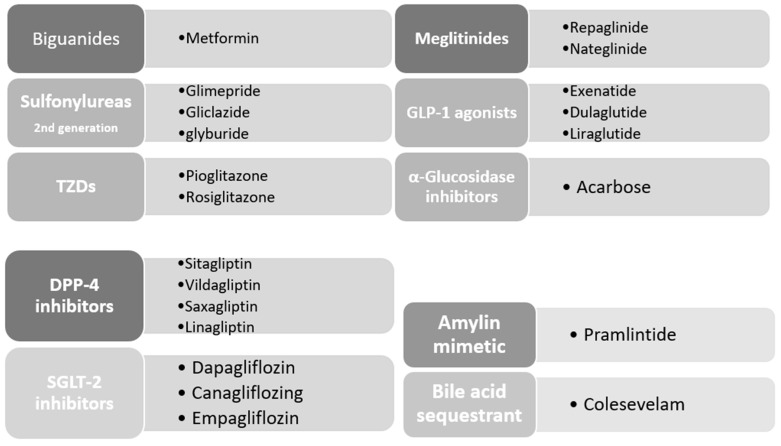
Many drugs have been approved to lower DM-associated hyperglycemia. These agents include, but are not limited to, biguanides, sulfonylureas, thiazolidinediones, GLP-1 agonists, dipeptidyl peptidase-4 (DPP-4) inhibitors, inhibitors of α-glucosidase, amylin mimetic drugs, bile acid binding resins and sodium–glucose co-transporter (SGLT) inhibitors (Figure 5).
Figure 5.
Classes of oral anti-diabetic agents and compounds approved by the American Diabetes Association (ADA).
7.1. Biguanides
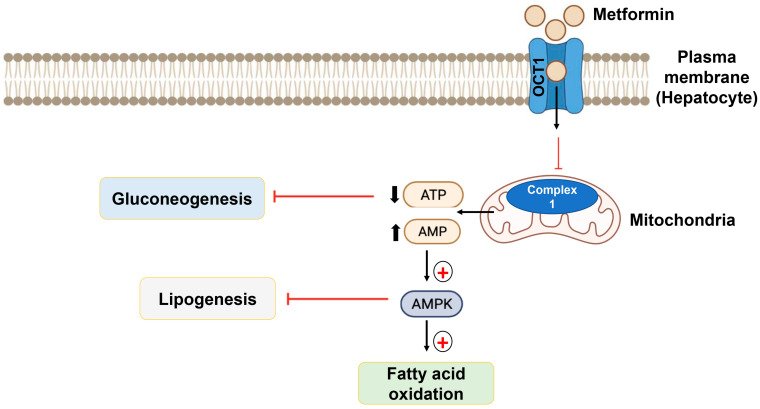
Metformin is the only agent of this group used today. It is the most commonly used agent for T2DM and is accepted as a first-line agent [138]. It operates through the activation of AMP-dependent protein kinase (AMPK), which is activated when cellular energy stores are depleted under normal conditions [139]. The activation of AMPK leads to fatty acid oxidation and inhibits gluconeogenesis in the liver. Moreover, metformin can stimulate glucagon-like peptide-1 (GLP-1) secretion, which improves insulin sensitivity by enhancing the expression of insulin receptors and improving tyrosine kinase activity [102,103,104,105]. Furthermore, metformin can also lower plasma lipid levels and reduce the incidence of cardiovascular disease by acting on peroxisome proliferator-activated receptors (PPAR-αs) [102]. These molecular effects of metformin account for both the hypoglycemic and weight-reducing actions of metformin (Figure 6).
Figure 6.
Mechanism of action of metformin. The binding of metformin with organic cation transporter-1 (OCT1) allows the metformin to reach the intracellular region, leading to the activation of AMPK, leading to the oxidation of fatty acid and the inhibition of gluconeogenesis in the liver.
Metformin can be used as a monotherapy and can also be found in combination with other hypoglycemic agents. Daily dosing varies depending on the formulation (immediate versus extended release), and the length of time on the medication but could range from 500 to 2550 mg daily. The recommended maximum dose of metformin is about 2550 mg daily [140,141].
The side effects caused by metformin mainly involve the gastrointestinal tract, including nausea, vomiting, diarrhea and abdominal discomfort [141]. Therefore, extended-release forms of metformin were developed to reduce the dosing frequency and eventually reduce these side effects. Moreover, the prolonged use of metformin is associated with folic acid and vitamin B12 deficiencies; as a result, monitoring the levels of both vitamins is needed, especially in the elderly [104,142]. Metformin should also be administered with caution and in low doses in patients with heart failure and renal failure as this category of patients have an increased risk of experiencing lactic acidosis, which is considered the most serious side effect of metformin [55]. The advantages and disadvantages of metformin are depicted in Table 4.
Table 4.
Advantages and disadvantages of metformin.
| Daily Dosage | Advantages | Side Effects | Contraindications |
|---|---|---|---|
|
|
|
|
7.2. Sulfonylureas
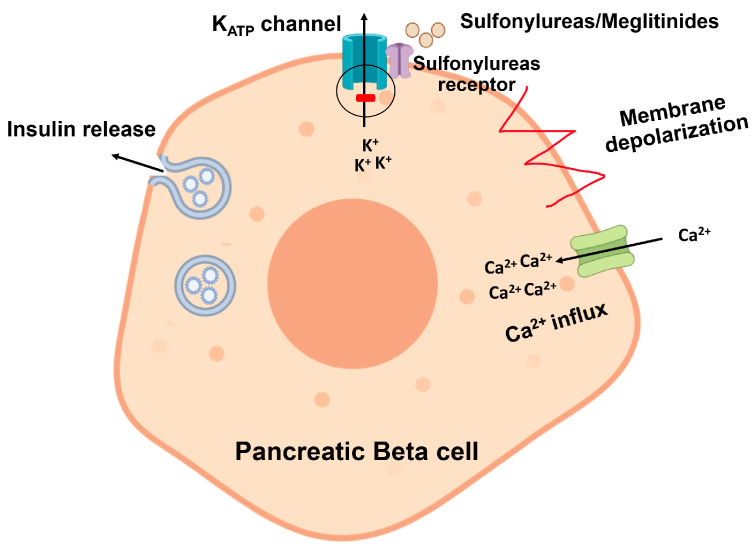
Sulfonylureas (SUs) are classified into first and second-generation agents. Due to their frequent dosing and higher risk of hypoglycemia, the first-generation drugs tolbutamide and tolzamide are no longer used clinically [54]. The second-generation agents, such as glibenclamide, gliclazide and glimepiride, are still in use, and some are available in extended-release formulations [142,143]. They exert their effect through the blockade of ATP-sensitive potassium channels found on the pancreatic β-cells, leading to cell depolarization, increasing cellular levels of calcium and enhancing the secretion of insulin, hence the name “insulin secretagogue” (Figure 7). In addition, SUs can also reduce the production of fatty acids and decrease insulin clearance 119. Due to their high efficacy in reducing HbA1c by up to 1–1.5% as and their cost-effectiveness, SUs are considered a second-line therapy and are currently used by 50–80% of diabetic patients worldwide [64,144]. However, prolonged use of these agents reduces their effectiveness. This may be due to progressive β-cell failure or an alteration in the drug’s metabolism.
Figure 7.
Sulfonylureas bind with the sulfonylurea receptor on the plasma membrane of pancreatic beta cells to block ATP-sensitive potassium channels. This leads to cell depolarization and subsequent increase in calcium-induced insulin release.
The major side effect seen with an SU is a weight gain of 1–3 kg. As a result, metformin is provided to patients on SU to reverse weight gain [109,142,145]. Hypoglycemia is also a common side effect, especially with glibenclamide and glimepiride; however, newer agents such as gliclazide have a lower tendency to cause this effect [141].
Sulfonylurea-induced hypoglycemia may be caused by decreased renal excretion, hepatic metabolism or displacement from protein-binding sites, which typically occurs in patients with renal/hepatic failure or when co-administered with CYP450 enzyme inhibitors such as aspirin and allopurinol [146]. The advantages and disadvantages of SUs are presented in Table 5.
Table 5.
Advantages and disadvantages of sulfonylureas.
| Daily Dosage | Advantages | Side Effects | Contraindications |
|---|---|---|---|
|
|
|
|
7.3. Meglitinides
Meglitinides, including repaglinide and nateglinide, belong to another class of insulin secretagogue agents that exert their action by blocking the ATP-sensitive potassium channels in pancreatic β-cells [110] (Figure 8). Unlike SUs, meglitinides have a rapid onset but a short duration of action. These features make them suitable for patients with inconsistent meal times and those who develop rapid postprandial hyperglycemia [100,101,147]. The advantages and disadvantages of meglitinides are shown in Table 6.
Figure 8.
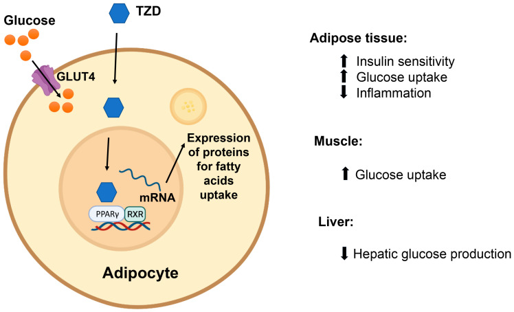
Thiazolidinediones (TZDs) activate PPARγ receptors in adipose tissue to enhance the uptake of circulating fatty acids into adipocytes.
Table 6.
Advantages and disadvantages of meglitinides.
| Daily Dosage | Advantages | Side Effects | Contraindications |
|---|---|---|---|
|
|
|
|
7.4. Thiazolidinediones
Two agents from this class are currently used clinically: pioglitazone and rosiglitazone. This pair of agents exerts its hypoglycemic effect by activating PPARγ receptors. PPARγ receptors are expressed primarily in adipose tissue, with lower expression in skeletal muscle, liver, pancreatic β-cells, the central nervous system (CNS) and vascular endothelial cells [55,101,147]. The primary effect of thiazolidinediones (TZDs) is believed to be through the activation of PPARγ receptors in adipose tissue, which promotes the uptake of circulating fatty acids into fat cells, thereby increasing insulin sensitivity (Figure 8).
Moreover, activation of this receptor in skeletal muscle and the liver also contribute to TZD action as they increase glucose uptake and reduce glucose production in both organs, respectively. TZD can cause a 0.5–1.4% reduction in HbA1c, and clinical trials showed a 10–15% reduction in plasma triglyceride levels [106,107]. In fact, this effect on the lipid profile is believed to be mediated through another isoform of PPAR receptors present in the liver, heart and skeletal muscles [148]. Furthermore, TZDs were also found to reduce the levels of inflammatory cytokines, such as tumor necrosis factor alpha, improving the function of pancreatic β-cells and increasing the levels of adiponectin, both of which are believed to contribute to their insulin-sensitizing effects [55].
The common side effects of TZDs include weight gain and edema. These are believed to occur because of the activation of PPARγ receptors in the CNS, which increases food intake [116]. Studies showed that TZDs can also increase the risk of bone fracture in women and caused a reduction in transaminases; therefore, they should be avoided in patients with liver disease. Rosiglitzone has also been associated with an increase in myocardial infarction incidence [149]. The advantages and disadvantages of TZDs are provided in Table 7.
Table 7.
Advantages and disadvantages of thiazolidinediones.
| Daily Dosage | Advantages | Side Effects | Contraindications |
|---|---|---|---|
|
|
|
|
7.5. Glucagon-like Peptide-1 (GLP-1) Agonists
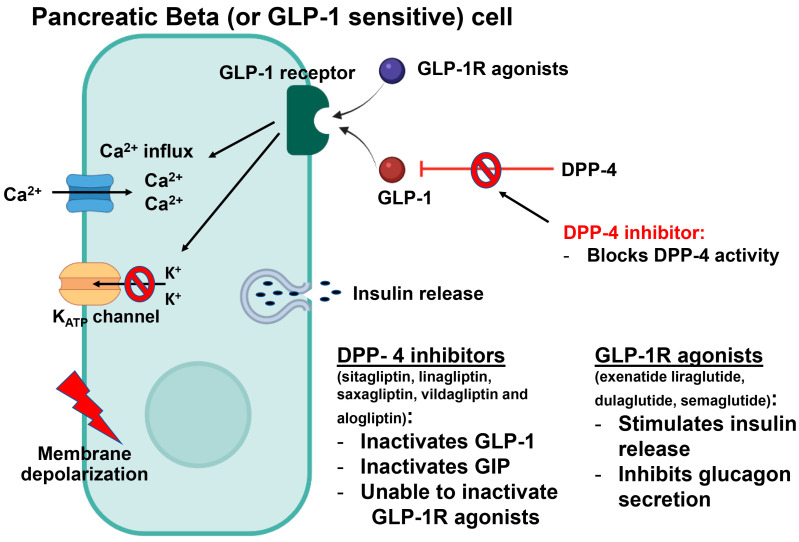
Glucagon-like peptide-1 (GLP-1) is an incretin secreted from the distal ileum in response to nutrients such as proteins and carbohydrates [150,151,152]. Following its release, GLP-1 binds to its receptor, GLP-1R, which is expressed on the pancreatic β-cells, thereby activating a cascade of intracellular events that increases the release of insulin, inhibits the release of glucagon, reduces food intake by causing satiety, delays food emptying and normalizes both postprandial and fasting insulin secretion [111] (Figure 9).
Figure 9.
GLP-1 binds to GLP-1R and activates a cascade of intracellular events, leading to insulin release and the inhibition of glucagon production.
Patients with DM were found to have a significant reduction in the levels of GLP-1, which is believed to occur due to a reduction in the expression of GLP-1 receptors in the pancreas [112,113] or an enhancement of DPP-4 activity [113]. Due to its potent insulinotropic effects, restoring the activity of GLP-1 arose as a potential target for researchers and pharmaceutical companies. As a result, several GLP-1 agonists have been developed and used clinically in the management of T2DM [119,153,154], including exenatide, liraglutide and dulaglutide [55,141]. These molecules are administered subcutaneously and have various pharmacokinetic properties accounting for the differences in dosing. The majority of side effects associated with the administration of these compounds involve the GI tract, and this includes diarrhea, nausea and vomiting. In addition, some patients reported abscesses, cellulitis formation and even tissue necrosis at the site of injection [153,155]. The risk for hypoglycemia is low unless they are used in combination with insulin or sulfonylureas. The advantages and disadvantages of incretins are provided in Table 8.
Table 8.
Advantages and disadvantages of incretins.
| Weekly Dosage | Advantages | Side Effects | Contraindications |
|---|---|---|---|
|
|
|
|
It is worth noting, however, that the stimulation of GLP-1R has many other effects outside of the pancreatic and the gastrointestinal systems. These effects range from neuroprotective action and nerve growth promotion to the ability to improve cardiovascular function [156].
7.6. Dipeptidyl Peptidase-4 (DPP-4) Inhibitors
DPP-4 is a serine protease that is expressed on endothelial cells and T-lymphocytes and in a free-circulating form. Its main function is the inactivation of the glucagon-like peptide-1 (GLP-1) and gastric inhibitory peptide (GIP) produced in the intestines [42,157]. These two hormones are known as incretins, and they play an essential metabolic role in augmenting the secretion of insulin, inhibiting glucagon secretion and reducing the absorption of nutrients [119,120]. By inhibiting this enzyme, DPP-4 inhibitors are considered oral hypoglycemic agents that are used widely in the management of DM (Figure 9).
Several DPP-4 inhibitors are available nowadays, such as sitagliptin, linagliptin, saxagliptin, vildagliptin and alogliptin. They can be used alone or in combination, and studies have shown a 0.48–0.6% reduction in HbA1c and >95% decrease in the activity of DPP-4 for 12 h [42,119].
Unlike the previously mentioned antidiabetic agents, DPP-4 inhibitors have no effect on insulin sensitivity or secretion; as a result, weight gain is not an adverse effect of gliptins [145,154,158]. Sitagliptin, saxagliptin and vildagliptin are excreted renally; therefore, dose adjustment is required for diabetic patients with moderate to severe renal disease. Linagliptin, on the other hand, is excreted by the enterohepatic system, so it can be used as the agent of choice in renal impairment [159].
Although they cause minimal to no weight gain and have a low incidence of hypoglycemia, DPP-4 inhibitors are associated with other side effects such as nasopharyngitis, upper respiratory tract infections and headaches. In addition, these agents were found to cause pancreatitis and hepatic dysfunction after prolonged use [160]. The advantages and disadvantages of incretins are depicted in Table 9.
Table 9.
Advantages and disadvantages of Dipeptidyl peptidase-4 (DPP-4) inhibitors.
| Daily Dosage | Advantages | Side Effects | Contraindications |
|---|---|---|---|
|
|
|
|
7.7. α-Glucosidase Inhibitors
Acarbose and miglitol are two agents of this class that are available and used clinically. α-glucosidase is an enzyme responsible for the breakdown of oligosaccharides into monosaccharides, and inhibiting it causes a reduction in intestinal glucose absorption by delaying the digestion of carbohydrates [2,5,59,117,118]. Moreover, these compounds were also reported to augment the release of GLP-1, which also contributes to their HbA1c-lowering activity (0.5–0.8%) [101,141]. The major side effects associated with this class are flatulence, diarrhea and abdominal pain [141]. The advantages and disadvantages of α-glucosidase inhibitors are provided in Table 10.
Table 10.
Advantages and disadvantages of α-Glucosidase inhibitors.
| Daily Dosage. | Advantages | Side Effects | Contraindications |
|---|---|---|---|
|
|
|
|
7.8. Amylin Mimetic
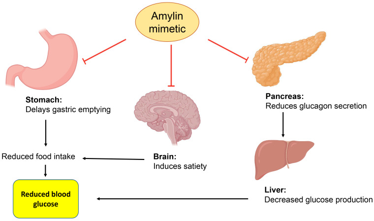
Amylin is a pancreatic hormone co-secreted with insulin from β-cells in the pancreas, and it acts by reducing the secretion of glucagon, delaying gastric emptying, and inducing satiety [114,115,116]. (Figure 10). Pramlintide is the only available amylin mimetic approved for use by the Food and Drug Administration. It is administered subcutaneously, and it is used in both T1DM and T2DM [161,162]. The advantages and disadvantages of amylin are provided in Table 11.
Figure 10.
Amylin delays gastric emptying, increases satiety, and reduces glucagon secretion.
Table 11.
Advantages and disadvantages of Amylin mimetics.
| Daily Dosage. | Advantages | Side Effects | Contraindications |
|---|---|---|---|
|
|
|
|
7.9. Bile Acid Binding Resins
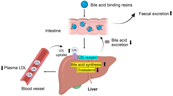
Colesevelam is the only agent in this class of hypoglycemic agents. Although it does not have a direct effect on insulin secretion and/or sensitivity, the glucose-lowering mechanism of bile acid sequestrants is mostly unknown [163,164]. It is known, however, that colesevelam can reverse dyslipidemia, which is recognized as an exacerbating factor in T2DM. Current data suggest that colesevelam alone can produce a 0.5% reduction in HbA1c and a 13–17% reduction in low-density lipoproteins (LDL) [165]. A lack of systemic side effects makes this a good adjunct medication for managing T2DM (Figure 11).
Figure 11.
Bile acid binding resins stimulate LDL-receptors on hepatocytes to enhance the uptake of LDL proteins from blood circulation.
7.10. Sodium–Glucose Co-Transporter (SGLT) Inhibitors
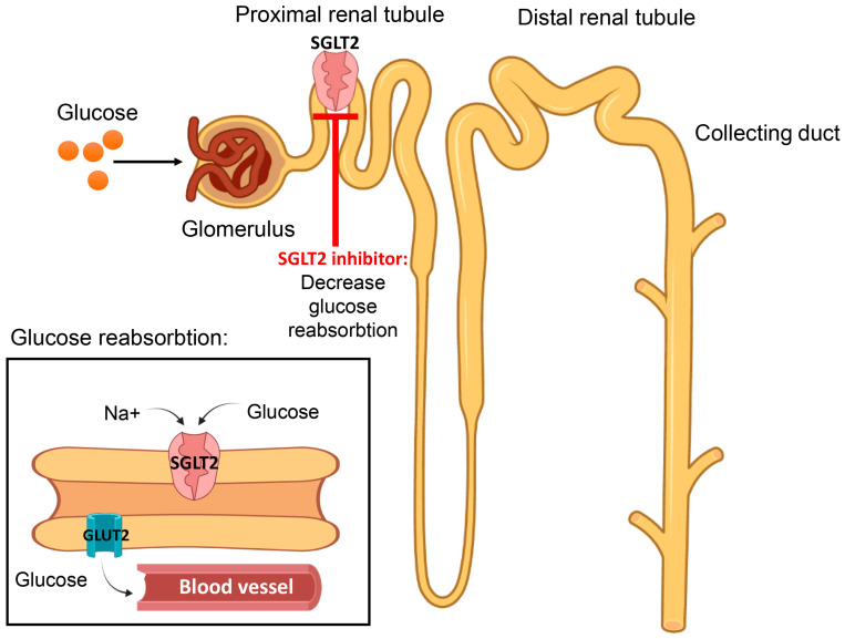
This is a newer class of antidiabetics that was introduced clinically in 2013, with canagliflozin being approved by the Food and Drug Administration (FDA) [166,167]. These molecules exert their action on the renal sodium–glucose co-transporter-2 (SGLT2) molecule, which is responsible for glucose reabsorption in the proximal renal tubules [108] (Figure 12).
Figure 12.
Sodium–glucose co-transporter (SGLT) inhibitors prevent the reabsorption of glucose from the proximal convoluted tubule, thereby reducing blood glucose level by increasing excretion.
This novel agent stimulates glucose excretion and has also been shown to have weight loss effects with minimal hypoglycemia [168,169]. In fact, canagliflozin was reported to cause a significant reduction in HbA1c of 0.77–1.03% [170], and dapagliflozin produced similar results after both short- and long-term treatments [171]. Another type of SGLT exists which is found in the intestines and the proximal convoluted tubules of the kidneys [172,173]. Although SGLT2 is responsible for the reabsorption of 90% of glucose filtered via glomeruli, diabetic patients with declining renal function may respond less to SGLT2 inhibitors, making SGLT1 inhibitors a better option for treatment [173]. Furthermore, dual SGLT1 and SGLT2 inhibitors such as sotagliflozin and licogliflozin are currently being investigated and are expected to have an agonistic hyoyglycemic effect while enhancing GLP-1 release from the intestines [173]. Currently, three types of SGLT2 inhibitors have been approved for use in the United States, including dapagliflozin, empagliflozin and the prototype SGLT2 inhibitor canagliflozin [169], while sotagliflozin is under investigation. In general, these agents have a good pharmacokinetic profile, including excellent oral bioavailability, a long half-life and limited renal excretion; however, they increase the risk for genital and urinary tract infections and orthostatic hypotension [159]. The advantages and disadvantages of SGLT2 inhibitors are provided in Table 12.
Table 12.
Advantages and disadvantages of SGLT inhibitors.
| Daily Dosage | Advantages | Side Effects | Contra-Indications |
|---|---|---|---|
|
|
|
|
8. Effectiveness of Different Classes of Anti-Diabetic Drugs on HbA1c
Table 13 presents the degree of HbA1c reduction by class of drug, which is an important measure for long-term glycemic control. The reports from the literature, as described in previous sections, show that metformin and second-generation sulfonylureas are the most effective agents for the reduction of HbA1c.
Table 13.
Effect of different classes of anti-diabetic drugs on HbA1c.
| Class of Drug | Expected Reduction in HbA1c | Contraindications |
|---|---|---|
|
|
|
|
|
|
|
|
|
|
|
|
|
|
|
|
|
|
|
|
|
|
|
|
|
|
|
|
|
|
9. Anti-Diabetic Drugs That Have Been Suspended
Despite their significant glucose-lowering activity, several agents have been discontinued by the Food and Drug administration (FDA) due to safety concerns. Troglitazone was the first TZD to be approved, but it was withdrawn due to the emergence of severe liver toxicity that resulted in 90 deaths [174]. Another agent that was put under heavy restrictions by the FDA is rosiglitazone. Rosiglitazone was associated with increased risk of heart conditions, including heart failure, stroke and death [175]. However, the restrictions on rosiglitazone were lifted in 2013 after a review of clinical data. First-generation Sus, including acetohexamide, chlorpropamide, tolazamide and tolbutamide, which were released in the 1960s, were replaced by second-generation SUs due to several side effects such as a disulfiram-like reaction and hepatotoxicity [176]. Moreover, two insulin combinations, ryzodeg and novolog, were recently withdrawn by the FDA due to concerns about cardiovascular side effects [177].
10. New Directions for the Prevention and Management of Diabetes Mellitus
Moreover, as prevention is known to be better than a cure, we can expect that genetic investigations can be utilized to screen individuals who are at risk of developing the disease and to detect genes responsible for the development of T2DM. In fact, genetic testing is already used in the diagnosis of MODY [178] and has been proposed for T2DM [179]. Using the genetic testing approaches [180] will not only aid in reducing the incidence of the illness but will also be crucial in the pharmacogenomic aspect of therapy via selecting the ideal treatment for each individual, which can optimize treatment outcomes. In addition to therapeutic advances, it is important to capitalize on the potential of lifestyle modifications in the management of T2DM.
11. Conclusions
Type 2 diabetes mellitus (T2DM) is a complex metabolic disorder that affects various organ systems and is multi-factorial in origin. Addressing the main issue of insulin resistance reduces the incidence of long-term complications of T2DM. Its management involves lifestyle modifications and pharmacotherapy with traditional and novel antidiabetic agents. Despite the variety of pharmacological agents currently available for the management of T2DM, research to discover novel targets that may broaden and individualize treatment options is ongoing. With our review, we hope to provide the latest updates on current and novel treatment regimens for T2DM, which may guide healthcare providers in managing this chronic disease.
Author Contributions
M.O.M.: conceptualization, data curation, and writing—original draft preparation; I.I.A.: reviewing and editing; J.O.A.: reviewing and editing; K.T.: reviewing and editing; H.K.: reviewing and editing; E.A.A.: conceptualization, reviewing and editing, data curation, and validation. All authors have read and agreed to the published version of the manuscript.
Institutional Review Board Statement
Not applicable.
Informed Consent Statement
Not applicable.
Conflicts of Interest
The authors declare no conflict of interest.
Funding Statement
This research was funded by United Arab Emirates University, grant numbers G00003627, G00002716 and G00003417.
Footnotes
Disclaimer/Publisher’s Note: The statements, opinions and data contained in all publications are solely those of the individual author(s) and contributor(s) and not of MDPI and/or the editor(s). MDPI and/or the editor(s) disclaim responsibility for any injury to people or property resulting from any ideas, methods, instructions or products referred to in the content.
References
- 1.IDF Diabetes Atlas . International Diabetes Federation Diabetes Atlas. 10th ed. International Diabetes Federation; Brussels, Belgium: 2021. [Google Scholar]
- 2.Chatterjee S., Khunti K., Davies M.J. Type 2 diabetes. Lancet. 2017;389:2239–2251. doi: 10.1016/S0140-6736(17)30058-2. [DOI] [PubMed] [Google Scholar]
- 3.Adeghate E. Diabetes mellitus-multifactorial in aetiology and global in prevalence. Arch. Physiol. Biochem. 2001;109:197–199. doi: 10.1076/apab.109.3.197.11588. [DOI] [PubMed] [Google Scholar]
- 4.Adeghate E., Schattner P., Dunn E. An Update on the Etiology and Epidemiology of Diabetes Mellitus. Ann. N. Y. Acad. Sci. 2006;1084:1–29. doi: 10.1196/annals.1372.029. [DOI] [PubMed] [Google Scholar]
- 5.Alberti K.G., Zimmet P.Z. Definition, diagnosis and classification of diabetes mellitus and its complications. Part 1: Diagnosis and classification of diabetes mellitus provisional report of a WHO consultation. Diabete Med. 1998;15:539–553. doi: 10.1002/(SICI)1096-9136(199807)15:7<539::AID-DIA668>3.0.CO;2-S. [DOI] [PubMed] [Google Scholar]
- 6.American Diabetes Association Diagnosis and Classification of Diabetes Mellitus. Diabetes Care. 2010;33((Suppl. 1)):S62–S69. doi: 10.2337/dc10-S062. [DOI] [PMC free article] [PubMed] [Google Scholar]
- 7.Diabetes Control and Complications Trial Research Group Effect of intensive diabetes treatment on the development and progression of long-term complications in adolescents with insulin-dependent diabetes mellitus: Diabetes Control and Complications Trial. J. Pediatr. 1994;125:177–188. doi: 10.1016/S0022-3476(94)70190-3. [DOI] [PubMed] [Google Scholar]
- 8.Wu Y., Ding Y., Tanaka Y., Zhang W. Risk factors contributing to type 2 diabetes and recent advances in the treatment and prevention. Int. J. Med. Sci. 2014;11:1185–1200. doi: 10.7150/ijms.10001. [DOI] [PMC free article] [PubMed] [Google Scholar]
- 9.Esposito K., Ciotola M., Maiorino M.I., Giugliano D. Lifestyle approach for type 2 diabetes and metabolic syndrome. Curr. Atheroscler. Rep. 2008;10:523–528. doi: 10.1007/s11883-008-0081-4. [DOI] [PubMed] [Google Scholar]
- 10.Gilmartin A.B.H., Ural S.H., Repke J.T. Gestational Diabetes Mellitus. Rev. Obstet. Gynecol. 2008;1:129–134. [PMC free article] [PubMed] [Google Scholar]
- 11.Couch S.C., Philipson E.H., Bendel R.B., Pujda L.M., A Milvae R., Lammi-Keefe C.J. Elevated Lipoprotein Lipids and Gestational Hormones in Women With Diet-Treated Gestational Diabetes Mellitus Compared to Healthy Pregnant Controls. J. Diabetes Its Complicat. 1998;12:1–9. doi: 10.1016/S1056-8727(97)00007-X. [DOI] [PubMed] [Google Scholar]
- 12.Zhu Y., Hedderson M.M., Quesenberry C.P., Feng J., Ferrara A. Central Obesity Increases the Risk of Gestational Diabetes Partially Through Increasing Insulin Resistance. Obesity. 2018;27:152–160. doi: 10.1002/oby.22339. [DOI] [PMC free article] [PubMed] [Google Scholar]
- 13.Amed S., Oram R. Maturity-Onset Diabetes of the Young (MODY): Making the Right Diagnosis to Optimize Treatment. Can. J. Diabetes. 2016;40:449–454. doi: 10.1016/j.jcjd.2016.03.002. [DOI] [PubMed] [Google Scholar]
- 14.Anık A., Çatlı G., Abacı A., Böber E. Maturity-onset diabetes of the young (MODY): An update. J. Pediatr. Endocrinol. Metab. 2015;28:251–263. doi: 10.1515/jpem-2014-0384. [DOI] [PubMed] [Google Scholar]
- 15.Timsit J., Saint-Martin C., Dubois-Laforgue D., Bellanne-Chantelot C. Searching for Maturity-Onset Diabetes of the Young (MODY): When and What for? Can. J. Diabetes. 2016;40:455–461. doi: 10.1016/j.jcjd.2015.12.005. [DOI] [PubMed] [Google Scholar]
- 16.Goran M.I., Bergman R.N., Cruz M.L., Watanabe R. Insulin resistance and associated compensatory responses in african-american and Hispanic children. Diabetes Care. 2002;25:2184–2190. doi: 10.2337/diacare.25.12.2184. [DOI] [PubMed] [Google Scholar]
- 17.Von Ah Morano A.E., Dorneles G.P., Peres A., Lira F.S. The role of glucose homeostasis on immune function in response to exercise: The impact of low or higher energetic conditions. J. Cell Physiol. 2020;235:3169–3188. doi: 10.1002/jcp.29228. [DOI] [PubMed] [Google Scholar]
- 18.Wen S., Wang C., Gong M., Zhou L. An overview of energy and metabolic regulation. Sci. China Life Sci. 2018;62:771–790. doi: 10.1007/s11427-018-9371-4. [DOI] [PubMed] [Google Scholar]
- 19.White M., Kahn C. The insulin signaling system. J. Biol. Chem. 1994;269:1–4. doi: 10.1016/S0021-9258(17)42297-6. [DOI] [PubMed] [Google Scholar]
- 20.Lee J., Pilch P.F. The insulin receptor: Structure, function, and signaling. Am. J. Physiol. 1994;266:C319–C334. doi: 10.1152/ajpcell.1994.266.2.C319. [DOI] [PubMed] [Google Scholar]
- 21.Hotamisligil G.S. Inflammation and endoplasmic reticulum stress in obesity and diabetes. Int. J. Obes. 2008;32:S52–S54. doi: 10.1038/ijo.2008.238. [DOI] [PMC free article] [PubMed] [Google Scholar]
- 22.Goodyear L.J., Giorgino F., Sherman L.A., Carey J., Smith R.J., Dohm G.L. Insulin receptor phosphorylation, insulin receptor substrate-1 phosphorylation, and phosphatidylinositol 3-kinase activity are decreased in intact skeletal muscle strips from obese subjects. J. Clin. Investig. 1995;95:2195–2204. doi: 10.1172/JCI117909. [DOI] [PMC free article] [PubMed] [Google Scholar]
- 23.Bjornholm M., Kawano Y., Lehtihet M., Zierath J.R. Insulin receptor substrate-1 phosphorylation and phosphatidylinositol 3-kinase activity in skeletal muscle from NIDDM subjects after in vivo insulin stimulation. Diabetes. 1997;46:524–527. doi: 10.2337/diab.46.3.524. [DOI] [PubMed] [Google Scholar]
- 24.Zaid H., Antonescu C.N., Randhawa V.K., Klip A. Insulin action on glucose transporters through molecular switches, tracks and tethers. Biochem. J. 2008;413:201–215. doi: 10.1042/BJ20080723. [DOI] [PubMed] [Google Scholar]
- 25.Klip A., McGraw T.E., James D.E. Thirty sweet years of GLUT4. J. Biol. Chem. 2019;294:11369–11381. doi: 10.1074/jbc.REV119.008351. [DOI] [PMC free article] [PubMed] [Google Scholar]
- 26.Shulman G.I. Cellular mechanisms of insulin resistance. J. Clin. Investig. 2000;106:171–176. doi: 10.1172/JCI10583. [DOI] [PMC free article] [PubMed] [Google Scholar]
- 27.Herzberg-Schäfer S.A., Heni M., Stefan N., Häring H.-U., Fritsche A.E. Impairment of GLP1-induced insulin secretion: Role of genetic background, insulin resistance and hyperglycaemia. Diabetes Obes. Metab. 2012;14((Suppl. 3)):85–90. doi: 10.1111/j.1463-1326.2012.01648.x. [DOI] [PubMed] [Google Scholar]
- 28.de Ferranti S., Mozaffarian D. The perfect storm: Obesity, adipocyte dysfunction, and metabolic consequences. Clin. Chem. 2008;54:945–955. doi: 10.1373/clinchem.2007.100156. [DOI] [PubMed] [Google Scholar]
- 29.Field A.E., Coakley E.H., Must A., Spadano J.L., Laird N., Dietz W.H., Rimm E., Colditz G. Impact of overweight on the risk of developing common chronic diseases during a 10-year period. Arch. Intern. Med. 2001;161:1581–1586. doi: 10.1001/archinte.161.13.1581. [DOI] [PubMed] [Google Scholar]
- 30.Greenberg A.S., McDaniel M.L. Identifying the links between obesity, insulin resistance and beta-cell function: Potential role of adipocyte-derived cytokines in the pathogenesis of type 2 diabetes. Eur. J. Clin. Investig. 2002;32((Suppl. 3)):24–34. doi: 10.1046/j.1365-2362.32.s3.4.x. [DOI] [PubMed] [Google Scholar]
- 31.Unger R.H. Lipotoxic diseases. Annu. Rev. Med. 2002;53:319–336. doi: 10.1146/annurev.med.53.082901.104057. [DOI] [PubMed] [Google Scholar]
- 32.Adeghate E. An update on the biology and physiology of resistin. Cell. Mol. Life Sci. 2004;61:2485–2496. doi: 10.1007/s00018-004-4083-2. [DOI] [PMC free article] [PubMed] [Google Scholar]
- 33.Adeghate E. Visfatin: Structure, function and relation to diabetes mellitus and other dysfunctions. Curr. Med. Chem. 2008;15:1851–1862. doi: 10.2174/092986708785133004. [DOI] [PubMed] [Google Scholar]
- 34.Scheen A.J. Pathophysiology of type 2 diabetes. Acta Clin. Belg. 2003;58:335–341. doi: 10.1179/acb.2003.58.6.001. [DOI] [PubMed] [Google Scholar]
- 35.Bastard J.-P., Maachi M., Lagathu C., Kim M.J., Caron M., Vidal H., Capeau J., Feve B. Recent advances in the relationship between obesity, inflammation, and insulin resistance. Eur. Cytokine Netw. 2006;17:4–12. [PubMed] [Google Scholar]
- 36.Dludla P.V., Mabhida S.E., Ziqubu K., Nkambule B.B., Mazibuko-Mbeje S.E., Hanser S., Basson A.K., Pheiffer C., Kengne A.P. Pancreatic β-cell dysfunction in type 2 diabetes: Implications of inflammation and oxidative stress. World J. Diabetes. 2023;14:130–146. doi: 10.4239/wjd.v14.i3.130. [DOI] [PMC free article] [PubMed] [Google Scholar]
- 37.Nauck M., Stockmann F., Ebert R., Creutzfeldt W. Reduced incretin effect in type 2 (non-insulin-dependent) diabetes. Diabetologia. 1986;29:46–52. doi: 10.1007/BF02427280. [DOI] [PubMed] [Google Scholar]
- 38.Fehmann H.C., Goke R., Goke B. Cell and molecular biology of the incretin hormones glucagon-like peptide-I and glucose-dependent insulin releasing polypeptide. Endocr. Rev. 1995;16:390–410. doi: 10.1210/edrv-16-3-390. [DOI] [PubMed] [Google Scholar]
- 39.Gautier J., Fetita S., Sobngwi E., Salaün-Martin C. Biological actions of the incretins GIP and GLP-1 and therapeutic perspectives in patients with type 2 diabetes. Diabetes Metab. 2005;31:233–242. doi: 10.1016/S1262-3636(07)70190-8. [DOI] [PubMed] [Google Scholar]
- 40.Buteau J. GLP-1 receptor signaling: Effects on pancreatic beta-cell proliferation and survival. Diabetes Metab. 2008;34((Suppl. 2)):S73–S77. doi: 10.1016/S1262-3636(08)73398-6. [DOI] [PubMed] [Google Scholar]
- 41.Lotfy M., Singh J., Rashed H., Tariq S., Zilahi E., Adeghate E. Mechanism of the beneficial and protective effects of exenatide in diabetic rats. J. Endocrinol. 2014;220:291–304. doi: 10.1530/JOE-13-0426. [DOI] [PubMed] [Google Scholar]
- 42.Lotfy M., Singh J., Kalász H., Tekes K., Adeghate E. Medicinal Chemistry and Applications of Incretins and DPP-4 Inhibitors in the Treatment of Type 2 Diabetes Mellitus. Open Med. Chem. J. 2011;5((Suppl. 2)):82–92. doi: 10.2174/1874104501105010082. [DOI] [PMC free article] [PubMed] [Google Scholar]
- 43.Surampudi P.N., John-Kalarickal J., Fonseca V.A. Emerging concepts in the pathophysiology of type 2 diabetes mellitus. Mt. Sinai J. Med. J. Transl. Pers. Med. 2009;76:216–226. doi: 10.1002/msj.20113. [DOI] [PubMed] [Google Scholar]
- 44.Westermark P., Johnson K.H., O’Brien T., Betsholtz C. Islet amyloid polypeptide? A novel controversy in diabetes research. Diabetologia. 1992;35:297–303. doi: 10.1007/BF00401195. [DOI] [PubMed] [Google Scholar]
- 45.You W., Yang J., Wang L., Liu Y., Wang W., Zhu L., Wang W., Yang J., Chen F. Case Report: A Chinese Family of Type A Insulin Resistance Syndrome With Diabetes Mellitus, With a Novel Heterozygous Missense Mutation of the Insulin Receptor Gene. Front. Endocrinol. 2022;13:895424. doi: 10.3389/fendo.2022.895424. [DOI] [PMC free article] [PubMed] [Google Scholar]
- 46.Kawahito S., Kitahata H., Oshita S. Problems associated with glucose toxicity: Role of hyperglycemia-induced oxidative stress. World J. Gastroenterol. 2009;15:4137–4142. doi: 10.3748/wjg.15.4137. [DOI] [PMC free article] [PubMed] [Google Scholar]
- 47.Lotfy M., Adeghate J., Kalasz H., Singh J., Adeghate E. Chronic complications of diabetes mellitus: A mini review. Curr. Diabetes Rev. 2017;13:3–10. doi: 10.2174/1573399812666151016101622. [DOI] [PubMed] [Google Scholar]
- 48.Yaribeygi H., Maleki M., Sathyapalan T., Jamialahmadi T., Sahebkar A. Pathophysiology of Physical Inactivity-Dependent Insulin Resistance: A Theoretical Mechanistic Review Emphasizing Clinical Evidence. J. Diabetes Res. 2021;2021:7796727. doi: 10.1155/2021/7796727. [DOI] [PMC free article] [PubMed] [Google Scholar]
- 49.Rehman K., Akash M.S. Mechanisms of inflammatory responses and development of insulin resistance: How are they interlinked? J. Biomed. Sci. 2016;23:87. doi: 10.1186/s12929-016-0303-y. [DOI] [PMC free article] [PubMed] [Google Scholar]
- 50.Cerf M.E. High fat programming of beta cell compensation, exhaustion, death and dysfunction. Pediatr. Diabetes. 2014;16:71–78. doi: 10.1111/pedi.12137. [DOI] [PubMed] [Google Scholar]
- 51.Samuel V.T., Shulman G.I. Mechanisms for Insulin Resistance: Common Threads and Missing Links. Cell. 2012;148:852–871. doi: 10.1016/j.cell.2012.02.017. [DOI] [PMC free article] [PubMed] [Google Scholar]
- 52.Lee S.-H., Park S.-Y., Choi C.S. Insulin Resistance: From Mechanisms to Therapeutic Strategies. Diabetes Metab. J. 2022;46:15–37. doi: 10.4093/dmj.2021.0280. [DOI] [PMC free article] [PubMed] [Google Scholar]
- 53.Ozcan U., Cao Q., Yilmaz E., Lee A.-H., Iwakoshi N.N., Özdelen E., Tuncman G., Görgün C., Glimcher L.H., Hotamisligil G.S. Endoplasmic Reticulum Stress Links Obesity, Insulin Action, and Type 2 Diabetes. Science. 2004;306:457–461. doi: 10.1126/science.1103160. [DOI] [PubMed] [Google Scholar]
- 54.Olokoba A.B., Obateru O.A., Olokoba L.B. Type 2 Diabetes Mellitus: A Review of Current Trends. Oman Med. J. 2012;27:269–273. doi: 10.5001/omj.2012.68. [DOI] [PMC free article] [PubMed] [Google Scholar]
- 55.Chaudhury A., Duvoor C., Reddy Dendi V.S., Kraleti S., Chada A., Ravilla R., Marco A., Shekhawat N.S., Montales M.T., Kuriakose K., et al. Clinical Review of Antidiabetic Drugs: Implications for Type 2 Diabetes Mellitus Management. Front. Endocrinol. 2017;8:6. doi: 10.3389/fendo.2017.00006. [DOI] [PMC free article] [PubMed] [Google Scholar]
- 56.Wannamethee S.G., Shaper A.G., Perry I.J. Smoking as a Modifiable Risk Factor for Type 2 Diabetes in Middle-Aged Men. Diabetes Care. 2001;24:1590–1595. doi: 10.2337/diacare.24.9.1590. [DOI] [PubMed] [Google Scholar]
- 57.Wei M., Gibbons L.W., Mitchell T.L., Kampert J.B., Blair S.N. Alcohol intake and incidence of type 2 diabetes in men. Diabetes Care. 2000;23:18–22. doi: 10.2337/diacare.23.1.18. [DOI] [PubMed] [Google Scholar]
- 58.Elhefnawy M.E., Ghadzi S.M.S., Harun S.N. Predictors Associated with Type 2 Diabetes Mellitus Complications over Time: A Literature Review. J. Vasc. Dis. 2022;1:13–23. doi: 10.3390/jvd1010003. [DOI] [Google Scholar]
- 59.Adeghate E.A., Kalász H., Al Jaberi S., Adeghate J., Tekes K. Tackling type 2 diabetes-associated cardiovascular and renal comorbidities: A key challenge for drug development. Expert Opin. Investig. Drugs. 2020;30:85–93. doi: 10.1080/13543784.2021.1865914. [DOI] [PubMed] [Google Scholar]
- 60.Ebady S.A., Arami M.A., Shafigh M.H. Investigation on the relationship between diabetes mellitus type 2 and cognitive impairment. Diabetes Res. Clin. Pract. 2008;82:305–309. doi: 10.1016/j.diabres.2008.08.020. [DOI] [PubMed] [Google Scholar]
- 61.Chávez-Reyes J., Escárcega-González C.E., Chavira-Suárez E., León-Buitimea A., Vázquez-León P., Morones-Ramírez J.R., Villalón C.M., Quintanar-Stephano A., Marichal-Cancino B.A. Susceptibility for Some Infectious Diseases in Patients With Diabetes: The Key Role of Glycemia. Front. Public Health. 2021;9:559595. doi: 10.3389/fpubh.2021.559595. [DOI] [PMC free article] [PubMed] [Google Scholar]
- 62.Akinpelu O.V., Mujica-Mota M., Daniel S.J. Is type 2 diabetes mellitus associated with alterations in hearing? A systematic review and meta-analysis. Laryngoscope. 2013;124:767–776. doi: 10.1002/lary.24354. [DOI] [PubMed] [Google Scholar]
- 63.Majety P., Orquera F.A.L., Edem D., Hamdy O. Pharmacological approaches to the prevention of type 2 diabetes mellitus. Front. Endocrinol. 2023;14:1118848. doi: 10.3389/fendo.2023.1118848. [DOI] [PMC free article] [PubMed] [Google Scholar]
- 64.Holman R.R., Paul S.K., Bethel M.A., Matthews D.R., Neil H.A. 10-year follow-up of intensive glucose control in type 2 diabetes. N. Engl. J. Med. 2008;359:1577–1589. doi: 10.1056/NEJMoa0806470. [DOI] [PubMed] [Google Scholar]
- 65.Al Jaberi S., Cohen A., Saeed Z., Ojha S., Singh J., Adeghate E. Obesity: Molecular Mechanisms, Epidemiology, Complications and Pharmacotherapy. Cell. Biochem. Mech. Obes. 2021;v23:249–266. [Google Scholar]
- 66.Tuso P. Prediabetes and lifestyle modification: Time to prevent a preventable disease. Perm. J. 2014;18:88–93. doi: 10.7812/TPP/14-002. [DOI] [PMC free article] [PubMed] [Google Scholar]
- 67.Deed G., Barlow J., Kawol D., Kilov G., Sharma A., Hwa L.Y. Diet and diabetes. Aust. Fam. Physician. 2015;44:192–196. [PubMed] [Google Scholar]
- 68.Brunetti L., Kalabalik J. Management of type-2 diabetes mellitus in adults: Focus on individualizing non-insulin therapies. P T Peer-Rev. J. Formul. Manag. 2012;37:687–696. [PMC free article] [PubMed] [Google Scholar]
- 69.Knowler W.C., Barrett-Connor E., Fowler S.E., Hamman R.F., Lachin J.M., Walker E.A., Nathan D.M., Diabetes Prevention Program Research Group Reduction in the incidence of type 2 diabetes with lifestyle intervention or metformin. N. Engl. J. Med. 2002;346:393–403. doi: 10.1056/NEJMoa012512. [DOI] [PMC free article] [PubMed] [Google Scholar]
- 70.Yamaoka K., Nemoto A., Tango T. Comparison of the Effectiveness of Lifestyle Modification with Other Treatments on the Incidence of Type 2 Diabetes in People at High Risk: A Network Meta-Analysis. Nutrients. 2019;11:1373. doi: 10.3390/nu11061373. [DOI] [PMC free article] [PubMed] [Google Scholar]
- 71.Fowler M.J. Diabetes Treatment, Part 1: Diet and Exercise. Clin. Diabetes. 2007;25:105. doi: 10.2337/diaclin.25.3.105. [DOI] [Google Scholar]
- 72.Marín-Aguilar F., Pavillard L.E., Giampieri F., Bullón P., Cordero M.D. Adenosine Monophosphate (AMP)-Activated Protein Kinase: A New Target for Nutraceutical Compounds. Int. J. Mol. Sci. 2017;18:288. doi: 10.3390/ijms18020288. [DOI] [PMC free article] [PubMed] [Google Scholar]
- 73.Taylor R., Ramachandran A., Yancy W.S., Jr., Forouhi N.G. Nutritional basis of type 2 diabetes remission. BMJ. 2021;374:n1449. doi: 10.1136/bmj.n1449. [DOI] [PMC free article] [PubMed] [Google Scholar]
- 74.Taylor R. Type 2 diabetes and remission: Practical management guided by pathophysiology. J. Intern. Med. 2020;289:754–770. doi: 10.1111/joim.13214. [DOI] [PMC free article] [PubMed] [Google Scholar]
- 75.American Diabetes Association. Lifestyle Management: Standards of Medical Care in Diabetes-2018. Diabetes Care. 2018;41((Suppl. 1)):S38–S50. doi: 10.2337/dc18-S004. [DOI] [PubMed] [Google Scholar]
- 76.Cespedes E.M., Hu F.B., Tinker L., Rosner B., Redline S., Garcia L., Hingle M., van Horn L., Howard B.V., Levitan E.B., et al. Multiple Healthful Dietary Patterns and Type 2 Diabetes in the Women’s Health Initiative. Am. J. Epidemiol. 2016;183:622–633. doi: 10.1093/aje/kwv241. [DOI] [PMC free article] [PubMed] [Google Scholar]
- 77.Esposito K., Maiorino M.I., Ciotola M., Di Palo C., Scognamiglio P., Gicchino M., Petrizzo M., Saccomanno F., Beneduce F., Ceriello A., et al. Effects of a Mediterranean-Style Diet on the Need for Antihyperglycemic Drug Therapy in Patients With Newly Diagnosed Type 2 Diabetes: A Randomized Trial. Ann. Intern Med. 2009;151:306–314. doi: 10.7326/0003-4819-151-5-200909010-00004. [DOI] [PubMed] [Google Scholar]
- 78.Ley S.H., Hamdy O., Mohan V., Hu F.B. Prevention and management of type 2 diabetes: Dietary components and nutritional strategies. Lancet. 2014;383:1999–2007. doi: 10.1016/S0140-6736(14)60613-9. [DOI] [PMC free article] [PubMed] [Google Scholar]
- 79.Campbell A.P. DASH Eating Plan: An Eating Pattern for Diabetes Management. Diabetes Spectr. 2017;30:76–81. doi: 10.2337/ds16-0084. [DOI] [PMC free article] [PubMed] [Google Scholar]
- 80.Franz M.J., MacLeod J., Evert A., Brown C., Gradwell E., Handu D., Reppert A., Robinson M. Academy of Nutrition and Dietetics Nutrition Practice Guideline for Type 1 and Type 2 Diabetes in Adults: Systematic Review of Evidence for Medical Nutrition Therapy Effectiveness and Recommendations for Integration into the Nutrition Care Process. J. Acad. Nutr. Diet. 2017;117:1659–1679. doi: 10.1016/j.jand.2017.03.022. [DOI] [PubMed] [Google Scholar]
- 81.Evert A.B., Boucher J.L., Cypress M., Dunbar S.A., Franz M.J., Mayer-Davis E.J., Neumiller J.J., Nwankwo R., Verdi C.L., Urbanski P., et al. Nutrition Therapy Recommendations for the Management of Adults With Diabetes. Diabetes Care. 2013;36:3821–3842. doi: 10.2337/dc13-2042. [DOI] [PMC free article] [PubMed] [Google Scholar]
- 82.Delahanty L.M., Nathan D.M., Lachin J.M., Hu F.B., Cleary P.A., Ziegler G.K., Wylie-Rosett J., Wexler D.J., Diabetes Control and Complications Trial/Epidemiology of Diabetes Association of diet with glycated hemoglobin during intensive treatment of type 1 diabetes in the Diabetes Control and Complications Trial. Am. J. Clin. Nutr. 2009;89:518–524. doi: 10.3945/ajcn.2008.26498. [DOI] [PMC free article] [PubMed] [Google Scholar]
- 83.DAFNE Study Group Training in flexible, intensive insulin management to enable dietary freedom in people with type 1 diabetes: Dose adjustment for normal eating (DAFNE) randomised controlled trial. BMJ. 2002;325:746. doi: 10.1136/bmj.325.7367.746. [DOI] [PMC free article] [PubMed] [Google Scholar]
- 84.Wheeler M.L., Dunbar S.A., Jaacks L.M., Karmally W., Mayer-Davis E.J., Wylie-Rosett J., Yancy W.S. Response to Comment on: Wheeler et al. Macronutrients, Food Groups, and Eating Patterns in the Management of Diabetes: A Systematic Review of the Literature, 2010. Diabetes Care. 2012;35:434–445. doi: 10.2337/dc11-2216. [DOI] [PMC free article] [PubMed] [Google Scholar]
- 85.Robertson L., Waugh N., Robertson A. Protein restriction for diabetic renal disease. Cochrane Database Syst. Rev. 2007;2007:Cd002181. doi: 10.1002/14651858.CD002181.pub2. [DOI] [PMC free article] [PubMed] [Google Scholar]
- 86.Trumbo P., Schlicker S., Yates A.A., Poos M., Food and Nutrition Board of the Institute of Medicine, The National Academies Dietary Reference Intakes for Energy, Carbohydrate, Fiber, Fat, Fatty Acids, Cholesterol, Protein and Amino Acids. J. Am. Diet. Assoc. 2002;102:1621–1630. doi: 10.1016/S0002-8223(02)90346-9. [DOI] [PubMed] [Google Scholar]
- 87.Estruch R., Ros E., Salas-Salvadó J., Covas M.-I., Corella D., Arós F., Gómez-Gracia E., Ruiz-Gutiérrez V., Fiol M., Lapetra J., et al. Primary Prevention of Cardiovascular Disease with a Mediterranean Diet. N. Engl. J. Med. 2013;368:1279–1290. doi: 10.1056/NEJMoa1200303. [DOI] [PubMed] [Google Scholar]
- 88.Ros E. Dietary cis-monounsaturated fatty acids and metabolic control in type 2 diabetes. Am. J. Clin. Nutr. 2003;78:617S–625S. doi: 10.1093/ajcn/78.3.617S. [DOI] [PubMed] [Google Scholar]
- 89.Kirwan J.P., Sacks J., Nieuwoudt S. The essential role of exercise in the management of type 2 diabetes. Clevel. Clin. J. Med. 2017;84:S15–S21. doi: 10.3949/ccjm.84.s1.03. [DOI] [PMC free article] [PubMed] [Google Scholar]
- 90.Dunstan D.W., Daly R.M., Owen N., Jolley D., de Courten M., Shaw J., Zimmet P. High-Intensity Resistance Training Improves Glycemic Control in Older Patients With Type 2 Diabetes. Diabetes Care. 2002;25:1729–1736. doi: 10.2337/diacare.25.10.1729. [DOI] [PubMed] [Google Scholar]
- 91.Dunstan D., Puddey I., Beilin L., Burke V., Morton A., Stanton K. Effects of a short-term circuit weight training program on glycaemic control in NIDDM. Diabetes Res. Clin. Pract. 1998;40:53–61. doi: 10.1016/S0168-8227(98)00027-8. [DOI] [PubMed] [Google Scholar]
- 92.Colberg S.R., Sigal R.J., Yardley J.E., Riddell M.C., Dunstan D.W., Dempsey P.C., Horton E.S., Castorino K., Tate D.F. Physical Activity/Exercise and Diabetes: A Position Statement of the American Diabetes Association. Diabetes Care. 2016;39:2065–2079. doi: 10.2337/dc16-1728. [DOI] [PMC free article] [PubMed] [Google Scholar]
- 93.Zanuso S., Jimenez A., Pugliese G., Corigliano G., Balducci S. Exercise for the management of type 2 diabetes: A review of the evidence. Acta Diabetol. 2009;47:15–22. doi: 10.1007/s00592-009-0126-3. [DOI] [PubMed] [Google Scholar]
- 94.Kadoglou N.P., Iliadis F., Angelopoulou N., Perrea D., Ampatzidis G., Liapis C., Alevizos M. The anti-inflammatory effects of exercise training in patients with type 2 diabetes mellitus. Eur. J. Prev. Cardiol. 2007;14:837–843. doi: 10.1097/HJR.0b013e3282efaf50. [DOI] [PubMed] [Google Scholar]
- 95.Boulé N.G., Kenny G.P., Haddad E., Wells G.A., Sigal R.J. Meta-analysis of the effect of structured exercise training on cardiorespiratory fitness in Type 2 diabetes mellitus. Diabetologia. 2003;46:1071–1081. doi: 10.1007/s00125-003-1160-2. [DOI] [PubMed] [Google Scholar]
- 96.Cuff D.J., Meneilly G.S., Martin A., Ignaszewski A., Tildesley H.D., Frohlich J.J. Effective exercise modality to reduce insulin resistance in women with type 2 diabetes. Diabetes Care. 2003;26:2977–2982. doi: 10.2337/diacare.26.11.2977. [DOI] [PubMed] [Google Scholar]
- 97.Schwingshackl L., Missbach B., Dias S., Koenig J., Hoffmann G. Impact of different training modalities on glycaemic control and blood lipids in patients with type 2 diabetes: A systematic review and network meta-analysis. Diabetologia. 2014;57:1789–1797. doi: 10.1007/s00125-014-3303-z. [DOI] [PubMed] [Google Scholar]
- 98.Gibala M.J., Little J.P., MacDonald M.J., Hawley J.A. Physiological adaptations to low-volume, high-intensity interval training in health and disease. J. Physiol. 2012;590:1077–1084. doi: 10.1113/jphysiol.2011.224725. [DOI] [PMC free article] [PubMed] [Google Scholar]
- 99.Jelleyman C., Yates T., O’Donovan G., Gray L.J., King J.A., Khunti K., Davies M.J. The effects of high-intensity interval training on glucose regulation and insulin resistance: A meta-analysis. Obes. Rev. 2015;16:942–961. doi: 10.1111/obr.12317. [DOI] [PubMed] [Google Scholar]
- 100.Tahrani A.A., Bailey C.J., Del Prato S., Barnett A.H. Management of type 2 diabetes: New and future developments in treatment. Lancet. 2011;378:182–197. doi: 10.1016/S0140-6736(11)60207-9. [DOI] [PubMed] [Google Scholar]
- 101.Lorenzati B., Zucco C., Miglietta S., Lamberti F., Bruno G. Oral Hypoglycemic Drugs: Pathophysiological Basis of Their Mechanism of ActionOral Hypoglycemic Drugs: Pathophysiological Basis of Their Mechanism of Action. Pharmaceuticals. 2010;3:3005–3020. doi: 10.3390/ph3093005. [DOI] [PMC free article] [PubMed] [Google Scholar]
- 102.Chatterjee S., Davies M.J. Current management of diabetes mellitus and future directions in care. Postgrad. Med. J. 2015;91:612–621. doi: 10.1136/postgradmedj-2014-133200. [DOI] [PubMed] [Google Scholar]
- 103.Leroith D., Biessels G.J., Braithwaite S.S., Casanueva F.F., Draznin B., Halter J.B., Hirsch I.B., McDonnell M., Molitch M.E.E., Murad M.H., et al. Treatment of diabetes in older adults: An endocrine society clinical practice guideline. J. Clin. Endocrinol. Metab. 2019;104:1520–1574. doi: 10.1210/jc.2019-00198. [DOI] [PMC free article] [PubMed] [Google Scholar]
- 104.American Diabetes Association Nutrition recommendations and principles for people with diabetes mellitus. Diabetes Care. 2000;23((Suppl. 1)):S43–S46. [PubMed] [Google Scholar]
- 105.Balakumar P., Maung-U K., Jagadeesh G. Prevalence and prevention of cardiovascular disease and diabetes mellitus. Pt 1Pharmacol. Res. 2016;113:600–609. doi: 10.1016/j.phrs.2016.09.040. [DOI] [PubMed] [Google Scholar]
- 106.Rosenfeld C.R. Insulin therapy in type 2 diabetes mellitus: History drives patient care toward a better future. J. Am. Osteopat. Assoc. 2013;113:S4–S5. [PubMed] [Google Scholar]
- 107.Jermendy G. Intensive insulin therapy in type 2 diabetes mellitus. Orv. Hetil. 2012;153:1487–1493. doi: 10.1556/OH.2012.29451. [DOI] [PubMed] [Google Scholar]
- 108.Wong J., Tabet E. The introduction of insulin in type 2 diabetes mellitus. Aust. Fam. Physician. 2015;44:278–283. [PubMed] [Google Scholar]
- 109.Nathan D.M. Diabetes: Advances in Diagnosis and Treatment. JAMA. 2015;314:1052–1062. doi: 10.1001/jama.2015.9536. [DOI] [PubMed] [Google Scholar]
- 110.Kaufman F.R. Type 1 diabetes mellitus. Pediatr. Rev. 2003;24:291–300. doi: 10.1542/pir.24.9.291. [DOI] [PubMed] [Google Scholar]
- 111.DeWitt D.E., Hirsch I.B. Outpatient insulin therapy in type 1 and type 2 diabetes mellitus: Scientific review. JAMA. 2003;289:2254–2264. doi: 10.1001/jama.289.17.2254. [DOI] [PubMed] [Google Scholar]
- 112.Setji T.L., Hong B.D., Feinglos M.N. Technosphere insulin: Inhaled prandial insulin. Expert Opin. Biol. Ther. 2015;16:111–117. doi: 10.1517/14712598.2016.1121230. [DOI] [PubMed] [Google Scholar]
- 113.Viollet B., Guigas B., Garcia N.S., Leclerc J., Foretz M., Andreelli F. Cellular and molecular mechanisms of metformin: An overview. Clin. Sci. 2012;122:253–270. doi: 10.1042/CS20110386. [DOI] [PMC free article] [PubMed] [Google Scholar]
- 114.Shin N.R., Lee J.C., Lee H.Y., Kim M.S., Whon T.W., Lee M.S., Bae J.W. An increase in the Akkermansia spp. population induced by metformin treatment improves glucose homeostasis in diet-induced obese mice. Gut. 2014;63:727–735. doi: 10.1136/gutjnl-2012-303839. [DOI] [PubMed] [Google Scholar]
- 115.Song R. Mechanism of Metformin: A Tale of Two Sites. Diabetes Care. 2016;39:187–189. doi: 10.2337/dci15-0013. [DOI] [PubMed] [Google Scholar]
- 116.Zhou G., Myers R., Li Y., Chen Y., Shen X., Fenyk-Melody J., Wu M., Ventre J., Doebber T., Fuji N., et al. Role of AMP-activated protein kinase in mechanism of metformin action. J. Clin. Investig. 2001;108:1167–1174. doi: 10.1172/JCI13505. [DOI] [PMC free article] [PubMed] [Google Scholar]
- 117.Ryan K.K., Li B., Grayson B.E., Matter E.K., Woods S.C., Seeley R.J. A role for central nervous system PPAR-gamma in the regulation of energy balance. Nat. Med. 2011;17:623–626. doi: 10.1038/nm.2349. [DOI] [PMC free article] [PubMed] [Google Scholar]
- 118.Park K.S., Ciaraldi T.P., Abrams-Carter L., Mudaliar S., Nikoulina S.E., Henry R.R. PPAR-gamma gene expression is elevated in skeletal muscle of obese and type II diabetic subjects. Diabetes. 1997;46:1230–1234. doi: 10.2337/diab.46.7.1230. [DOI] [PubMed] [Google Scholar]
- 119.Kosiborod M., Gause-Nilsson I., Xu J., Sonesson C., Johnsson E. Efficacy and safety of dapagliflozin in patients with type 2 diabetes and concomitant heart failure. J. Diabetes Complicat. 2017;31:1215–1221. doi: 10.1016/j.jdiacomp.2017.02.001. [DOI] [PubMed] [Google Scholar]
- 120.Proks P., Reimann F., Green N., Gribble F., Ashcroft F. Sulfonylurea Stimulation of Insulin Secretion. Diabetes. 2002;51((Suppl. 1)):S368–S376. doi: 10.2337/diabetes.51.2007.S368. [DOI] [PubMed] [Google Scholar]
- 121.Becker M., Galler A., Raile K. Meglitinide Analogues in Adolescent Patients With HNF1A-MODY (MODY 3) Pediatrics. 2014;133:e775–e779. doi: 10.1542/peds.2012-2537. [DOI] [PubMed] [Google Scholar]
- 122.Seino Y., Fukushima M., Yabe D. GIP and GLP-1, the two incretin hormones: Similarities and differences. J. Diabetes Investig. 2010;1:8–23. doi: 10.1111/j.2040-1124.2010.00022.x. [DOI] [PMC free article] [PubMed] [Google Scholar]
- 123.Lynn F.C., Pamir N., Ng E.H., McIntosh C.H., Kieffer T.J., Pederson R.A. Defective Glucose-Dependent Insulinotropic Polypeptide Receptor Expression in Diabetic Fatty Zucker Rats. Diabetes. 2001;50:1004–1011. doi: 10.2337/diabetes.50.5.1004. [DOI] [PubMed] [Google Scholar]
- 124.Lynn F.C., Thompson S.A., Pospisilik J.A., Ehses J.A., Hinke S.A., Pamir N., Mclntosh C.H.S., Pederson R.A. A novel pathway for regulation of glucose-dependent insulinotropic polypeptide (GIP) receptor expression in beta cells. FASEB J. 2003;17:91–93. doi: 10.1096/fj.02-0243fje. [DOI] [PubMed] [Google Scholar]
- 125.Boyle C.N., Lutz T.A., Le Foll C. Amylin—Its role in the homeostatic and hedonic control of eating and recent developments of amylin analogs to treat obesity. Mol. Metab. 2017;8:203–210. doi: 10.1016/j.molmet.2017.11.009. [DOI] [PMC free article] [PubMed] [Google Scholar]
- 126.Schmitz O., Brock B., Rungby J. Amylin Agonists: A Novel Approach in the Treatment of Diabetes. Diabetes. 2004;53:S233–S238. doi: 10.2337/diabetes.53.suppl_3.S233. [DOI] [PubMed] [Google Scholar]
- 127.Adeghate E., Kalász H. Amylin Analogues in the Treatment of Diabetes Mellitus: Medicinal Chemistry and Structural Basis of its Function. Open Med. Chem. J. 2011;5:78–81. doi: 10.2174/1874104501105010078. [DOI] [PMC free article] [PubMed] [Google Scholar]
- 128.Gopal S.S., Lakshmi M.J., Sharavana G., Sathaiah G., Sreerama Y.N., Baskaran V. Lactucaxanthin-a potential anti-diabetic carotenoid from lettuce (Lactuca sativa) inhibits alpha-amylase and alpha-glucosidase activity in vitro and in diabetic rats. Food Funct. 2017;8:1124–1131. doi: 10.1039/C6FO01655C. [DOI] [PubMed] [Google Scholar]
- 129.Adeghate E., Kalasz H., Veress G., Tekes K. Medicinal Chemistry of Drugs Used in Diabetic Cardiomyopathy. Curr. Med. Chem. 2010;17:517–551. doi: 10.2174/092986710790416281. [DOI] [PubMed] [Google Scholar]
- 130.Deacon C.F., Mannucci E., Ahrén B. Glycaemic efficacy of glucagon-like peptide-1 receptor agonists and dipeptidyl peptidase-4 inhibitors as add-on therapy to metformin in subjects with type 2 diabetes-a review and meta analysis. Diabetes Obes. Metab. 2012;14:762–767. doi: 10.1111/j.1463-1326.2012.01603.x. [DOI] [PubMed] [Google Scholar]
- 131.Kobayashi K., Yokoh H., Sato Y., Takemoto M., Uchida D., Kanatsuka A., Kuribayashi N., Terano T., Hashimoto N., Sakurai K., et al. Efficacy and safety of the dipeptidyl peptidase-4 inhibitor sitagliptin compared with alpha-glucosidase inhibitor in Japanese patients with type 2 diabetes inadequately controlled on sulfonylurea alone (SUCCESS-2): A multicenter, randomized, open-label, non-inferiority trial. Diabetes Obes. Metab. 2014;16:761–765. doi: 10.1111/dom.12264. [DOI] [PubMed] [Google Scholar]
- 132.Shahani S., Shahani L. Use of insulin in diabetes: A century of treatment. Hong Kong Med. J. 2015;21:553–559. doi: 10.12809/hkmj154557. [DOI] [PubMed] [Google Scholar]
- 133.Steineck I., Ranjan A., Norgaard K., Schmidt S. Sensor-Augmented Insulin Pumps and Hypoglycemia Prevention in Type 1 Diabetes. J. Diabetes Sci. Technol. 2016;11:50–58. doi: 10.1177/1932296816672689. [DOI] [PMC free article] [PubMed] [Google Scholar]
- 134.American Diabetes Association Standards of medical care in diabetes—2014. Diabetes Care. 2014;37((Suppl. 1)):S14–S80. doi: 10.2337/dc14-S014. [DOI] [PubMed] [Google Scholar]
- 135.Leahy J.L. Insulin therapy in type 2 diabetes mellitus. Endocrinol. Metab. Clin. North Am. 2012;41:119–144. doi: 10.1016/j.ecl.2012.03.004. [DOI] [PubMed] [Google Scholar]
- 136.Ilkova H., Glaser B., Tunçkale A., Bagriaçik N., Cerasi E. Induction of Long-Term Glycemic Control in Newly Diagnosed Type 2 Diabetic Patients by Transient Intensive Insulin Treatment. Diabetes Care. 1997;20:1353–1356. doi: 10.2337/diacare.20.9.1353. [DOI] [PubMed] [Google Scholar]
- 137.Li Y., Xu W., Liao Z., Yao B., Chen X., Huang Z., Hu G., Weng J. Induction of long-term glycemic control in newly diagnosed type 2 diabetic patients is associated with improvement of beta-cell function. Diabetes Care. 2004;27:2597–2602. doi: 10.2337/diacare.27.11.2597. [DOI] [PubMed] [Google Scholar]
- 138.Inzucchi S.E., Bergenstal R.M., Buse J.B., Diamant M., Ferrannini E., Nauck M., Peters A.L., Tsapas A., Wender R., Matthews D.R. Management of hyperglycemia in type 2 diabetes, 2015: A patient-centered approach: Update to a position statement of the American Diabetes Association and the European Association for the Study of Diabetes. Diabetes Care. 2015;38:140–149. doi: 10.2337/dc14-2441. [DOI] [PubMed] [Google Scholar]
- 139.Rena G., Hardie D.G., Pearson E.R. The mechanisms of action of metformin. Diabetologia. 2017;60:1577–1585. doi: 10.1007/s00125-017-4342-z. [DOI] [PMC free article] [PubMed] [Google Scholar]
- 140.Lim P.C., Chong C.P. What’s next after metformin? focus on sulphonylurea: Add-on or combination therapy. Pharm. Pract. 2015;13:606. doi: 10.18549/PharmPract.2015.03.606. [DOI] [PMC free article] [PubMed] [Google Scholar]
- 141.Marín-Peñalver J.J., Martín-Timón I., Sevillano-Collantes C., Del Cañizo-Gómez F.J. Update on the treatment of type 2 diabetes mellitus. World J. Diabetes. 2016;7:354–395. doi: 10.4239/wjd.v7.i17.354. [DOI] [PMC free article] [PubMed] [Google Scholar]
- 142.Tran L., Zielinski A., Roach A.H., Jende J.A., Householder A.M., Cole E.E., Atway S.A., Amornyard M., Accursi M.L., Shieh S.W., et al. Pharmacologic treatment of type 2 diabetes: Oral medications. Ann. Pharmacother. 2015;49:540–556. doi: 10.1177/1060028014558289. [DOI] [PubMed] [Google Scholar]
- 143.Eldor R., Raz I. Diabetes therapy--focus on Asia: Second-line therapy debate: Insulin/secretagogues. Diabetes/Metab. Res. Rev. 2012;28:85–89. doi: 10.1002/dmrr.2358. [DOI] [PubMed] [Google Scholar]
- 144.Lau D.C., Teoh H. Impact of Current and Emerging Glucose-Lowering Drugs on Body Weight in Type 2 Diabetes. Can. J. Diabetes. 2015;39:S148–S154. doi: 10.1016/j.jcjd.2015.09.090. [DOI] [PubMed] [Google Scholar]
- 145.Nauck M.A., Meininger G., Sheng D., Terranella L., Stein P.P. Sitagliptin Study 024 Group Efficacy and safety of the dipeptidyl peptidase-4 inhibitor, sitagliptin, compared with the sulfonylurea, glipizide, in patients with type 2 diabetes inadequately controlled on metformin alone: A randomized, double-blind, non-inferiority trial. Diabetes Obes. Metab. 2007;9:194–205. doi: 10.1111/j.1463-1326.2006.00704.x. [DOI] [PubMed] [Google Scholar]
- 146.Scott L.J. Repaglinide: A review of its use in type 2 diabetes mellitus. Drugs. 2012;72:249–272. doi: 10.2165/11207600-000000000-00000. [DOI] [PubMed] [Google Scholar]
- 147.Skliros N.P., Vlachopoulos C., Tousoulis D. Treatment of diabetes: Crossing to the other side. Hell. J. Cardiol. 2016;57:304–310. doi: 10.1016/j.hjc.2016.07.002. [DOI] [PubMed] [Google Scholar]
- 148.Jung Y., Cao Y., Paudel S., Yoon G., Cheon S.H., Bae G.U., Jin L.T., Kim Y.K., Kim S.-N. Antidiabetic effect of SN158 through PPARalpha/gamma dual activation in ob/ob mice. Chem. Biol. Interact. 2017;268:24–30. doi: 10.1016/j.cbi.2017.02.014. [DOI] [PubMed] [Google Scholar]
- 149.Vieira R., Souto S.B., Sánchez-López E., López Machado A., Severino P., Jose S., Santini A., Fortuna A., García M.L., Silva A.M., et al. Sugar-Lowering Drugs for Type 2 Diabetes Mellitus and Metabolic Syndrome—Review of Classical and New Compounds: Part-I. Pharmaceuticals. 2019;12:152. doi: 10.3390/ph12040152. [DOI] [PMC free article] [PubMed] [Google Scholar]
- 150.Scheen A.J. Dulaglutide for the treatment of type 2 diabetes. Expert Opin. Biol. Ther. 2017;17:485–496. doi: 10.1080/14712598.2017.1296131. [DOI] [PubMed] [Google Scholar]
- 151.Wysham C.H., Lin J., Kuritzky L. Safety and efficacy of a glucagon-like peptide-1 receptor agonist added to basal insulin therapy versus basal insulin with or without a rapid-acting insulin in patients with type 2 diabetes: Results of a meta-analysis. Postgrad. Med. 2017;129:1–10. doi: 10.1080/00325481.2017.1297669. [DOI] [PubMed] [Google Scholar]
- 152.Zhou M., Mok M.T., Sun H., Chan A.W., Huang Y., Cheng A.S., Xu G. The anti-diabetic drug exenatide, a glucagon-like peptide-1 receptor agonist, counteracts hepatocarcinogenesis through cAMP-PKA-EGFR-STAT3 axis. Oncogene. 2017;36:4135–4149. doi: 10.1038/onc.2017.38. [DOI] [PubMed] [Google Scholar]
- 153.Cao L., Li D., Feng P., Li L., Xue G.-F., Li G., Hölscher C. A novel dual GLP-1 and GIP incretin receptor agonist is neuroprotective in a mouse model of Parkinson’s disease by reducing chronic inflammation in the brain. Neuroreport. 2016;27:384–391. doi: 10.1097/WNR.0000000000000548. [DOI] [PubMed] [Google Scholar]
- 154.Jermendy G. Incretin-based antidiabetic treatment and diseases of the pancreas (pancreatitis, pancreas carcinoma) Orv. Hetil. 2016;157:523–528. doi: 10.1556/650.2016.30409. [DOI] [PubMed] [Google Scholar]
- 155.Garber A., Henry R., Ratner R., Garcia-Hernandez P.A., Rodriguez-Pattzi H., Olvera-Alvarez I., Hale P.M., Zdravkovic M., Bode B. Liraglutide versus glimepiride monotherapy for type 2 diabetes (LEAD-3 Mono): A randomised, 52-week, phase III, double-blind, parallel-treatment trial. Lancet. 2009;373:473–481. doi: 10.1016/S0140-6736(08)61246-5. [DOI] [PubMed] [Google Scholar]
- 156.Zhao X., Wang M., Wen Z., Lu Z., Cui L., Fu C., Xue H., Liu Y., Zhang Y. GLP-1 Receptor Agonists: Beyond Their Pancreatic Effects. Front. Endocrinol. 2021;12:721135. doi: 10.3389/fendo.2021.721135. [DOI] [PMC free article] [PubMed] [Google Scholar]
- 157.Ishii H., Hayashino Y., Akai Y., Yabuta M., Tsujii S. Dipeptidyl peptidase-4 inhibitors as preferable oral hypoglycemic agents in terms of treatment satisfaction: Results from a multicenter, 12-week, open label, randomized controlled study in Japan (PREFERENCE 4 study) J. Diabetes Investig. 2017;9:137–145. doi: 10.1111/jdi.12659. [DOI] [PMC free article] [PubMed] [Google Scholar]
- 158.Nicholson G., Hall G.M. Diabetes mellitus: New drugs for a new epidemic. Br. J. Anaesth. 2011;107:65–73. doi: 10.1093/bja/aer120. [DOI] [PubMed] [Google Scholar]
- 159.Forst T., Uhlig-Laske B., Ring A., Graefe-Mody U., Friedrich C., Herbach K., Woerle H.-J., Dugi K.A. Linagliptin (BI 1356), a potent and selective DPP-4 inhibitor, is safe and efficacious in combination with metformin in patients with inadequately controlled Type 2 diabetes. Diabete Med. 2010;27:1409–1419. doi: 10.1111/j.1464-5491.2010.03131.x. [DOI] [PubMed] [Google Scholar]
- 160.Amori R.E., Lau J., Pittas A.G. Efficacy and safety of incretin therapy in type 2 diabetes: Systematic review and meta-analysis. JAMA. 2007;298:194–206. doi: 10.1001/jama.298.2.194. [DOI] [PubMed] [Google Scholar]
- 161.Liu M., Hoskins A., Verma N., Bers D.M., Despa S., Despa F. Amylin and diabetic cardiomyopathy—Amylin-induced sarcolemmal Ca2+ leak is independent of diabetic remodeling of myocardium. Biochim. Biophys. Acta-Mol. Basis Dis. 2018;1864:1923–1930. doi: 10.1016/j.bbadis.2017.10.024. [DOI] [PMC free article] [PubMed] [Google Scholar]
- 162.Nyholm B., Brock B., Ørskov L., Schmitz O. Amylin receptor agonists: A novel pharmacological approach in the management of insulin-treated diabetes mellitus. Expert Opin. Investig. Drugs. 2001;10:1641–1652. doi: 10.1517/13543784.10.9.1641. [DOI] [PubMed] [Google Scholar]
- 163.Hansen M., Sonne D.P., Mikkelsen K.H., Gluud L.L., Vilsbøll T., Knop F.K. Effect of bile acid sequestrants on glycaemic control: Protocol for a systematic review with meta-analysis of randomised controlled trials. BMJ Open. 2012;2:e001803. doi: 10.1136/bmjopen-2012-001803. [DOI] [PMC free article] [PubMed] [Google Scholar]
- 164.Hansen M., Sonne D.P., Mikkelsen K.H., Gluud L.L., Vilsbøll T., Knop F.K. Bile acid sequestrants for glycemic control in patients with type 2 diabetes: A systematic review with meta-analysis of randomized controlled trials. J. Diabetes Complicat. 2017;31:918–927. doi: 10.1016/j.jdiacomp.2017.01.011. [DOI] [PubMed] [Google Scholar]
- 165.Brunetti L., Hermes-DeSantis E.R. The Role of Colesevelam Hydrochloride in Hypercholesterolemia and Type 2 Diabetes Mellitus. Ann. Pharmacother. 2010;44:1196–1206. doi: 10.1345/aph.1M728. [DOI] [PubMed] [Google Scholar]
- 166.Steen O., Goldenberg R.M. The Role of Sodium-Glucose Cotransporter 2 Inhibitors in the Management of Type 2 Diabetes. Can. J. Diabetes. 2017;41:517–523. doi: 10.1016/j.jcjd.2017.08.241. [DOI] [PubMed] [Google Scholar]
- 167.Weir M.R. The kidney and type 2 diabetes mellitus: Therapeutic implications of SGLT2 inhibitors. Postgrad. Med. 2016;128:290–298. doi: 10.1080/00325481.2016.1147926. [DOI] [PubMed] [Google Scholar]
- 168.Bailey C.J., Tahrani A.A., Barnett A.H. Future glucose-lowering drugs for type 2 diabetes. Lancet Diabetes Endocrinol. 2016;4:350–359. doi: 10.1016/S2213-8587(15)00462-3. [DOI] [PubMed] [Google Scholar]
- 169.Scheen A.J. Pharmacodynamics, Efficacy and Safety of Sodium–Glucose Co-Transporter Type 2 (SGLT2) Inhibitors for the Treatment of Type 2 Diabetes Mellitus. Drugs. 2014;75:33–59. doi: 10.1007/s40265-014-0337-y. [DOI] [PubMed] [Google Scholar]
- 170.Stenlöf K., Cefalu W.T., Kim K., Alba M., Usiskin K., Tong C., Canovatchel W., Meininger G. Efficacy and safety of canagliflozin monotherapy in subjects with type 2 diabetes mellitus inadequately controlled with diet and exercise. Diabetes Obes. Metab. 2013;15:372–382. doi: 10.1111/dom.12054. [DOI] [PMC free article] [PubMed] [Google Scholar]
- 171.Bailey C.J., Gross J.L., Hennicken D., Iqbal N., Mansfield T.A., List J.F. Dapagliflozin add-on to metformin in type 2 diabetes inadequately controlled with metformin: A randomized, double-blind, placebo-controlled 102-week trial. BMC Med. 2013;11:43. doi: 10.1186/1741-7015-11-43. [DOI] [PMC free article] [PubMed] [Google Scholar]
- 172.Bagnasco A., Di Giacomo P., Mora R.D.R.D., Catania G., Turci C., Rocco G., Sasso L. Factors influencing self-management in patients with type 2 diabetes: A quantitative systematic review protocol. J. Adv. Nurs. 2013;70:187–200. doi: 10.1111/jan.12178. [DOI] [PubMed] [Google Scholar]
- 173.Adeghate E., Mohsin S., Adi F., Ahmed F., Yahya A., Kalász H., Tekes K., Adeghate E.A. An update of SGLT1 and SGLT2 inhibitors in early phase diabetes-type 2 clinical trials. Expert Opin. Investig. Drugs. 2019;28:811–820. doi: 10.1080/13543784.2019.1655539. [DOI] [PubMed] [Google Scholar]
- 174.Babai S., Auclert L., Le-Louët H. Safety data and withdrawal of hepatotoxic drugs. Therapies. 2021;76:715–723. doi: 10.1016/j.therap.2018.02.004. [DOI] [PubMed] [Google Scholar]
- 175.Qato D.M., Trivedi A.N., Mor V., Dore D. Disparities in Discontinuing Rosiglitazone Following the 2007 FDA Safety Alert. Med. Care. 2016;54:406–413. doi: 10.1097/MLR.0000000000000502. [DOI] [PMC free article] [PubMed] [Google Scholar]
- 176.Halegoua-De Marzio D., Navarro V.J. Chapter 29-Hepatotoxicity of Cardiovascular and Antidiabetic Drugs. In: Kaplowitz N., DeLeve L.D., editors. Drug-Induced Liver Disease. 3rd ed. Academic Press; Boston, MA, USA: 2013. pp. 519–540. [Google Scholar]
- 177.Dahlén A.D., Dashi G., Maslov I., Attwood M.M., Jonsson J., Trukhan V., Schiöth H.B. Trends in Antidiabetic Drug Discovery: FDA Approved Drugs, New Drugs in Clinical Trials and Global Sales. Front. Pharmacol. 2022;12:807548. doi: 10.3389/fphar.2021.807548. [DOI] [PMC free article] [PubMed] [Google Scholar]
- 178.Gardner D.S., Tai E.S. Clinical features and treatment of maturity onset diabetes of the young (MODY) Diabetes Metab. Syndr. Obes. Targets Ther. 2012;5:101–108. doi: 10.2147/DMSO.S23353. [DOI] [PMC free article] [PubMed] [Google Scholar]
- 179.Sathananthan A., Vella A. Personalized pharmacotherapy for Type 2 diabetes mellitus. Pers. Med. 2009;6:417–422. doi: 10.2217/pme.09.3. [DOI] [PMC free article] [PubMed] [Google Scholar]
- 180.Semiz S., Dujic T., Causevic A. Pharmacogenetics and personalized treatment of type 2 diabetes. Biochem. Med. 2013;23:154–171. doi: 10.11613/BM.2013.020. [DOI] [PMC free article] [PubMed] [Google Scholar]


