Abstract
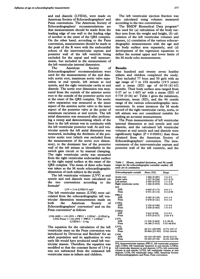
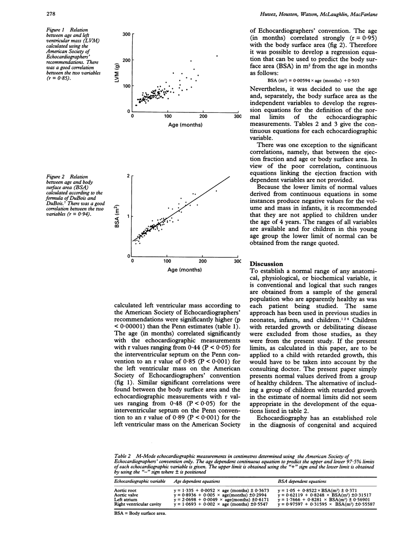
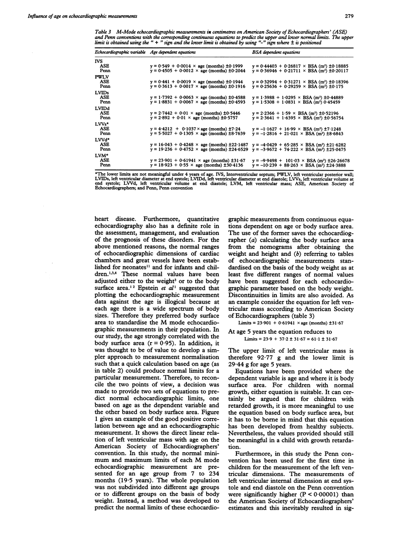
BACKGROUND--M Mode echocardiograms can be measured by two different conventions. In addition, normal limits of echocardiographic measurements have customarily been stratified according to age or body surface area. There is therefore a need to develop a more easily managed approach to calculating normal limits of measurements for the two conventions, one of which, the Penn convention, has not previously been used for echocardiographic measurements in children. METHODS--M mode echocardiograms were recorded in 127 healthy subjects aged from 7 months to 19.5 years. Measurements were made from paper recordings according to the recommendations of the American Society of Echocardiographers and those of the Penn convention. RESULTS--Age and body surface area were found to be highly correlated; but for completeness separate age dependent and body surface area dependent equations for the normal limits of M mode echocardiographic variables were developed. CONCLUSION--A set of age dependent equations and a set of body surface area dependent equations are presented for easy calculation of upper and lower limits of normal M mode echocardiographic variables in infants and children.
Full text
PDF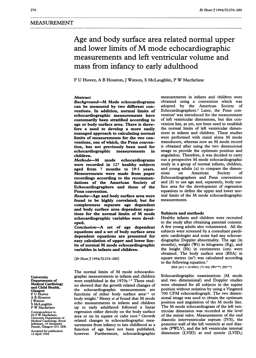
Selected References
These references are in PubMed. This may not be the complete list of references from this article.
- Allen H. D., Goldberg S. J., Sahn D. J., Schy N., Wojcik R. A quantitative echocardiographic study of champion childhood swimmers. Circulation. 1977 Jan;55(1):142–145. doi: 10.1161/01.cir.55.1.142. [DOI] [PubMed] [Google Scholar]
- Devereux R. B., Reichek N. Echocardiographic determination of left ventricular mass in man. Anatomic validation of the method. Circulation. 1977 Apr;55(4):613–618. doi: 10.1161/01.cir.55.4.613. [DOI] [PubMed] [Google Scholar]
- Epstein M. L., Goldberg S. J., Allen H. D., Konecke L., Wood J. Great vessel, cardiac chamber, and wall growth patterns in normal children. Circulation. 1975 Jun;51(6):1124–1129. doi: 10.1161/01.cir.51.6.1124. [DOI] [PubMed] [Google Scholar]
- Hagan A. D., Deely W. J., Sahn D., Friedman W. F. Echocardiographic criteria for normal newborn infants. Circulation. 1973 Dec;48(6):1221–1226. doi: 10.1161/01.cir.48.6.1221. [DOI] [PubMed] [Google Scholar]
- Henry W. L., Ware J., Gardin J. M., Hepner S. I., McKay J., Weiner M. Echocardiographic measurements in normal subjects. Growth-related changes that occur between infancy and early adulthood. Circulation. 1978 Feb;57(2):278–285. doi: 10.1161/01.cir.57.2.278. [DOI] [PubMed] [Google Scholar]
- Lundström N. R. Clinical applications of echocardiography in infants and children. I. Investigation of infants and children without heart disease. Acta Paediatr Scand. 1974 Jan;63(1):23–32. doi: 10.1111/j.1651-2227.1974.tb04345.x. [DOI] [PubMed] [Google Scholar]
- Pombo J. F., Troy B. L., Russell R. O., Jr Left ventricular volumes and ejection fraction by echocardiography. Circulation. 1971 Apr;43(4):480–490. doi: 10.1161/01.cir.43.4.480. [DOI] [PubMed] [Google Scholar]
- Sahn D. J., DeMaria A., Kisslo J., Weyman A. Recommendations regarding quantitation in M-mode echocardiography: results of a survey of echocardiographic measurements. Circulation. 1978 Dec;58(6):1072–1083. doi: 10.1161/01.cir.58.6.1072. [DOI] [PubMed] [Google Scholar]
- Troy B. L., Pombo J., Rackley C. E. Measurement of left ventricular wall thickness and mass by echocardiography. Circulation. 1972 Mar;45(3):602–611. doi: 10.1161/01.cir.45.3.602. [DOI] [PubMed] [Google Scholar]


