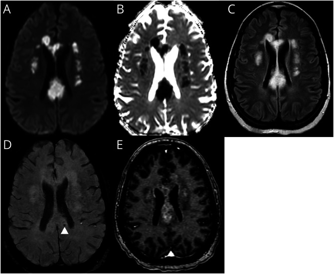
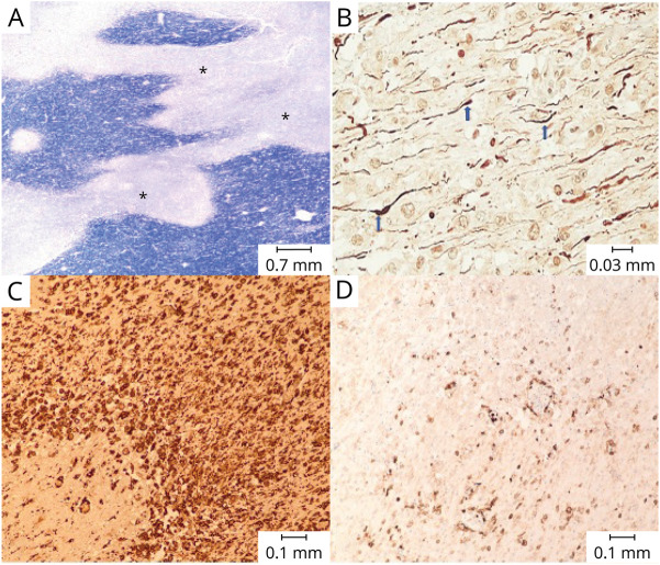
A 41-year-old woman with type 1 diabetes admitted with SARS-CoV-2 PCR confirmed respiratory failure developed altered mental status. EEG was unrevealing, and CSF showed an elevated protein (110 mg/dL) and normal glucose (159 mg/dL), without pleocytosis or oligoclonal bands and normal IgG index. MRI demonstrated FLAIR hyperintensities in the corpus callosum and periventricular white matter (Figure 1). She was treated with plasmapheresis for presumed SARS-CoV-2–related acute demyelinating encephalomyelitis (ADEM) but succumbed to cardiopulmonary arrest. Postmortem histology revealed irregular zones of demyelination with axonal sparing and perivascular inflammatory infiltrate, consistent with ADEM (Figure 2). There was no inflammation within the vessel walls as is seen in vasculitis. SARS-CoV-2 ADEM has variable clinical presentations. Involvement of deep white matter and the corpus callosum has been previously reported, as well as hemorrhagic leukoencephalopathy, although only minimal microhemorrhage was present for this patient.1 ADEM can be difficult to diagnose, and outcomes are often poor.2
Figure 1. MRI of the Brain With Contrast.
Diffusion-weighted image (A) with Apparent Diffusion Coefficient (B) showing confluent areas of restricted diffusion with associated FLAIR hyperintensity (C) involving the corpus callosum and bilateral corona radiata. Microhemorrhages (D) in the corpus callosum. Postcontrast images (E) demonstrate heterogeneous enhancement.
Figure 2. Postmortem Histology From the Splenium of the Corpus Callosum.
(A) Areas of pallor representing demyelination (*) on Luxol Fast Blue stain. (B) Relative preservation of axons (blue arrows), shown by neurofilament immunohistochemistry. (C) Dense macrophage infiltration as evidenced by CD163 immunohistochemistry. (D) Scattered lymphocytic infiltration as demonstrated by leukocyte common antigen immunohistochemistry.
Footnotes
Teaching slides links.lww.com/WNL/C651
Author Contributions
R. Lalla: drafting/revision of the manuscript for content, including medical writing for content; major role in the acquisition of data; study concept or design; analysis or interpretation of data. R. Narasimhan: drafting/revision of the manuscript for content, including medical writing for content; major role in the acquisition of data. M. Abdalkader: drafting/revision of the manuscript for content, including medical writing for content; major role in the acquisition of data. D. Virmani: study concept or design. K. Suchdev: drafting/revision of the manuscript for content, including medical writing for content. B. Moore: drafting/revision of the manuscript for content, including medical writing for content; major role in the acquisition of data; study concept or design; analysis or interpretation of data. A. Cervantes-Arslanian: drafting/revision of the manuscript for content, including medical writing for content; major role in the acquisition of data; study concept or design; analysis or interpretation of data.
Study Funding
The authors report no targeted funding.
Disclosure
The authors report no disclosures relevant to the manuscript. Go to Neurology.org/N for full disclosures.
References
- 1.Reichard RR, Kashani KB, Boire NA, Constantopoulos E, Guo Y, Lucchinetti CF. Neuropathology of COVID-19: a spectrum of vascular and acute disseminated encephalomyelitis (ADEM)-like pathology. Acta Neuropathol. 2020;140(1):1-6. [DOI] [PMC free article] [PubMed] [Google Scholar]
- 2.Wang Y, Wang Y, Huo L, Li Q, Chen J, Wang H. SARS-CoV-2-associated acute disseminated encephalomyelitis: a systematic review of the literature. J Neurol. 2022;269(3):1071-1092. [DOI] [PMC free article] [PubMed] [Google Scholar]


