

Abstract
There is a lack of data in the mainstream literature regarding the interactions between gingival fibroblasts, as a component of the local niche, and tumor precursors of B-lymphocytes. Although it is known that the development of tumors and tumor precursors depends on the local environment’s characteristics. In order to experimentally evaluate the apoptosis of pro-B type lymphocytes, induced as a result of the known activation of orphan nuclear receptor 4A1 (NR4A1), through Cytosporone B (Csn-B, 10 μM), in the presence or absence of exosomes derived from gingival fibroblasts, we administered as a treatment: 1 μM R-7050 [functional inhibitor of tumor necrosis factor alpha (TNFα)], 1 μM Z-IETD-FMK (functional inhibitor of caspase 8), 1 μM GSK690693 (functional inhibitor of Akt 1/2/3 pathways) and, last but not least, 1 μM scutellarin [functional inhibitor of receptor activator of nuclear factor-kappa B ligand (RANKL)] and therefore of the signal transducer and activator of transcription 3 (STAT3) pathway. Firstly, it is really clear that the presence of exosomes in the pro-B lymphocytes culture medium amplified the apoptotic effects of 10 μM Csn-B. The inhibition of tumoral precursors development, namely the pro-B type, might be highly dependent on the inhibition of Akt 1/2/3 pathways, the first and most important consequence being apoptosis induced by the activation of NR4A1 orphan nuclear receptors.
Keywords: pro-B lymphocyte , Cytosporone B , apoptosis , gingival fibroblast , Akt
Introduction
Random variable, diversity, and joining (VDJ) genes early recombination in T- and B-cell growth allows the flexible immune system to acknowledge a large variety of evolving pathogens using antigen (Ag) receptors. However, the capacity of such randomly produced T-cell receptor (TCR) as well as B-cell receptor (BCR) to identify as well as to react to self-Ags needs layers of tolerance mechanisms to alleviate the risk of serious autoimmunity. Since they were originally duplicated more than 30 years earlier, the nuclear receptor 4A (NR4A) household of nuclear hormonal agent receptors has actually been implicated in numerous important facets of immune tolerance, including the negative option of thymocytes, outer T-cell resistance, governing regulatory T-cells (Treg), as well as most just recently in outer B-cell resistance. In some research, there is evidence of crucial understandings from several laboratories as well as our own team into the feature and mechanisms through which this tiny course of primary feedback genes promotes self-tolerance and also immune homeostasis to balance the need for host defense against the intrinsic threats positioned by the adaptive body immune system [1]. Oxidized lipids, and also inflammatory cytokines, are believed to play a causal function in atherosclerosis through the focusing of gene expression in macrophages, as well as in various other cells. Previous work has linked nuclear receptors, peroxisome proliferator-activated receptors, and liver X receptors in the control of lipid-dependent gene expression and inflammation. Right here, we show that the expression of a third team of nuclear receptors, the NR4A ligand-independent orphan receptors, is highly inducible in macrophages by various inflammatory stimuli. Lipopolysaccharide (LPS), cytokine, or oxidized lipid therapy of macrophages activates the transcriptional induction of neuron-derived clone 77 (Nur77)/nuclear receptor 4A1 (NR4A1), nuclear receptor-related 1 (Nurr1)/nuclear receptor 4A2 (NR4A2), and neuron-derived orphan receptor 1 (NOR1)/nuclear receptor 4A3 (NR4A3) expression. Several lines of evidence implicate the nuclear factor kappa-light-chain-enhancer of activated B-cells (NF-κB) signaling pathway as a primary mediator of inducible NR4A expression in macrophages. Evaluation of murine and human Nur77 promoters revealed two highly conserved NF-κB action motifs. Abnormality of these elements prevented LPS-dependent expression of the Nur77 promoter in short-term transfection assays. Furthermore, the induction of Nur77 expression by LPS was drastically compromised in fibroblasts lacking the three NF-κB subunits, NF-κB subunit 1 (NFKB1), c-Rel, as well as RelA. Consistent with its ability to be induced by oxidized lipids, Nur77 was revealed in macrophages in human atherosclerotic lesions. These results implicate nuclear NR4A receptors as potential transcriptional arbiters of macrophage-dependent inflammatory signals [2]. Distinguishing real Ag-stimulated lymphocytes from bystanders triggered by the inflammatory scene has been tough. Nur77 is an immediate very early genetic response, whose expression is quickly upregulated by TCR signaling in murine T-cells and human thymocytes. Nur77/NR4A1–green fluorescent protein (GFP) transgenes serve as details TCR and also BCR signaling reporters in murine transgenic designs. In this research, there is demonstrated that endogenous Nur77 healthy protein expression can serve as a reporter of TCR and also BCR certain signaling in human peripheral blood mononuclear cells. Nur77 protein amounts were examined by immunofluorescence as well as flow cytometry in T- and also B-cells, separated from human peripheral blood mononuclear cells, acquired from healthy contributors that had been boosted by their respective Ag receptors. We demonstrate that endogenous Nur77 is a more specific reporter of Ag-specific signaling occasions than the typically used cluster of differentiation (CD)69 activation pen in both human T- and B-cells. This is reflective of the disparity in signaling pathways that control the expression of Nur77 and also CD69. Assessing endogenous Nur77 healthy protein expression has excellent perspectives to identify Ag-activated lymphocytes in human disease [3]. Ag excitement (signal 1) activates B-cell proliferation and tops B-cells to hire, involve, and also react to T-helper (Th) cells (signal 2). Failure to obtain signal 2 within a defined time window results in B-cell apoptosis, yet the devices that enforce reliance on co-stimulation are incompletely recognized. NR4A1–3 encodes a tiny family of orphan nuclear receptors that are swiftly generated by B-cell Ag receptor excitement. Below, we reveal that NR4A1 and also NR4A3 play partly repetitive functions to limit B-cell reactions to Ag in the lack of co-stimulation and also do so, in part, by quelching the expression of basic leucine zipper transcription factor, ATF-like (BATF) and also, subsequently, MYC (a family of regulator genes and proto-oncogenes). The NR4A household also limits B-cell accessibility to T-cell aid by quelching expression of the T-cell chemokines [C–C motif chemokine ligand 3 (CCL3)], as well as CCL4, as well as CD86, and intercellular adhesion molecule-1 (ICAM-1). Such NR4A-mediated policy plays an important role, especially under problems of competition for limiting Th cells [4]. Upon Ag direct exposure, ignorant B-cells, revealing BCR, and able to bind Ag, can go through robust spreading as well as differentiation that can cause the production of antibody (Ab)-secreting as well as memory B-cells. The variables determining whether a specific naïve B-cell will certainly proliferate following the Ag experience remain uncertain. In this research study, we discovered that polyclonal naïve murine B-cell populations, specific for a variety of Ags, express high levels of the orphan nuclear receptor Nur77, which is recognized to be upregulated downstream of BCR signaling because of cross-reactivity with self-Ags in vivo. Likewise, a portion of naïve human B-cells certain for clinically-relevant Ags stemmed from breathing syncytial virus, and also human immunodeficiency virus-1 (HIV-1) additionally displayed an immunoglobulin Mlowimmunoglobulin D+ (IgMLOWIgD+) phenotype, which is connected with self-Ag cross-reactivity. Functionally, naïve B-cells expressing moderate degrees of Nur77 are more than likely to proliferate in vivo complying with Ag injection. With each other, our information shows that BCR cross-reactivity with self-Ag is a common function of populaces of ignorant B-cells details for foreign Ags as well as a moderate degree of cross-reactivity primes specific cells for optimal proliferative reactions following Ag direct exposure [5]. If the involvement of orphan receptors of NR4A1 type in acute myeloid leukemia seems more obvious, the involvement in acute lymphoblastic one is far from being elucidated. On the other hand, there is not much data in the mainstream publications regarding the interactions between gingival fibroblasts (as a component of the local niche) and tumor precursors of B-lymphocytes, although it is known that their evolution is dependent on the characteristics of the local environment.
Aim
The aim of the study was to follow the effects of some exosomes/microvesicles from activated fibroblasts on pro-B lymphocyte development in culture, especially apoptosis. The development of tumoral or tumor precursor cells is known to be closely related to the niche.
Materials and Methods
Cell cultures, exosomes, and treatments
The gingival fibroblasts were obtained by applying the techniques and protocols previously described [6, 7], approved at that time by the Research Ethics Committee of the Grigore T. Popa University of Medicine and Pharmacy, Iaşi, Romania. The fibroblasts thus obtained were kept at -80ºC and were then multiplied according to the experimental needs. Dulbecco’s complete medium, L-Glutamine, Streptomycin sulfate, and Amphotericin B, as well as decomplemented 10% fetal bovine serum (FBS), were used as development conditions, ensuring at the same time a minimum of standard conditions: 37ºC and 5% carbon dioxide (CO2). To obtain exosomes from gingival fibroblasts, their culture medium was first centrifuged at 300×g and then consecutively at 2000×g and 10 000×g (4ºC, for 30 minutes each). Finally, we applied centrifugation at 100 000×g, at 4ºC, and for a duration of 60 minutes [8]. The obtained sediment was resuspended in phosphate-buffered saline. The concentration of exosomes/microvesicles was quantified by measuring the optical density by ultraviolet/visible (UV/VIS) spectrophotometry, being otherwise administered in a constant concentration of 0.01%, relative to each milliliter of culture medium for type B pro-lymphocytes. For the complete development of the experimental plan, another cell line, pro-B lymphocyte type, dependent on interleukin (IL)-1, was used. For the multiplication of these cells in culture, Roswell Park Memorial Institute (RPMI) 1640 medium was used, also supplemented with L-Glutamine, Penicillin, and Streptomycin (antibiotics), decomplemented 10% FBS, and, last but not least, 5% CO2 (but also 37oC) [9]. A series of preliminary studies demonstrated that this cell line was surface positive for calnexin, but not for CD19-type molecules, thus proving that they did not belong to pre-B lymphocyte cell lines (flow cytometry). The viability of pro-B lymphocytes, when exosomes derived from gingival fibroblasts were present (exosomes+) or absent (exosomes-) from the culture medium of this cell type, was quantified using Trypan Blue (0.2%) and phase contrast microscopy (Nikon Eclipse TE300) [9]. In order to experimentally evaluate the apoptosis of pro-B type lymphocytes, induced as a result of the known activation of NR4A1-type orphan receptors [10 μM Cytosporone B (Csn-B)], in the presence or absence of exosomes derived from gingival fibroblasts, we administered as a treatment: 1 μM R-7050 [functional inhibitor of tumor necrosis factor alpha (TNFα)], 1 μM Z-IETD-FMK (functional inhibitor of caspase 8), 1 μM GSK690693 (functional inhibitor of Akt 1/2/3 pathways) and, last but not least, 1 μM scutellarin [functional inhibitor of receptor activator of nuclear factor-kappa B ligand (RANKL)] and therefore of the signal transducer and activator of transcription 3 (STAT3) pathway.
Pro-B lymphocyte apoptosis evaluation
Apoptosis of pro-B type lymphocytes was quantified by flow cytometry (FACSCalibur™, Becton Dickinson) and annexin V (1 μM), coupled with R-phycoerythrin, a classical marker for programmed cell death programs. 488 nm (excitation) and 578 nm (emission) wavelengths were used. The pre-evaluated acquisition parameters were as follows: FL1 360 V, FL2 310 V, the FL1/FL2 ratio of 4.7%, and then the FL2/FL1 ratio of 52%. In addition, for the morphological highlighting of apoptosis, we used phase-contrast microscopy for all pro-B lymphocytes subjected to experiments (the same Nikon Eclipse TE300 type microscope, appropriate ×60 objectives, and the software associated with the setup).
Statistical analyses
All the results obtained experimentally were evaluated by statistical methods represented by one-way analysis of variance (ANOVA), completed in case of need with the classical Student–Newman–Keuls test.
Results
Effects of functional modulation of TNFα
The results obtained through the present experiments (Figure 1) highlight the fact that between the programmed cell death, induced by the agonist Csn-B (at a concentration of 10 μM) in the case of pro-B type cells and in the combined presence of exosomes derived from gingival fibroblasts as well as of R-7050, after 48 hours of treatment, as compared to the control cells, there are differences considered to be statistically significant (the averages being comparatively 83.57% and, respectively, 10% in the case of those without treatment). It can also be observed that this means an average increase of approximately 731% (p7lt;0.001) in the presence of R-7050. On the other hand, we can observe that between the apoptotic effects of Csn-B, induced on pro-B exosomes+ and pro-B exosomes- lymphocytes, in the presence of R-7050 over a period of 48 hours, there are also a series of statistically significant differences, respectively of 54.87% on average (p<0.001). Moreover, it is also worth noting that there are statistically significant differences between the apoptotic effects of Csn-B on pro-B exosomes+ cells, compared in the presence or absence of R-7050, which represent 34.53% on average, the effects being more evident when R-7050 is present in the culture medium (p<0.05).
Figure 1.
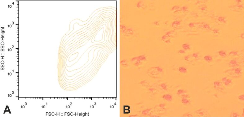
Representative flow cytometry (A) and phase contrast microscopy (B) when pro-B lymphocytes are treated with exosomes derived from gingival fibroblasts and with R-7050, an inhibitor of tumor necrosis factor alpha (TNFα).
Involvement of caspase 8
The experimental results (Figure 2) also suggest that between the apoptosis induced by the Csn-B agonist (administered at a concentration of 10 μM) in the case of pro-B lymphocytes and the simultaneous presence of exosomes released from gingival fibroblasts, as well as of Z-IETD-FMK (1 μM), after exactly 48 hours of treatment, when the comparison is made with control cells, there are differences considered to be statistically significant (means being 66.81% and, respectively, 10% in the case of those without any treatment). It can therefore be observed that there is an average increase of approximately 568% (p<0.001) in the presence of Z-IETD-FMK. Consecutively, however, we can also observe that between the apoptotic effects of the Csn-B activator, manifested at the level of pro-B lymphocytes in the presence or absence of microvesicles released from gingival fibroblasts in culture, in the presence of Z-IETD-FMK for 48 hours, there are no differences with statistical significance, these being only 7.55% on average (p>0.05). And even more, it is clear that between the effects induced by Csn-B at the level of pro-B lymphocytes in the presence of microvesicles and, at the same time, compared in the presence or absence of Z-IETD-FMK, there are relative differences, which can be considered significant statistically, of only 11.27% on average, the effects being somewhat more evident only when Z-IETD-FMK is present (p<0.05).
Figure 2.

Representative flow cytometry (A) and pro-B lymphocytes in phase contrast (B) in the presence of caspase 8 inhibitor, Z-IETD-FMK, and exosomes released into the environment by gingival fibroblasts.
Functional inhibition of Akt 1/2/3 pathways
Figure 3 shows that the presence of gingival fibroblast exosomes and GSK690693 (1 μM) for 48 hours has a stimulatory effect on the apoptosis of pro-B lymphocytes, induced by treatment with Csn-B (10 μM), obtaining truly statistically significant differences (p<0.001) and an average amplification of approximately 801%, compared to control pro-B lymphocytes (90.13% and 10%, respectively). Another statistically significant difference can be highlighted between the effects obtained in the presence or absence from the culture medium of exosomes produced by gingival fibroblasts, but in the presence of GSK690693, the average being otherwise 76.14% (p<0.05). Considering the results further, it also becomes evident that between the effects found at the level of B-lymphocyte apoptosis in the presence of fibroblast exosomes, but in the presence or absence of GSK690693, there are clear and statistically significant differences, of 45.01% on average (p<0.05).
Figure 3.

Flow cytometry (A) and phase contrast microscopy (B) when pro-B cells are treated with exosomes derived from gingival fibroblasts and with GSK690693, an inhibitor of Akt 1/2/3 pathways
RANKL and STAT3 pathways modulation
Also important are the results (Figure 4) that associate Csn-B (10 μM)-induced apoptosis amplification effects together with exosomes released in the culture medium by gingival fibroblasts in the case of pro-B cells, the comparison with control cells being statistically significant (averages being 63.47% and, in contrast, only 10%). Practically, these results equate to an amplification of approximately 534% (p<0.001) in the presence of scutellarin and exosomes. If we consider the absolute presence of scutellarin, in a 48-hour interval, we find statistically significant differences of 48.22% on average (p<0.01) between the conditions of the presence or absence of fibroblast exosomes in the pro-B lymphocyte culture medium. Finally, it is also worth noting that in the presence of exosomes, but in the presence or absence of scutellarin from the culture medium, we cannot highlight statistically significant differences, these being only 0.85% on average (p>0.05).
Figure 4.

Representative flow cytometry (A) and pro-B lymphocytes in phase contrast (B) in the presence of RANKL/STAT3 inhibitor, scutellarin, and exosomes released into the environment by gingival fibroblasts. RANKL: Receptor activator of nuclear factor-kappa B ligand; STAT3: Signal transducer and activator of transcription 3.
Discussions
It is extremely well known that the most common cells in periodontal connective tissue are fundamentally fibroblasts and that periodontal fibroblasts produce and, at the same time, essentially modify the extracellular matrix and play a particularly important role in maintaining the integrity of the tissue that contains them and of its homeostasis [10]. The Nur77 immediate-early gene, which encodes an orphan nuclear receptor, is rapidly caused by various stress and anxiety stimuli, including growth necrosis element [tumor necrosis factor (TNF)]. Nur77 has been implicated in moderating apoptosis, especially in T-cells and lump cells. We report right here that Nur77 can play a role in annoying apoptosis in TNF signaling. Nur77 expression is highly caused by TNF. Remarkably, unlike many anti-apoptotic particles, this generated expression of Nur77 is mainly independent of NF-κB. Ectopic expression of Nur77 can secure wild-type, TNF receptor-associated factor 2 (TRAF2)–/–, and also RelA–/– (NF-κB p65 subunit) cells from apoptosis induced by TNF, whereas expression of a dominant-negative type of Nur77 (DN-Nur77) accelerates TNF-mediated cell death in the mutant cells. In mouse beginning fibroblasts, Nur77 continues to be in the center in feedback to TNF as well as is not translocated to the mitochondria, where it was reported to mediate apoptosis. Our results recommend that Nur77 is a survival effector protein in the context of TNF-mediated signaling [11]. Fas (apoptosis Ag 1)-apoptosis inhibitory molecule (FAIM) has actually been demonstrated to provide resistance to Fas-induced apoptosis of lymphocytes and also hepatocytes in vitro and also in vivo. Below, we reveal that FAIM has been up-regulated in thymocytes upon TCR involvement, when FAIM–/– thymocytes are extremely at risk for TCR-mediated apoptosis with increased activation of caspase 8 as well as caspase 9. Furthermore, injection of anti-CD3 antibodies causes augmented exhaustion of CD4+CD8+ T-cells in the thymus of FAIM–/– mice, as compared to wild-type control, suggesting that FAIM contributes to thymocyte apoptosis. Cross-linking of the TCR on FAIM–/– thymocytes leads to a raised protein degree of the Nur77 orphan nuclear receptor, which contributes to thymocyte apoptosis. Interestingly, in the lack of FAIM, there is lowered ubiquitination and destruction of the Nur77 protein. FAIM–/– thymocytes additionally display a malfunctioning TCR-induced activation of Akt whose activity we currently reveal is required for Nur77 ubiquitination. More evaluations using FAIM-deficient main thymocytes as well as FAIM-overexpressing DO-11.10 T-cells suggest that FAIM acts upstream of Akt throughout TCR signaling as well as influences the localization of Akt to lipid boatings, thus affecting its activation. Taken together, our study specified a TCR-induced FAIM/Akt/Nur77 signaling axis that is crucial for modulating the apoptosis of establishing thymocytes [12]. Apoptosis signal-regulating kinase 1 (ASK1) is a mitogen-activated protein kinase kinase kinase (MAPKKK) that turns on downstream c-Jun N-terminal kinase (JNK) and p38 mitogen-activated healthy protein kinase (MAPK) to relay fatality signals into cells in response to numerous environmental anxieties. Nevertheless, whether ASK1 contributes to TCR-mediated apoptosis of thymocytes is vague. Right here, we reveal that ASK1 is triggered upon TCR stimulation as well as plays an essential role in TCR-mediated apoptosis of thymocytes by activating downstream JNK and p38 signaling cascade. Mechanistically, ASK1–JNK/p38 signaling leads to the upregulation of Nur77, a critical proapoptotic protein involved in the TCR-mediated apoptosis of thymocytes. In addition, we demonstrate that the activation of ASK1 is adversely regulated by Akt upon TCR stimulation. Therefore, our results determine a formerly unappreciated signaling mechanism entailing ASK1 in TCR-mediated apoptosis of thymocytes [13]. Therapy of LPS-activated microglia with 6-Mercaptopurine (6-MP) considerably undermined TNFα development. In 6-MP pretreated microglia, LPS-induced MAPK signaling, I kappa B-alpha destruction, NF-κB p65 nuclear translocation, as well as artificial insemination p65 deoxyribonucleic acid (DNA) binding task were not impaired. Nonetheless, 6-MP suppressed the transactivation task of NF-κB and also TNFα marker by inhibiting phosphorylation and also acetylation of p65 on Ser276 and also Lys310, respectively. Chromatin immunoprecipitation (ChIP) technique evaluations disclosed that 6-MP wetted LPS-induced histone H3 acetylation of chromatin surrounding the TNFα marker, inevitably causing a reduction in p65/coactivator-mediated transcription of TNFα genetics. Additionally, 6-MP boosted Nur77 orphan nuclear receptor expression. Making use of the ribonucleic acid (RNA) disturbance technique, we further demonstrated that Nur77 upregulation contributes to a 6-MP-mediated repressive effect on TNFα production. Furthermore, 6-MP likewise hampered TNFα messenger RNA (mRNA) translation via avoidance of LPS-activated phosphatidylinositol-3-kinase (PI3K)/protein kinase B (Akt)/mammalian target of rapamycin (mTOR) signaling cascade [14]. B-lymphocytes, a vital cluster of cells making up the immune system, can protect against abnormal biological factors. Heme oxygenase-1 (HO-1) plays crucial functions in cell spreading and also immune status, however, its impacts on the advancement and growth of B-lymphocytes are still unidentified. Here, the matter of B-lymphocytes in HO-1 gene knockout (HO-1+/–) mice was dramatically lower than that of the HO-1 gene wild-type (HO-1WT) mice. At the same time, the cell count of HO-1+/– mice did not recuperate after irradiation for one week, because of the G0/G1 stage apprehension of pro-B cells and the enhanced apoptosis of pre-B cells. Up-regulation of HO-1 by lentivirus attenuated the pro-B cell cycle apprehension, as well as pre-B cell apoptosis. A protein-to-protein communication network and Western blot were utilized to understand the molecular device by which HO-1 knockout obstructed B-lymphocyte growth. The PI3K/Akt signaling pathway moderated the regulatory impacts of HO-1 on B-lymphocytes. HO-1 is a critical transcriptional repressor for B-cell development [15]. The exact molecular mechanism underlying the patterns of very early B-cell lymphopoiesis is vague. The phospholipase C gamma (PLCγ) signaling pathway is critical for Ag receptor-mediated lymphocyte activation, yet its function in cytokine signaling is unknown. Below, we show that PLCγ1/PLCγ2 dual shortage in mice blocks early B-cell advancement at the pre-pro-B cell phase and makes B-cell progenitors less competent to IL-7. PLCγ pathway restraint obstructs IL-7-induced activation of mTOR, but not signal transducer and activator of transcription 5 (STAT5). The PLCγ pathway activates mTOR through the 1,2-diacyl-sn-glycerol (DAG)/protein kinase C (PKC) signaling branch, independent of the conventional Akt/tuberous sclerosis complex (TSC)/Ras homolog enriched in brain (Rheb) signaling axis. Restraint of PLCγ/PKC-induced mTOR activation harms the IL-7-mediated B-cell advancement. PLCγ1/PLCγ2 double-deficient B-cell progenitors have actually reduced the expression of genes related to the B-cell family tree, IL-7 signaling, and cell cycle. Hence, the IL-7 receptor manages very early B-lymphopoiesis via activation of mTOR through PLCγ/DAG/PKC signaling, not using Akt/Rheb signaling [16].
Conclusions
We followed the effects of Csn-B on the apoptosis of pro-B lymphocytes in the presence of exosomes derived from gingival fibroblasts in culture, together with 1 μM R-7050 (functional inhibitor of TNFα), 1 μM Z-IETD-FMK (functional inhibitor of caspase 8), 1 μM GSK690693 (functional inhibitor of Akt 1/2/3 pathways) and, last but not least, 1 μM scutellarin (functional inhibitor of RANKL and therefore of the STAT3 pathway). Firstly, it is really clear that the presence of exosomes in the pro-B lymphocytes culture medium amplified the apoptotic effects of 10 μM Csn-B. The amplification of the effects was as follows: R-7050 > GSK690693 > Z-IETD-FMK > scutellarin. If we take as base exosomes, the amplification of the apoptotic effects is more important and related to GSK690693 than R-7050 (and so consecutively, to inhibition of Akt 1/2/3 pathways as compared to inhibition of TNFα functioning). Thus, the inhibition of tumoral precursors development, namely the pro-B type, might be highly dependent on the inhibition of Akt 1/2/3 pathways, the first and most important consequence being apoptosis induced by the activation of NR4A1 orphan nuclear receptors.
Conflict of interests
The authors declare that they have no conflict of interests.
References
- 1.Hiwa R, Brooks JF, Mueller JL, Nielsen HV, Zikherman J. NR4A nuclear receptors in T and B lymphocytes: gatekeepers of immune tolerance. Immunol Rev. 2022;307(1):116–133. doi: 10.1111/imr.13072. [DOI] [PubMed] [Google Scholar]
- 2.Pei L, Castrillo A, Chen M, Hoffmann A, Tontonoz P. Induction of NR4A orphan nuclear receptor expression in macrophages in response to inflammatory stimuli. J Biol Chem. 2005;280(32):29256–29262. doi: 10.1074/jbc.M502606200. [DOI] [PubMed] [Google Scholar]
- 3.Ashouri JF, Weiss A. Endogenous Nur77 is a specific indicator of antigen receptor signaling in human T and B cells. J Immunol. 2017;198(2):657–668. doi: 10.4049/jimmunol.1601301. [DOI] [PMC free article] [PubMed] [Google Scholar]
- 4.Tan C, Hiwa R, Mueller JL, Vykunta V, Hibiya K, Noviski M, Huizar J, Brooks JF, Garcia J, Heyn C, Li Z, Marson A, Zikherman J. NR4A nuclear receptors restrain B cell responses to antigen when second signals are absent or limiting. Nat Immunol. 2020;21(10):1267–1279. doi: 10.1038/s41590-020-0765-7. [DOI] [PMC free article] [PubMed] [Google Scholar]
- 5.Steach HR, DeBuysscher BL, Schwartz A, Boonyaratanakornkit J, Baker ML, Tooley MR, Pease NA, Taylor JJ. Cross-reactivity with self-antigen tunes the functional potential of naive B cells specific for foreign antigens. J Immunol. 2020;204(3):498–509. doi: 10.4049/jimmunol.1900799. [DOI] [PMC free article] [PubMed] [Google Scholar]
- 6.Goriuc A, Foia LG, Minea B, Luchian AI, Surdu AE, Toma V, Costuleanu M, Mârţu I. Drug-induced gingival hyperplasia - experimental model. Rom J Morphol Embryol. 2017;58(4):1371–1376. [PubMed] [Google Scholar]
- 7.Barnea TV, Sava A, Gentimir C, Goriuc A, Boişteanu O, Chelaru L, Iancu RI, Avram CA, Acatrinei DD, Bogza EG, Răducanu OC, Cioloca DP, Vasincu D, Costuleanu M. Genetic polymorphisms of TNFA and IL-1A and generalized aggressive periodontitis. Rom J Morphol Embryol. 2015;56(2):459–464. [PubMed] [Google Scholar]
- 8.Lobb RJ, Becker M, Wen SW, Wong CSF, Wiegmans AP, Leimgruber A, Möller A. Optimized exosome isolation protocol for cell culture supernatant and human plasma. J Extracell Vesicles. 2015;4:27031–27031. doi: 10.3402/jev.v4.27031. [DOI] [PMC free article] [PubMed] [Google Scholar]
- 9.Gentimir C, Tatarciuc D, Zaharia CA, Chelaru L, Costin A, Jitareanu A, Bejinariu C, Costuleanu M. Biochemical effects of some tyrosine kinase inhibitors on pro-B cells-induced apoptosis. Rev Chim (Bucharest) 2017;68(8):1864–1867. [Google Scholar]
- 10.Hassell TM, Hefti AF. Drug-induced gingival overgrowth: old problem, new problem. Crit Rev Oral Biol Med. 1991;2(1):103–137. doi: 10.1177/10454411910020010201. [DOI] [PubMed] [Google Scholar]
- 11.Suzuki S, Suzuki N, Mirtsos C, Horacek T, Lye E, Noh SK, Ho A, Bouchard D, Mak TW, Yeh WC. Nur77 as a survival factor in tumor necrosis factor signaling. Proc Natl Acad Sci U S A. 2003;100(14):8276–8280. doi: 10.1073/pnas.0932598100. [DOI] [PMC free article] [PubMed] [Google Scholar]
- 12.Huo J, Xu S, Lam KP. Fas apoptosis inhibitory molecule regulates T cell receptor-mediated apoptosis of thymocytes by modulating Akt activation and Nur77 expression. J Biol Chem. 2010;285(16):11827–11835. doi: 10.1074/jbc.M109.072744. [DOI] [PMC free article] [PubMed] [Google Scholar]
- 13.Huo J, Xu S, Lam KP. ASK1 Mediates Nur77 expression in T-cell receptor mediated thymocyte apoptosis. Cells. 2020;9(3):585–585. doi: 10.3390/cells9030585. [DOI] [PMC free article] [PubMed] [Google Scholar]
- 14.Huang HY, Chang HF, Tsai MJ, Chen JS, Wang MJ. Mercaptopurine attenuates tumor necrosis factor-α production in microglia through Nur77-mediated transrepression and PI3K/Akt/mTOR signaling-mediated translational regulation. J Neuroinflammation. 2016;13(1):78–78. doi: 10.1186/s12974-016-0543-5. [DOI] [PMC free article] [PubMed] [Google Scholar]
- 15.Zhou Z, Ma D, Liu P, Wang P, Wei D, Yu K, Li P, Fang Q, Wang J. Deletion of HO-1 blocks development of B lymphocytes in mice. Cell Signal. 2019;63:109378–109378. doi: 10.1016/j.cellsig.2019.109378. [DOI] [PubMed] [Google Scholar]
- 16.Yu M, Chen Y, Zeng H, Zheng Y, Fu G, Zhu W, Broeckel U, Aggarwal P, Turner A, Neale G, Guy C, Zhu N, Chi H, Wen R, Wang D. PLCγ-dependent mTOR signalling controls IL-7-mediated early B cell development. Nat Commun. 2017;8(1):1457–1457. doi: 10.1038/s41467-017-01388-5. [DOI] [PMC free article] [PubMed] [Google Scholar]