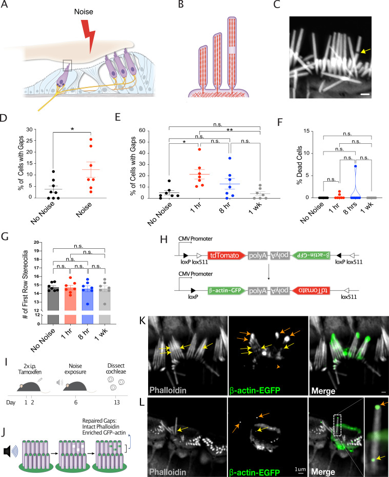
Figure 1. Noise-induced lesions in the F-actin cores of stereocilia is repaired by actin remodeling.
(A) Cartoon showing cross section of the organ of Corti. (B) Cartoon depicting side view of an inner hair cell (IHC), with a gap in stereocilia F-actin. (C) Representative image of an IHC with a gap in stereocilia F-actin, indicated by arrow. Scale bar: 1 μm. (D) Increased percentage of cells with phalloidin-negative gaps in IHC stereocilia F-actin following 1 hr noise exposure (*, p=0.0306). No Noise: n=8 organs of Corti, 4 mice; Noise: n=7 organs of Corti, 4 mice. (E) Percentage of cells with gaps initially increases 1 hr following 2 hr noise exposure (p=0.004) but then decreases (p=0.003) to levels not significantly different than in unexposed mice (p=0.74). n=7 organs of Corti, 4 mice per group. (F) Percentage of dead IHCs per cochlea does not significantly change within 1 week of noise exposure. No Noise vs 1 hr - n.s., p=0.974, No Noise vs 8hr - n.s., p=0.521, No Noise vs 1 week - n.s., p>0.999. n=7 organs of Corti, 4 mice per group. (G) Number of tallest row stereocilia per hair cell does not significantly change at any measured point within 1 week of noise exposure. No Noise vs 1 hr - n.s., p>0.999, No Noise vs 8 hr - n.s., p=0.969, No Noise vs 1 week - n.s., p=0.969. (H) Diagram of Cre-mediated inversion in FLEx-β-actin-EGFP mice following tamoxifen injection. Expression of tdTomato is turned off and EGFP-β-actin expression is turned on. (I) Experimental schematic for the observation of the localization of newly synthesized EGFP-tagged β-actin. Mice are injected on days 1 and 2 with tamoxifen and exposed to noise on day 6. Cochleae were dissected and processed on day 13. (J) Cartoon demonstrating the expected localization of EGFP-tagged β-actin in repaired gaps. (K, L) Representative examples from >8 experiments of likely repaired gaps. Yellow arrows point to sites of enriched EGFP-tagged β-actin along stereocilia length with intact phalloidin staining in a Cre-recombined cell following 1 week of recovery from noise exposure. Orange arrows indicate EGFP-labeled stereocilia tips. Due to low recombination rates, the surrounding cells do not express EGFP-β-actin. All error bars represent the standard error of the mean (SEM).

