Abstract
Purpose
This systematic review and meta-analysis aimed to explore the relationship of endometrial thickness (EMT) with obstetric and neonatal outcomes in assisted reproductive cycles.
Methods
PubMed, EMBASE, Cochrane Library and Web of Science were searched for eligible studies through April 2023. Obstetric outcomes include placenta previa, placental abruption, hypertensive disorders of pregnancy (HDP), gestational diabetes mellitus (GDM) and cesarean section (CS). Neonatal outcomes include birthweight, low birth weight (LBW), gestational age (GA), preterm birth (PTB), small for gestational age (SGA) and large for gestational age (LGA). The effect size was estimated as odds ratio (OR) or mean difference (MD) with 95% confidence interval (CI) using a random-effects model. Inter-study heterogeneity was assessed by the chi-square homogeneity test. One-study removal method was used to determine the sensitivity of the meta-analysis.
Results
Nineteen studies involving 76,404 cycles were included. The pooled results revealed significant differences between the thin endometrium group and the normal group in placental abruption (OR = 2.45, 95% CI: 1.11–5.38, P = 0.03; I2 = 0%), HDP (OR = 1.72, 95% CI: 1.44–2.05, P < 0.0001; I2 = 0%), CS (OR = 1.33, 95% CI: 1.06–1.67, P = 0.01; I2 = 77%), GA (MD = -1.27 day, 95% CI: -2.41– -1.02, P = 0.03; I2 = 73%), PTB (OR = 1.56, 95% CI: 1.34–1.81, P < 0.0001; I2 = 33%), birthweight (MD = -78.88 g, 95% CI: -115.79– -41.98, P < 0.0001; I2 = 48%), LBW (OR = 1.84, 95% CI: 1.52–2.22, P < 0.00001; I2 = 3%) and SGA (OR = 1.41, 95% CI: 1.17–1.70, P = 0.0003; I2 = 15%). No statistical differences were found in placenta previa, GDM, and LGA.
Conclusion
Thin endometrium was associated with lower birthweight or GA and higher risks of placental abruption, HDP, CS, PTB, LBW and SGA. Therefore, these pregnancies need special attention and close follow-up by obstetricians. Due to the limited number of included studies, further studies are needed to confirm the results.
Supplementary Information
The online version contains supplementary material available at 10.1186/s12958-023-01105-6.
Keywords: Endometrial thickness, Obstetric outcome, Neonatal outcome, Meta-analysis
Introduction
Since the birth of the first test-tube baby, assisted reproductive technology (ART) has brought hope to many infertile families. However, with the deepening of research, emerging studies have found that there are potential safety issues in ART pregnancy [1–4], such as low birth weight (LBW), preterm birth (PTB) and hypertensive disorders of pregnancy (HDP). At present, the mechanisms remain unclear and complex.
A thin endometrium is of great concern in ART cycles. Endometrial thickness (EMT) can be measured through a convenient way by transvaginal ultrasound and less than 7 or 8 mm is generally considered to be thin [5]. Although patients with thin endometrium can achieve and maintain a pregnancy spontaneously, these patients are reported to have significantly lower biochemical pregnancy, implantation and live birth rates during the process of ART [6, 7]. Furthermore, recent studies have revealed an association of thin endometrium with adverse obstetric and neonatal outcomes. However, no consensus has been reached and the relevance is still controversial [8–29]. Many factors may lead to this controversy, such as the type of embryo transfer, different cut-off values of EMT, and the number of cases reported in the study. Therefore, we conducted this systematic review and meta-analysis to determine associations between EMT and ART cycle outcomes to shed further light on this question.
Materials and methods
Protocol and registration
We conducted and reported our review based on the Preferred Reporting Items for Systematic Reviews and Meta-Analysis Statement (PRISMA2020) [30]. The study protocol is accessible at https://www.crd.york.ac.uk/PROSPERO/ (registration number CRD42021273323) while we excluded ectopic pregnancy in this study.
Data sources, search strategy and selection criteria
The electronic databases PubMed, Cochrane Library, Embase and Web of Science were searched until April 2023 for articles which evaluated effect of EMT on obstetric and neonatal outcomes in assisted reproduction. The selection criteria were described according to Patients, Intervention, Comparison and Outcomes (PICO) statements. Briefly, we included infertile women who had singleton livebirths after undergoing in vitro fertilization/ intracytoplasmic sperm injection (IVF/ICSI) or intra-uterine insemination (IUI) cycles. Patients were divided into the thin (intervention) and normal (comparison) groups based on the EMT cut-off values referring to the original studies. EMT was defined as the maximal distance between one interface of endometrium– myometrium to the other and measured according to corresponding cycles (Table 1). The outcomes included obstetric outcomes (placenta previa, placental abruption, HDP, gestational diabetes mellitus [GDM] and cesarean section [CS]) as well as neonatal outcomes (birthweight, LBW, gestational age [GA], PTB, small for gestational age [SGA] and large for gestational age [LGA]) defined according to International Classification of Diseases (ICD)-10 codes. Studies were excluded if: (1) studies were published as a letter, abstract or case report; (2) studies were not published in English; (3) samples were duplicated; and (4) samples were less than 20.
Table 1.
Main characteristics of included studies in the systematic review and meta-analysis
| Study | Country | Study design | No. of cycles | Mean age | Cut-off value | Treatment | Type of embryo transfer | Cycle protocol | Day of EMT measurement | NOS scorea |
|---|---|---|---|---|---|---|---|---|---|---|
| Hu 2021 [12] | China | Retrospective cohort | 5220 | 30.2 | 8.0 mm | IVF/ICSI | Frozen | artificial cycle, natural cycle, ovulation induction cycle | hCG trigger or progesterone initiation day | 9 |
| Guo 2020 [10] | China | Retrospective cohort | 3157 | 31.5 | 7.5 mm | IVF/ICSI | Fresh | GnRH agonist, GnRH antagonist, mild stimulation, natural cycle | hCG trigger day | 8 |
| He 2019 [11] | China | Retrospective cohort | 1139 | 30.5 | 8.0 mm | IVF/ICSI | Fresh and frozen | NA | hCG trigger or progesterone initiation day | 7 |
| Borges 2019 [8] | Brazil | Retrospective cohort | 402 | 34.1 | NA | ICSI | Fresh | GnRH antagonist | NA | 8 |
| Oron 2018 [17] | Israel | Retrospective cohort | 864 | 32.5 | 7.5 mm | IVF/ICSI | Fresh | GnRH agonist, GnRH antagonist, natural cycle | hCG trigger day | 7 |
| Kaser 2015 [15] | US | Case–control | 199 | NA | 9.7 mm | IVF/ICSI | Fresh and frozen |
Fresh: NA Frozen: artificial cycle |
NA | 9 |
| Chung 2006 [9] | US | Case–control | 435 | 31.9 | 10 mm | IVF/ICSI | NA | NA | The last recorded thickness prior to oocyte retrieval | 7 |
| Liu 2021 [20] | China | Retrospective cohort | 9266 | 30.9 | 8.0 mm | IVF/ICSI | Fresh | GnRH agonist, GnRH antagonist, other | hCG trigger day | 8 |
| Huang 2020 [13] | China | Retrospective cohort | 1016 | 30.2 | 7.6 mm | IUI | NA | Letrozole + HMG | hCG trigger day | 9 |
| Ribeiro 2018 [18] | Belgium | Retrospective cohort | 939 | NA | 7.0 mm | IVF/ICSI | Fresh | GnRH antagonist | hCG trigger day | 8 |
| Jing 2019 [14] | China | Retrospective cohort | 5251 | 30.9 | 9.0 mm | IVF/ICSI | Frozen | Artificial cycle, natural cycle | the day before embryo thawing | 9 |
| Zhang 2019 [19] | China | Retrospective cohort | 6181 | 32.0 | 8.0 mm | IVF/ICSI | Frozen | Artificial cycle, natural cycle | hCG trigger or progesterone initiation day | 9 |
| Moffat 2017 [16] | Switzerland | Retrospective cohort | 764 | 34.4 | NA | IVF/ICSI | Fresh | GnRH agonist, GnRH antagonist | hCG trigger day | 9 |
| Rombauts 2014 [21] | Australia | Retrospective cohort | 4537 | 34.4 | 9.0 mm | IVF/ICSI | Fresh and frozen |
Fresh: GnRH agonist, GnRH antagonist Frozen: artificial cycle, natural cycle |
hCG trigger or progesterone initiation day | 8 |
| Huang 2021 [23] | China | Retrospective cohort | 1755 | 30.0 | 8.0 mm | IVF/ICSI | Frozen | GnRH agonist, GnRH antagonist | hCG trigger or progesterone initiation day | 8 |
| Liu 2021a [25] | China | Retrospective cohort | 9273 | 31.2 | 8.0 mm | IVF/ICSI | Fresh | GnRH agonist, GnRH antagonist, other | hCG trigger day | 7 |
| Zhang 2022 [28] | China | Retrospective cohort | 13,458 | 30.5 | 8.0 mm | IVF/ICSI | Frozen | Natural cycle, artificial cycle | hCG trigger or progesterone initiation day | 7 |
| Zheng 2022 [29] | China | Retrospective cohort | 4313 | NA | 8.0 mm | IVF/ICSI | Frozen | Artificial cycle, natural cycle | progesterone initiation day | 7 |
| He 2022 [22] | China | Retrospective cohort | 8235 | 29.4 | 7.5 mm | IVF/ICSI | Frozen | Natural cycle, artificial cycle | hCG trigger or progesterone initiation day | 9 |
aStudy with scores greater than 7 was regarded as high quality. GnRH gonadotrophin releasing hormone, HMG human menopausal gonadotrophin, ICSI intracytoplasmic sperm injection, IUI intrauterine insemination, IVF in vitro fertilization, NA not available, NOS Newcastle–Ottawa Scale
The following keywords and their synonyms were used for literature search: [(‘endometrial thickness’) and (‘IVF’ or ‘ICSI’ or ‘infertility treatment’ or ‘IUI’ or ‘assisted reproductive technology’) and (‘pregnancy complications’ or ‘infant, newborn, diseases’ or ‘neonatal outcome’)] (see Supplementary File 1 for full strategy). Titles and abstracts of all identified studies were screened and the full paper of the preselected articles was scrutinized by two researchers (Z.F. and J.Q.M.). Any disagreement was settled by a third author (J.L.H.) to make the final decision.
Data collection and quality assessment
Two independent authors (Z.F. and J.Q.M.) extracted data from eligible studies by using standardized extraction forms. The following variables were collected: first author’s surname, publication year, country, study design, number of cycles, mean age, cut-off value of EMT, treatment, type of embryo transfer, cycle protocol, and obstetric and neonatal outcomes in the corresponding EMT groups. If 2 × 2 tables could be constructed, the study was selected for meta-analysis. If not, the study was selected for systematic review. In the 2 × 2 tables, the number of cycles with obstetric complications or reported neonatal outcomes for different EMT cut-off values was recorded. Authors were contacted by email if information was missing. Any disagreement between the two researchers was resolved through discussion, or in case of persistent disagreement, by consultation with a third author (J.L.H.).
Study quality was assessed by two researchers (Z.F. and J.Q.M.) using the Newcastle–Ottawa Scale (NOS) [31] based on selection, comparability and exposure (case–control study) or outcomes (cohort study).The score of a study below 6 signifies low quality, 6 and 7 represents moderate quality, while 8 and 9 means good quality. Any inconsistencies between the two authors were adjudicated by an additional author (X.H.W.) referring to the original article.
Statistical analysis
The pooled data for investigated outcomes were calculated using the random-effects model, considering that the underlying effects varied across the studies included [32, 33]. The incidences of placenta previa, placental abruption, HDP, GDM, PTB, LBW, SGA, LGA and CS were assigned as dichotomous data, and the odds ratios (ORs) with 95% confidence intervals (CIs) were calculated. The GA and birthweight were assigned as continuous variables, and mean differences (MDs) were calculated between the groups to determine the effect size [34].
Chi-squared homogeneity test and Higgins index (I2) were applied to evaluate the heterogeneity of articles. Heterogeneity was regarded as: none (I2 < 25%), low (25% ≤ I2 < 50%), moderate (25% ≤ I2 < 75%), or high (I2 > 75%). To assess the impact of a single study on the outcome, one-study removal method was used to determine the sensitivity of the meta-analysis [35]. Since not enough studies (fewer than ten) were included in the pooled analysis, the assessment of publication bias was not conducted according to Cochrane Handbook recommendations [14]. Subgroup analyses for HDP, GDM, SGA, PTB and LBW were conducted based on type of embryo.
RevMan software (Review Manager, version 5.4) was used for all statistical analyses conducted in this study. All tests were two tailed and a P-value of less than 0.05 was deemed statistically significant.
Result
Literature search and selection
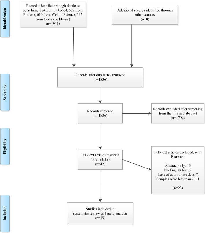
The search strategy identified a total of 1911 articles. After removing duplicates, 1836 abstracts were reviewed, and 42 full-text articles were further assessed. Thirteen studies were published as conference abstract, two studies were not English, and seven studies only reported live birth rate without obstetric or neonatal outcomes. In addition, the study by Martel et al. [36] was excluded as only 7 patients were enrolled in the thin endometrium group. Finally, 19 articles were appropriate to be included in this systematic review and meta-analysis (Fig. 1) [8–23, 25, 28, 29].
Fig. 1.
The flow diagram of study selection
The characteristics of all 19 included studies are presented in Table 1. Among them, 17 were retrospective cohort studies and 2 were case–control studies. The study sample size ranged from 199 to 13,458 cycles, for a total of 76,404 cycles. Studies were published between 2006 and 2022, and participants were mainly from China. EMT was divided into dichotomous variables in 15 studies [10–14, 17–21], which were thus included in meta-analysis. Most studies defined thin endometrium as EMT below 8 mm [11, 12, 19, 20, 23, 25, 28, 29], while different cut-off values of 7.0, 7.5, 7.6 and 9.0 mm were used in other studies [10, 13, 14, 17, 21, 22]. Some of the data in Liu et al. [20] were partially duplicated with those in Guo et al. [10], and we retained the study containing a larger sample size during the analysis. The remaining four studies analyzed EMT as a continuous variable and were included in the systematic review as data could not be extracted. Overall, the included studies were at low risk of bias with a NOS score of 7 (six studies), 8 (six studies) or 9 (seven studies), with details shown in Table S1.
Systematic review
Borges and colleagues analyzed the effect of EMT on birthweight of 402 newborns and showed that EMT was positively correlated with birthweight (β = 28.351, P = 0.044) and was significantly lower in the SGA group compared to the normal group [8]. Similarly, by analyzing 764 fresh cycles, Moffat et al. found that the EMT could predict neonatal birthweight [16].The study conducted by Chung and colleagues analyzed the effect of EMT on PTB, LBW and intrauterine fetal demise by comparing 159 cases and 276 controls [9]. The study showed a two-fold overall increased risk in the EMT ≤ 10 mm group compared to the EMT > 12 mm group (OR = 2.04, 95% CI: 1.09–3.83). With each millimeter increase in EMT, the risk of adverse perinatal outcome could decrease by 12%.
In addition, Kaser et al. conducted a case–control study analyzing 50 placenta accreta cases and 149 controls [15]. The study demonstrated that in cryopreserved embryo transfer cycles, the accreta patients had a significantly lower EMT than non-accreta patients.
Meta-analysis of obstetric outcomes
Placenta previa
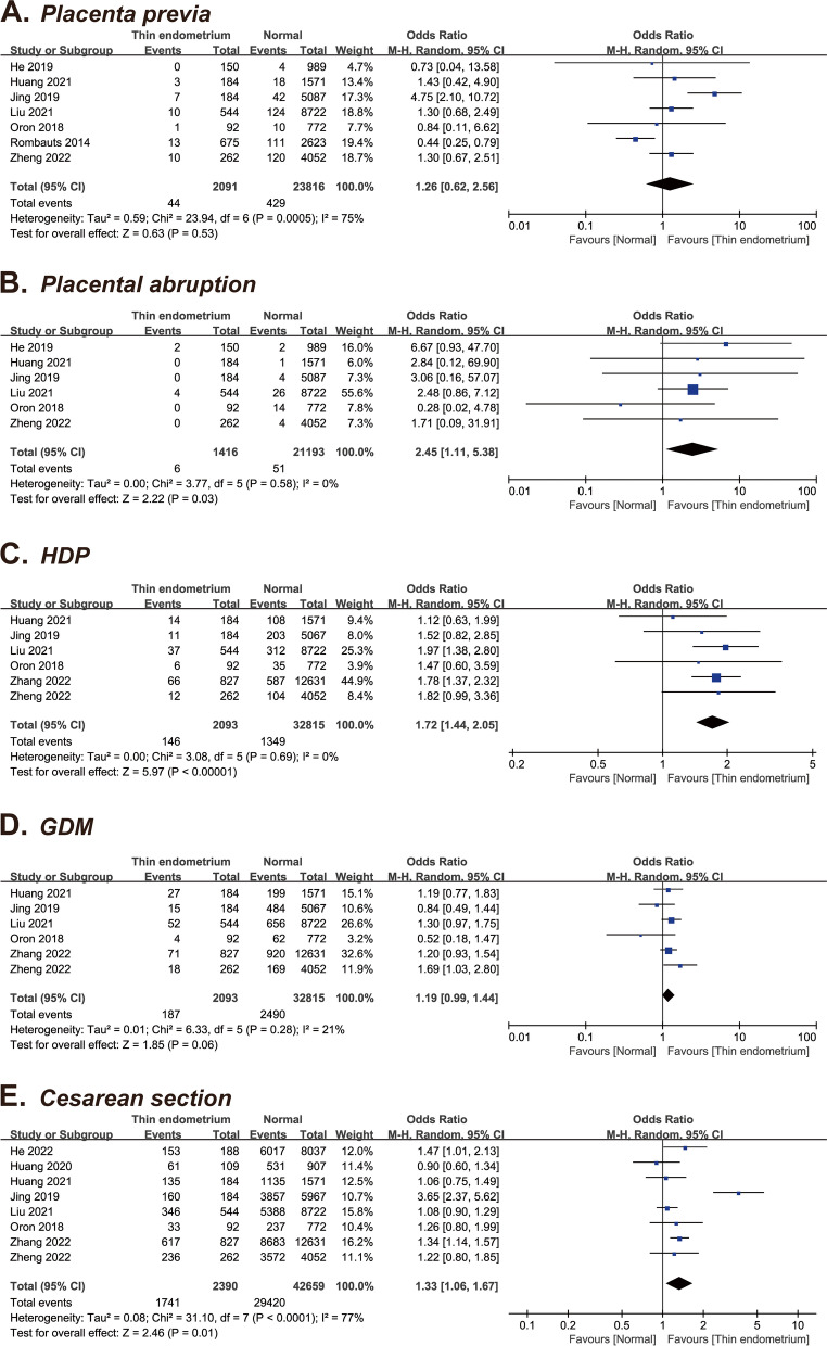
Seven studies [11, 14, 17, 20, 21, 23, 29], including 25,907 patients, reported on placenta previa rate. Pooled analysis revealed that thin endometrium was not associated with the risk of placenta previa (OR = 1.26, 95% CI: 0.62–2.56, P = 0.53; I2 = 75%) (Fig. 2A).
Fig. 2.
Forest plots of obstetric outcomes for thin versus normal endometrium. A Placenta previa; B Placental abruption; C Hypertensive disorders of pregnancy; D Gestational diabetes mellitus; E Cesarean section
Placental abruption
Six studies [11, 14, 17, 20, 23, 29], including 22,609 patients, reported on placental abruption. The overall OR for placental abruption was 2.45 (95% CI: 1.11–5.38, P = 0.03; I2 = 0%), suggesting no significant difference between the thin and normal endometrium groups (Fig. 2B).
Hypertensive disorders of pregnancy
Six studies [14, 17, 20, 23, 28, 29], including 34,908 patients, were pooled in this meta-analysis. Overall, the risk of HDP was significantly higher in the thin endometrium group than in the normal EMT group (OR = 1.72, 95% CI: 1.44–2.05, P < 0.0001; I2 = 0%) (Fig. 2C).
Gestational diabetes mellitus
Six studies [14, 17, 20, 23, 28, 29], including 34,908 patients, were combined in this analysis. Overall, no difference was noted in the GDM risk between the thin endometrium group and the normal endometrium groups (OR = 1.19, 95% CI: 0.99–1.44, P = 0.06; I2 = 21%) (Fig. 2D).
Cesarean section
Eight studies [13, 14, 17, 20, 22, 23, 28, 29], including 45,049 patients, were pooled in this analysis. Compared with the normal EMT group, the thin endometrium group showed a higher incidence of cesarean section (OR = 1.33, 95% CI: 1.06–1.67, P = 0.01) and the heterogeneity was high (I2 = 77%) (Fig. 2E).
Meta-analysis of neonatal outcomes
Gestational age
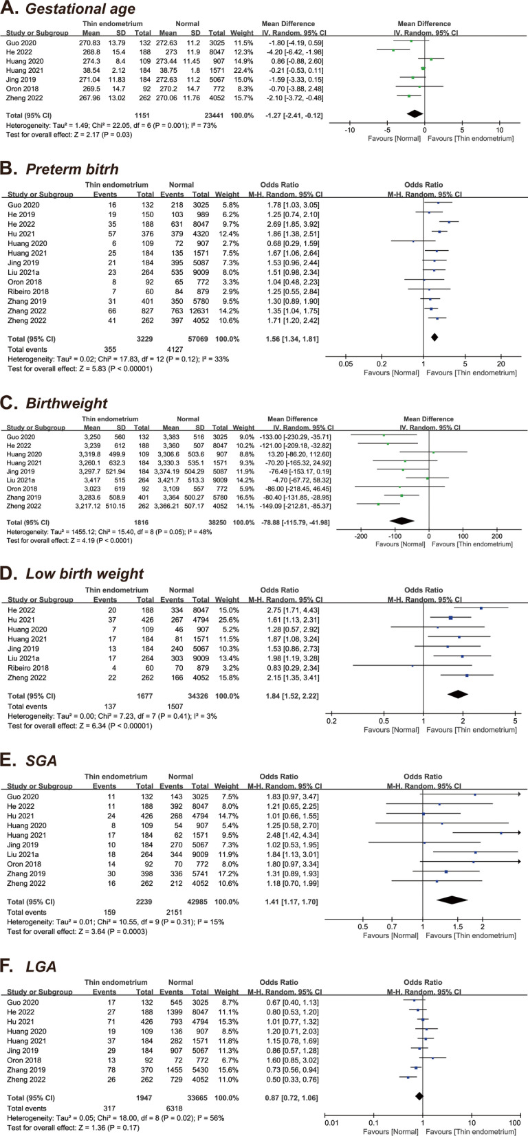
Seven studies [10, 13, 14, 17, 22, 23, 29], including 24,592 patients, were part of this analysis. Overall, decrease was noted in the GA between the thin endometrium and the normal EMT groups (MD = -1.27 days, 95% CI: -2.41–0.12, P = 0.03), with a high heterogeneity (I2 = 73%) (Fig. 3A).
Fig. 3.

Forest plots of neonatal outcomes for thin versus normal endometrium. A Gestational age; B Preterm birth; C Birthweight; D Low birth weight; E Small for gestational age; F Large for gestational age
Preterm birth
Thirteen studies [10–14, 17–19, 22, 23, 25, 28, 29], including 60,298 patients, provided information on PTB, which allowed quantitative pooled analysis. A significantly higher risk of PTB was found in the thin endometrium group relative to the normal endometrium group (OR = 1.56, 95% CI: 1.34–1.81, P < 0.0001; I2 = 33%) (Fig. 3B).
Birthweight
Nine studies [10, 13, 14, 17, 19, 22, 23, 25, 29], which included 40,066 patients, provided data on the birthweight. Overall, the thin endometrium group showed a significantly lower birthweight than the normal endometrium group (MD = -78.88 g, 95% CI: -115.79– -41.98, P < 0.0001; I2 = 48%) (Fig. 3C).
Low birthweight
Eight studies [12–14, 18, 22, 23, 25, 29], including 36,003 patients, were analyzed. Overall, the risk of LBW was significantly higher in the thin endometrium group than in the normal EMT group (OR = 1.84, 95% CI: 1.52–2.22, P < 0.00001; I2 = 3%) (Fig. 3D).
Small for gestational age
Ten studies [10, 12–14, 17, 19, 22, 23, 25, 28, 29], including 45,224 patients, were pooled in this analysis. When comparing the thin endometrium group to the normal EMT group, the OR for SGA was 1.41 (95% CI: 1.17–1.70, P = 0.0003) with low heterogeneity (I2 = 0%) (Fig. 3E).
Large for gestational age
Nine studies [10, 12–14, 17, 19, 22, 23, 29], including 35,612 patients, evaluated the LGA outcome. No significant difference was noted in the LGA incidence between the thin endometrium group and the normal EMT group (OR = 0.87, 95% CI: 0.72–1.06, P = 0.17; I2 = 56%) (Fig. 3F).
Sensitivity analysis
On excluding the study by Jing et al. or Oron et al., pooled analysis revealed that thin endometrium was associated with significantly higher risk of GDM (excluded Jing et al.: OR = 1.25, 95% CI: 1.04–1.49, P = 0.02; excluded Oron et al.: OR = 1.23, 95% CI: 1.05–1.44, P = 0.01). Contrarily, removal of any other individual studies did not modify the pooled estimates significantly in other obstetric and neonatal outcomes.
Discussion
Principal findings
The results of this meta-analysis showed that the HDP, CS, PTB, LBW and SGA risks were significantly higher in the thin endometrium group while neonatal birthweight and GA were significantly lower in the thin endometrium group. There were no significant differences in other maternal and perinatal outcomes between the two groups.
Interpretation of the findings
Previous meta-analyses have been conducted to investigate the association between EMT and pregnancy outcomes following IVF/ICSI treatment. It was generally concluded that thin endometrium could lead to lower rates of implantation, clinical pregnancy, ongoing pregnancy and live birth [5, 7]. Previous meta-analysis showed thin endometrium leads to a higher incidence of HDP and a lower birth weight, while a thick endometrium had no influence on pregnancy, maternal, or perinatal outcomes [24], compared to this study, we included more studies and outcomes. In the present study, we demonstrate the first-time systematic evidence that decreased EMT is also linked with increased obstetric and neonatal complications, indicating that long-term healthcare should be provided for these women even in cases of successful pregnancy.
In the subgroup analysis, outcomes were conducted based on type of embryo. Previous studies have shown that FET was associated with higher risk of HDP, LGA while lower risk of placenta previa, placental abruption, LBW, PTB and SGA [26]. We found that the subgroup analysis did not change the original results except for LBW of fresh cycles, this may be a bias due to the small number of studies (Figure S1, S2, S3, S4 and S5).
In sensitivity analyses, most findings remained coincident when one study was removed at a time, implying the reliability of our meta-analyses. Nonetheless, removal of the study by Jing et al. or Oron et al. resulted in a significant change of the pooled estimate of GDM [14, 17]. Both studies detected no difference in GDM rate between groups. However, the cut-off of EMT was 9.0 mm in Jing’s study, which may cause bias compared to other studies. Therefore, this inconsistent result may be attributed to the limited sample size or cut-off values that differed from other studies, and further large cohorts should be performed for confirmation.
Biological plausibility
HDP is a common pregnancy complication and a major contributor to PTB, LBW and SGA. However, the etiology of HDP has not yet been fully elucidated [37]. HDP is usually associated with uteroplacental hypoperfusion and ischemia, a common pathophysiologic mechanism also shared by placental abruption and intrauterine growth restriction. During the formation of the placenta, extravillous trophoblast cells invade the inner third of the uterine myometrium, replace the spiral artery endothelium, cause the collapse of vascular smooth muscles and thus remodel the blood vessels in this area. After vascular remodeling, a low-resistance blood flow connection is established between the spiral artery and the uterine radial artery, consequently increasing the circulating blood volume in the intervillous space and the placenta to provide nutrients for the growth and development of the fetus [38]. However, this process may be disordered in patients with decreased EMT. Indeed, an important feature of thin endometrium is the increased resistance of the uterine radial artery, which affects the normal placental blood supply [39]. In addition, studies have found that factors related to angiogenesis, such as leukemia inhibitory factor (LIF), vascular endothelial growth factor (VEGF) and β3 integrin are insufficient or even completely absent [40]. In this regard, endothelial cells could lack stimulation of pro-angiogenic factors during the remodeling process, resulting in reduced blood supply and leading to HDP or adverse neonatal outcomes [41].
Another mechanism may lie in the difference of oxygen tension between thin and normal endometrium. Under normal circumstances, the spiral arteries would constrict after ovulation, and the blood flow on the endometrial surface is reduced [42], creating a local low oxygen concentration that is conducive to successful embryo implantation. However, in patients with thin endometrium, the placenta and the fetus may be closer to the basal endometrium with greater blood flow and an oxygen-rich environment [43]. As a result, more free radicals are generated, thus possibly leading to impaired fetal growth [44].
In addition to the above, decreased EMT may be associated with certain ART process, thus leading to higher risks. For example, a previous meta-analysis showed that artificial cycles could lead to higher HDP and PTB rate [27]. In most of included studies, the endometrial preparation protocols were significantly different between normal and thin endometrium groups. Therefore, this may lead to a difference in obstetric and neonatal outcomes.
Strengths and limitations
To our knowledge, this systematic review and meta-analysis is the first study to comprehensively evaluate the relationship between thin endometrium and adverse obstetric and neonatal outcomes. Of the 14 included studies, 11 were published in recent five years. Hence, the effects of publication year and associated technical change on the pooled analysis were greatly reduced. Sensitivity analyses were performed by one-study removal method to assess the robustness of pooled data. Moreover, this meta-analysis was performed strictly according to the PRISMA 2020 statement and therefore, the quality of the methodology and reporting is high.
This study has some limitations. First and most importantly, all included studies are retrospective cohort studies or case–control studies with inherent bias. Analysis was based on crude data instead of adjusted data, and lack of prospective studies may lead to overestimation or underestimation of results. Secondly, due to different cut-off values of included studies, we cannot find a unified EMT to analyze. Thirdly, this study was based on small number of studies in each outcome, which limited our further conduction of subgroup analyses according to type of embryo transfer and number of cycles. Other unavoidable biases, such as the inclusion of only articles published in English and the exclusion of conference abstracts, may also have affected the results.
Conclusion
Our meta-analysis showed that patients with thin endometrium may face higher risks of certain adverse obstetric and neonatal outcomes compared to those with normal endometrium. This finding could provide useful information for both clinicians and infertile patients. Effective treatment should be provided to these women to increase EMT during ART cycles, while continuous monitoring and follow-up are needed throughout pregnancy. Given the present limitations, more prospective cohort studies with larger sample size are warranted to confirm the conclusions.
Supplementary Information
Additional file 1: Table S1. Quality assessment of included studies by the Newcastle–Ottawa scale.
Additional file 2: Figure S1. Subgroup analyses for HDP based on type of embryo.
Additional file 3: Figure S2. Subgroup analyses for GDM based on type of embryo.
Additional file 4: Figure S3. Subgroup analyses for SGA based on type of embryo.
Additional file 5: Figure S4. Subgroup analyses for PTB based on type of embryo.
Additional file 6: Figure S5. Subgroup analyses for LBW based on type of embryo.
Acknowledgements
We would like to thank Prof. Rombauts from the Department of Obstetrics and Gynaecology, Monash University for kindly providing raw research data concerning his article.
Abbreviations
- ART
Assisted reproductive technology
- CI
Confidence interval
- CS
Cesarean section
- EMT
Endometrial thickness
- GA
Gestational age
- GDM
Gestational diabetes mellitus
- HDP
Hypertensive disorders of pregnancy
- IUI
Intra-uterine insemination
- IVF/ICSI
In vitro fertilization/ intracytoplasmic sperm injection
- LBW
Low birth weight
- LIF
Leukemia inhibitory factor
- LGA
Large for gestational age
- MD
Mean difference
- NOS
Newcastle–Ottawa Scale
- OR
Odds ratio
- PICO
Patients, Intervention, Comparison and Outcomes
- PRISMA
Preferred Reporting Items for Systematic Reviews and Meta-Analysis Statement
- PTB
Preterm birth
- SGA
Small for gestational age
- VEGF
Vascular endothelial growth factor
Authors’ contributions
Z.F. and J.L.H. worked on study concept and design, acquisition of data, analysis and interpretation of data and drafting the article. J.Q.M. worked on data collection and critical revision of the article. L.M.Y. worked on critical revision of the article. X.H.W. worked on the analysis and interpretation of data and the final draft. The author(s) read and approved the final manuscript.
Funding
This study was supported by the National Natural Science Foundation of China (No. 81871182).
Availability of data and materials
The original data from the survey is available.
Declarations
Ethics approval and consent to participate
Not applicable.
Consent for publication
Not applicable.
Competing interests
The authors declare no competing interests.
Footnotes
Publisher’s Note
Springer Nature remains neutral with regard to jurisdictional claims in published maps and institutional affiliations.
Zheng Fang and Jialyu Huang contributed equally to this work.
References
- 1.Hou W, Shi G, Ma Y, Liu Y, Lu M, Fan X, Sun Y. Impact of preimplantation genetic testing on obstetric and neonatal outcomes: a systematic review and meta-analysis. Fertil Steril. 2021;116:990–1000. doi: 10.1016/j.fertnstert.2021.06.040. [DOI] [PubMed] [Google Scholar]
- 2.Pandey S, Shetty A, Hamilton M, Bhattacharya S, Maheshwari A. Obstetric and perinatal outcomes in singleton pregnancies resulting from IVF/ICSI: a systematic review and meta-analysis. Hum Reprod Update. 2012;18:485–503. doi: 10.1093/humupd/dms018. [DOI] [PubMed] [Google Scholar]
- 3.Zhao J, Yan Y, Huang X, Li Y. Do the children born after assisted reproductive technology have an increased risk of birth defects? A systematic review and meta-analysis. J Matern Fetal Neonatal Med. 2020;33:322–333. doi: 10.1080/14767058.2018.1488168. [DOI] [PubMed] [Google Scholar]
- 4.Omani-Samani R, Alizadeh A, Almasi-Hashiani A, Mohammadi M, Maroufizadeh S, Navid B, KhedmatiMorasae E, Amini P. Risk of preeclampsia following assisted reproductive technology: systematic review and meta-analysis of 72 cohort studies. J Matern Fetal Neonatal Med. 2020;33:2826–2840. doi: 10.1080/14767058.2018.1560406. [DOI] [PubMed] [Google Scholar]
- 5.Kasius A, Smit JG, Torrance HL, Eijkemans MJC, Mol BW, Opmeer BC, Broekmans FJM. Endometrial thickness and pregnancy rates after IVF: a systematic review and meta-analysis. Hum Reprod Update. 2014;20:530–541. doi: 10.1093/humupd/dmu011. [DOI] [PubMed] [Google Scholar]
- 6.Weiss NS, van Vliet MN, Limpens J, Hompes PGA, Lambalk CB, Mochtar MH, van der Veen F, Mol BWJ, van Wely M. Endometrial thickness in women undergoing IUI with ovarian stimulation. How thick is too thin? A systematic review and meta-analysis. Hum Reprod. 2017;32:1009–1018. doi: 10.1093/humrep/dex035. [DOI] [PubMed] [Google Scholar]
- 7.Gao G, Cui X, Li S, Ding P, Zhang S, Zhang Y. Endometrial thickness and IVF cycle outcomes: a meta-analysis. Reprod Biomed Online. 2020;40:124–133. doi: 10.1016/j.rbmo.2019.09.005. [DOI] [PubMed] [Google Scholar]
- 8.Borges E, Jr, Zanetti BF, Braga D, Setti AS, Figueira RCS, Iaconelli A., Jr Ovarian response to stimulation and suboptimal endometrial development are associated with adverse perinatal outcomes in intracytoplasmic sperm injection cycles. JBRA Assist Reprod. 2019;23:123–129. doi: 10.5935/1518-0557.20190001. [DOI] [PMC free article] [PubMed] [Google Scholar]
- 9.Chung K, Coutifaris C, Chalian R, Lin K, Ratcliffe SJ, Castelbaum AJ, Freedman MF, Barnhart KT. Factors influencing adverse perinatal outcomes in pregnancies achieved through use of in vitro fertilization. Fertil Steril. 2006;86:1634–1641. doi: 10.1016/j.fertnstert.2006.04.038. [DOI] [PubMed] [Google Scholar]
- 10.Guo Z, Xu X, Zhang L, Zhang L, Yan L, Ma J. Endometrial thickness is associated with incidence of small-for-gestational-age infants in fresh in vitro fertilization–intracytoplasmic sperm injection and embryo transfer cycles. Fertil Steril. 2020;113:745–752. doi: 10.1016/j.fertnstert.2019.12.014. [DOI] [PubMed] [Google Scholar]
- 11.He L, Zhang Z, Li H, Li Y, Long L, He W. Correlation between endometrial thickness and perinatal outcome for pregnancies achieved through assisted reproduction technology. J Perinat Med. 2019;48:16–20. doi: 10.1515/jpm-2019-0159. [DOI] [PubMed] [Google Scholar]
- 12.Hu KL, Kawai A, Hunt S, Li W, Li X, Zhang R, Hu Y, Gao H, Zhu Y, Xing L, et al. Endometrial thickness in the prediction of neonatal adverse outcomes in frozen cycles for singleton pregnancies. Reprod Biomed Online. 2021;43:553–560. doi: 10.1016/j.rbmo.2021.04.014. [DOI] [PubMed] [Google Scholar]
- 13.Huang J, Lin J, Lu X, Gao H, Song N, Cai R, Kuang Y. Association between endometrial thickness and neonatal outcomes in intrauterine insemination cycles: a retrospective analysis of 1,016 live-born singletons. Reprod Biol Endocrinol. 2020;18:48. doi: 10.1186/s12958-020-00597-w. [DOI] [PMC free article] [PubMed] [Google Scholar]
- 14.Jing S, Li X, Zhang S, Gong F, Lu G, Lin G. The risk of placenta previa and cesarean section associated with a thin endometrial thickness: a retrospective study of 5251 singleton births during frozen embryo transfer in China. Arch Gynecol Obstet. 2019;300:1227–1237. doi: 10.1007/s00404-019-05295-6. [DOI] [PubMed] [Google Scholar]
- 15.Kaser DJ, Melamed A, Bormann CL, Myers DE, Missmer SA, Walsh BW, Racowsky C, Carusi DA. Cryopreserved embryo transfer is an independent risk factor for placenta accreta. Fertil Steril. 2015;103:1176–1184.e1172. doi: 10.1016/j.fertnstert.2015.01.021. [DOI] [PubMed] [Google Scholar]
- 16.Moffat R, Beutler S, Schötzau A, De Geyter M, De Geyter C. Endometrial thickness influences neonatal birth weight in pregnancies with obstetric complications achieved after fresh IVF–ICSI cycles. Arch Gynecol Obstet. 2017;296:115–122. doi: 10.1007/s00404-017-4411-z. [DOI] [PubMed] [Google Scholar]
- 17.Oron G, Hiersch L, Rona S, Prag-Rosenberg R, Sapir O, Tuttnauer-Hamburger M, Shufaro Y, Fisch B, Ben-Haroush A. Endometrial thickness of less than 7.5 mm is associated with obstetric complications in fresh IVF cycles: a retrospective cohort study. Reprod BioMed Online. 2018;37:341–348. doi: 10.1016/j.rbmo.2018.05.013. [DOI] [PubMed] [Google Scholar]
- 18.Ribeiro VC, Santos-Ribeiro S, De Munck N, Drakopoulos P, Polyzos NP, Schutyser V, Verheyen G, Tournaye H, Blockeel C. Should we continue to measure endometrial thickness in modern-day medicine? The effect on live birth rates and birth weight. Reprod Biomed Online. 2018;36:416–426. doi: 10.1016/j.rbmo.2017.12.016. [DOI] [PubMed] [Google Scholar]
- 19.Zhang J, Liu H, Mao X, Chen Q, Si J, Fan Y, Xiao Y, Wang Y, Kuang Y. Effect of endometrial thickness on birthweight in frozen embryo transfer cycles: an analysis including 6181 singleton newborns. Human Reprod (Oxford, England) 2019;34:1707–1715. doi: 10.1093/humrep/dez103. [DOI] [PubMed] [Google Scholar]
- 20.Liu X, Wang J, Fu X, Li J, Zhang M, Yan J, Gao S, Ma J. Thin endometrium is associated with the risk of hypertensive disorders of pregnancy in fresh IVF/ICSI embryo transfer cycles: a retrospective cohort study of 9,266 singleton births. Reprod Biol Endocrinol. 2021;19:55. doi: 10.1186/s12958-021-00738-9. [DOI] [PMC free article] [PubMed] [Google Scholar]
- 21.Rombauts L, Motteram C, Berkowitz E, Fernando S. Risk of placenta praevia is linked to endometrial thickness in a retrospective cohort study of 4537 singleton assisted reproduction technology births. Hum Reprod. 2014;29:2787–2793. doi: 10.1093/humrep/deu240. [DOI] [PubMed] [Google Scholar]
- 22.He T, Li M, Li W, Meng P, Xue X, Shi J. Endometrial thickness is associated with low birthweight in frozen embryo transfer cycles: a retrospective cohort study of 8,235 singleton newborns. Front Endocrinol (Lausanne) 2022;13:929617. doi: 10.3389/fendo.2022.929617. [DOI] [PMC free article] [PubMed] [Google Scholar]
- 23.Huang J, Lin J, Xia L, Tian L, Xu D, Liu P, Zhu J, Wu Q. Decreased endometrial thickness is associated with higher risk of neonatal complications in women with polycystic ovary syndrome. Front Endocrinol (Lausanne) 2021;12:766601. doi: 10.3389/fendo.2021.766601. [DOI] [PMC free article] [PubMed] [Google Scholar]
- 24.Liao Z, Liu C, Cai L, Shen L, Sui C, Zhang H, Qian K. The effect of endometrial thickness on pregnancy, maternal, and perinatal outcomes of women in fresh cycles after IVF/ICSI: a systematic review and meta-analysis. Front Endocrinol (Lausanne) 2021;12:814648. doi: 10.3389/fendo.2021.814648. [DOI] [PMC free article] [PubMed] [Google Scholar]
- 25.Liu X, Wu H, Fu X, Li J, Zhang M, Yan J, Ma J, Gao S. Association between endometrial thickness and birth weight in fresh IVF/ICSI embryo transfers: a retrospective cohort study of 9273 singleton births. Reprod Biomed Online. 2021;43:1087–1094. doi: 10.1016/j.rbmo.2021.08.021. [DOI] [PubMed] [Google Scholar]
- 26.Sha T, Yin X, Cheng W, Massey IY. Pregnancy-related complications and perinatal outcomes resulting from transfer of cryopreserved versus fresh embryos in vitro fertilization: a meta-analysis. Fertil Steril. 2018;109(330–342):e339. doi: 10.1016/j.fertnstert.2017.10.019. [DOI] [PubMed] [Google Scholar]
- 27.Wu H, Zhou P, Lin X, Wang S, Zhang S. Endometrial preparation for frozen-thawed embryo transfer cycles: a systematic review and network meta-analysis. J Assist Reprod Genet. 2021;38:1913–1926. doi: 10.1007/s10815-021-02125-0. [DOI] [PMC free article] [PubMed] [Google Scholar]
- 28.Zhang M, Li J, Fu X, Zhang Y, Zhang T, Wu B, Han X, Gao S. Endometrial thickness is an independent risk factor of hypertensive disorders of pregnancy: a retrospective study of 13,458 patients in frozen-thawed embryo transfers. Reprod Biol Endocrinol. 2022;20:93. doi: 10.1186/s12958-022-00965-8. [DOI] [PMC free article] [PubMed] [Google Scholar]
- 29.Zheng Y, Chen B, Dai J, Xu B, Ai J, Jin L, Dong X. Thin endometrium is associated with higher risks of preterm birth and low birth weight after frozen single blastocyst transfer. Front Endocrinol (Lausanne) 2022;13:1040140. doi: 10.3389/fendo.2022.1040140. [DOI] [PMC free article] [PubMed] [Google Scholar]
- 30.Page MJ, McKenzie JE, Bossuyt PM, Boutron I, Hoffmann TC, Mulrow CD, Shamseer L, Tetzlaff JM, Akl EA, Brennan SE, et al. The PRISMA 2020 statement: an updated guideline for reporting systematic reviews. BMJ. 2021;372:n71. doi: 10.1136/bmj.n71. [DOI] [PMC free article] [PubMed] [Google Scholar]
- 31.GA Wells BS, D O'Connell, J Peterson, V Welch, M Losos, P Tugwell: The Newcastle-Ottawa scale (NOS) for assessing the quality of nonrandomised studies in meta-analyses. . Available: www.ohrica/programs/clinical_epidemiology/oxfordasp. 2011.
- 32.DerSimonian R, Laird N. Meta-analysis in clinical trials. Control Clin Trials. 1986;7:177–188. doi: 10.1016/0197-2456(86)90046-2. [DOI] [PubMed] [Google Scholar]
- 33.Ades AE, Lu G, Higgins JP. The interpretation of random-effects meta-analysis in decision models. Med Decis Making. 2005;25:646–654. doi: 10.1177/0272989X05282643. [DOI] [PubMed] [Google Scholar]
- 34.Higgins JP, Thompson SG, Deeks JJ, Altman DG. Measuring inconsistency in meta-analyses. BMJ. 2003;327:557–560. doi: 10.1136/bmj.327.7414.557. [DOI] [PMC free article] [PubMed] [Google Scholar]
- 35.Kendrick D, Kumar A, Carpenter H, Zijlstra GAR, Skelton DA, Cook JR, Stevens Z, Belcher CM, Haworth D, Gawler SJ, et al. Exercise for reducing fear of falling in older people living in the community. Cochrane Database Syst Rev. 2014;2014:009848. doi: 10.1002/14651858.CD009848.pub2. [DOI] [PMC free article] [PubMed] [Google Scholar]
- 36.Martel RA, Blakemore JK, Grifo JA. The effect of endometrial thickness on live birth outcomes in women undergoing hormone-replaced frozen embryo transfer. F S Rep. 2021;2:150–155. doi: 10.1016/j.xfre.2021.04.002. [DOI] [PMC free article] [PubMed] [Google Scholar]
- 37.Ramlakhan KP, Johnson MR, Roos-Hesselink JW. Pregnancy and cardiovascular disease. Nat Rev Cardiol. 2020;17:718–731. doi: 10.1038/s41569-020-0390-z. [DOI] [PubMed] [Google Scholar]
- 38.Fisher SJ. Why is placentation abnormal in preeclampsia? Am J Obstet Gynecol. 2015;213:S115–122. doi: 10.1016/j.ajog.2015.08.042. [DOI] [PMC free article] [PubMed] [Google Scholar]
- 39.Takasaki A, Tamura H, Miwa I, Taketani T, Shimamura K, Sugino N. Endometrial growth and uterine blood flow: a pilot study for improving endometrial thickness in the patients with a thin endometrium. Fertil Steril. 2010;93:1851–1858. doi: 10.1016/j.fertnstert.2008.12.062. [DOI] [PubMed] [Google Scholar]
- 40.Miwa I, Tamura H, Takasaki A, Yamagata Y, Shimamura K, Sugino N. Pathophysiologic features of "thin" endometrium. Fertil Steril. 2009;91:998–1004. doi: 10.1016/j.fertnstert.2008.01.029. [DOI] [PubMed] [Google Scholar]
- 41.Alfer J, Happel L, Dittrich R, Beckmann MW, Hartmann A, Gaumann A, Buck VU, Classen-Linke I. Insufficient Angiogenesis: Cause of Abnormally Thin Endometrium in Subfertile Patients? Geburtshilfe Frauenheilkd. 2017;77:756–764. doi: 10.1055/s-0043-111899. [DOI] [PMC free article] [PubMed] [Google Scholar]
- 42.Mizutani S, Matsumoto K, Kato Y, Mizutani E, Mizutani H, Iwase A, Shibata K. New insights into human endometrial aminopeptidases in both implantation and menstruation. Biochim Biophys Acta Proteins Proteom. 2020;1868:140332. doi: 10.1016/j.bbapap.2019.140332. [DOI] [PubMed] [Google Scholar]
- 43.Casper RF. It's time to pay attention to the endometrium. Fertil Steril. 2011;96:519–521. doi: 10.1016/j.fertnstert.2011.07.1096. [DOI] [PubMed] [Google Scholar]
- 44.Al-Gubory KH, Garrel C. Antioxidative signalling pathways regulate the level of reactive oxygen species at the endometrial-extraembryonic membranes interface during early pregnancy. Int J Biochem Cell Biol. 2012;44:1511–1518. doi: 10.1016/j.biocel.2012.06.017. [DOI] [PubMed] [Google Scholar]
Associated Data
This section collects any data citations, data availability statements, or supplementary materials included in this article.
Supplementary Materials
Additional file 1: Table S1. Quality assessment of included studies by the Newcastle–Ottawa scale.
Additional file 2: Figure S1. Subgroup analyses for HDP based on type of embryo.
Additional file 3: Figure S2. Subgroup analyses for GDM based on type of embryo.
Additional file 4: Figure S3. Subgroup analyses for SGA based on type of embryo.
Additional file 5: Figure S4. Subgroup analyses for PTB based on type of embryo.
Additional file 6: Figure S5. Subgroup analyses for LBW based on type of embryo.
Data Availability Statement
The original data from the survey is available.


