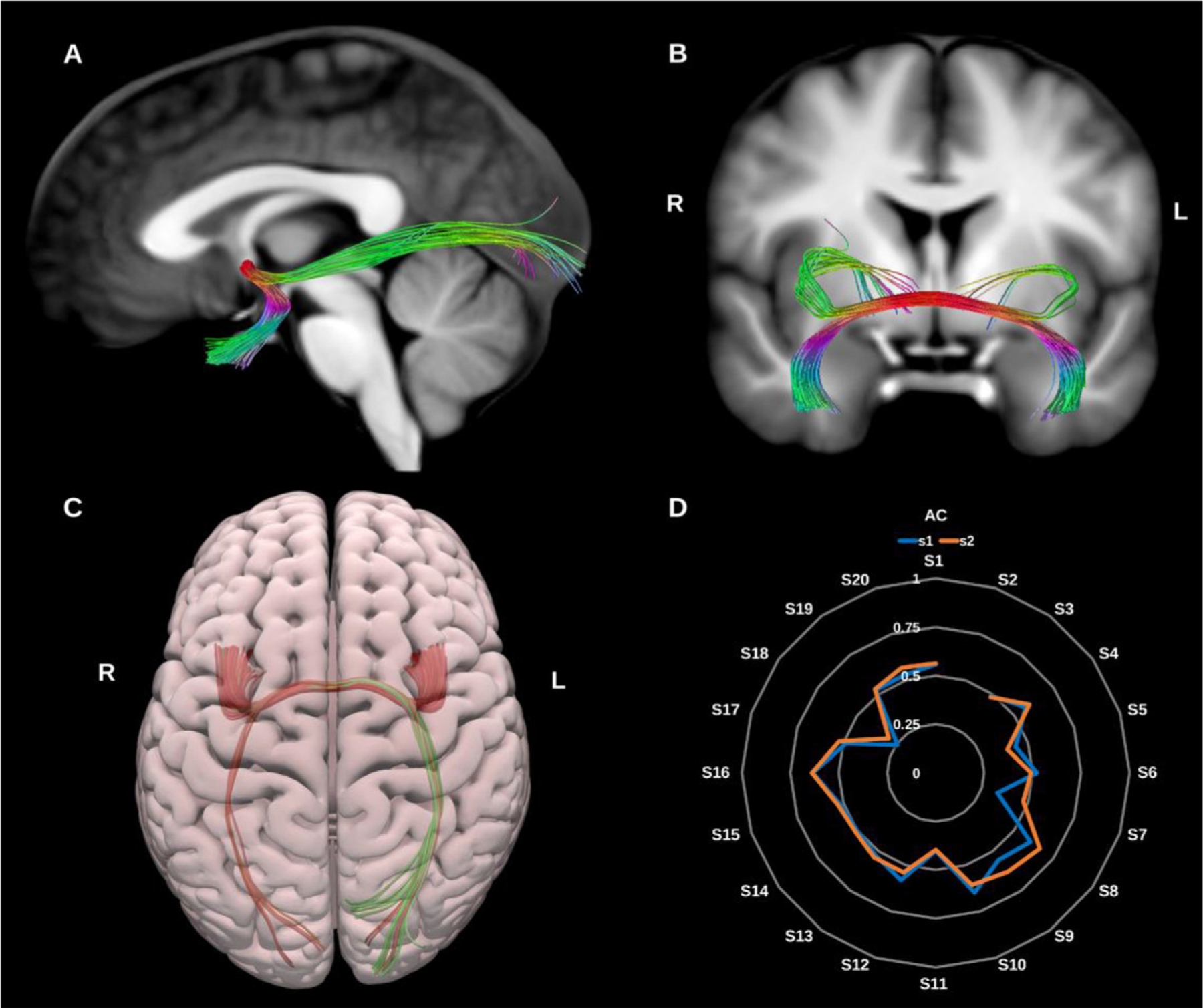
Fig. 12.

(A) and (B) Anterior commissure (AC) overlaid in color coding on sagittal and coronal slices of the T1-weighted images. (C) 3D superior projection of the semitransparent MNI pial surface with the AC shown in directional color coding. (D) Radar plot of the wDSC scores (vertical range) of the AC reconstructed using first session (blue) and second session (orange). L = left, R = right, S = subject, MNI = Montreal Neurological Institute, wDSC = weighted dice similarity coefficient.
