

Abstract
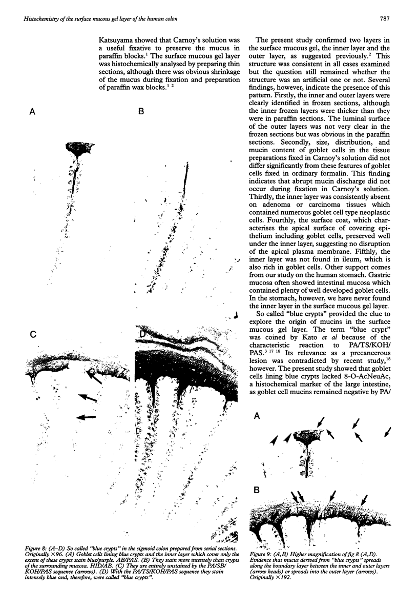
BACKGROUND AND AIMS: Histochemical analysis of the surface mucous gel layer of the human colon is difficult, as it dissolves in fixatives. This study was undertaken to explore the surface mucous gel layer on the normal mucosa and neoplastic tissues of the large intestine. In addition, the distribution of different mucins secreted from goblet cells was studied with a series of histochemical stains for mucins. METHODS: Twenty four surgically resected specimens were fixed in Carnoy's solution and embedded in paraffin. In four cases, the surface mucous gel layer was also studied in frozen sections. Serial sections were stained by a battery of histochemical techniques characterising mucins. RESULTS AND CONCLUSION: The surface mucous gel layer consisted of the inner and outer layers. The first covered the luminal surface of the mucosa, consisted of mucins, and showed a vertical striped pattern. The second overlaid the first, showed a lateral striped pattern, and was contaminated with bacteria and other substances. Their thickness in paraffin sections varied considerably among the sites in the large intestine, but was the thickest in the rectum and measured 12.7 (SEM 6.0) microns and 88.8 (SEM 80.1) microns respectively. Mucins forming the inner layer were obviously derived from goblet cells underlying it.
Full text
PDF


Images in this article


Selected References
These references are in PubMed. This may not be the complete list of references from this article.
- Bickel M., Kauffman G. L., Jr Gastric gel mucus thickness: effect of distention, 16,16-dimethyl prostaglandin e2, and carbenoxolone. Gastroenterology. 1981 Apr;80(4):770–775. [PubMed] [Google Scholar]
- Bollard J. E., Vanderwee M. A., Smith G. W., Tasman-Jones C., Gavin J. B., Lee S. P. Preservation of mucus in situ in rat colon. Dig Dis Sci. 1986 Dec;31(12):1338–1344. doi: 10.1007/BF01299812. [DOI] [PubMed] [Google Scholar]
- Culling C. F., Reid P. E., Clay M. G., Dunn W. L. The histochemical demonstration of O-acylated sialic acid in gastrointestinal mucins. Their association with the potassium hydroxide-periodic acid-schiff effect. J Histochem Cytochem. 1974 Aug;22(8):826–831. doi: 10.1177/22.8.826. [DOI] [PubMed] [Google Scholar]
- Culling C. F., Reid P. E., Dunn W. L. A new histochemical method for the identification and visualization of both side chain acylated and nonacylated sialic acids. J Histochem Cytochem. 1976 Dec;24(12):1225–1230. doi: 10.1177/24.12.187689. [DOI] [PubMed] [Google Scholar]
- Fuller C. E., Davies R. P., Williams G. T., Williams E. D. Crypt restricted heterogeneity of goblet cell mucus glycoprotein in histologically normal human colonic mucosa: a potential marker of somatic mutation. Br J Cancer. 1990 Mar;61(3):382–384. doi: 10.1038/bjc.1990.83. [DOI] [PMC free article] [PubMed] [Google Scholar]
- Garland C. D., Nash G. V., McMeekin T. A. The preservation of mucus and surface-associated microorganisms using acrolein vapour fixation. J Microsc. 1982 Dec;128(Pt 3):307–312. doi: 10.1111/j.1365-2818.1982.tb04633.x. [DOI] [PubMed] [Google Scholar]
- Hayama M., Ono K., Katsuyama T. An improved method for identifying 8-O-acetyl-N-acetylneuraminic acid. Stain Technol. 1985 Jul;60(4):201–205. doi: 10.3109/10520298509113913. [DOI] [PubMed] [Google Scholar]
- Iida F. Mucous barrier and peptic ulcer of the stomach. Gastroenterol Jpn. 1976;11(3):175–181. doi: 10.1007/BF02777701. [DOI] [PubMed] [Google Scholar]
- Jass J. R., Roberton A. M. Colorectal mucin histochemistry in health and disease: a critical review. Pathol Int. 1994 Jul;44(7):487–504. doi: 10.1111/j.1440-1827.1994.tb02599.x. [DOI] [PubMed] [Google Scholar]
- Kato Y., Sugano H. [Morphological studies on precancerous lesions of the stomach and large intestine]. Gan To Kagaku Ryoho. 1983 Feb;10(2 Pt 2):443–458. [PubMed] [Google Scholar]
- Kerss S., Allen A., Garner A. A simple method for measuring thickness of the mucus gel layer adherent to rat, frog and human gastric mucosa: influence of feeding, prostaglandin, N-acetylcysteine and other agents. Clin Sci (Lond) 1982 Aug;63(2):187–195. doi: 10.1042/cs0630187. [DOI] [PubMed] [Google Scholar]
- Morris G. P., Harding R. K., Wallace J. L. A functional model for extracellular gastric mucus in the rat. Virchows Arch B Cell Pathol Incl Mol Pathol. 1984;46(3):239–251. doi: 10.1007/BF02890313. [DOI] [PubMed] [Google Scholar]
- Muto T., Kamiya J., Sawada T., Agawa S., Morioka Y., Utsunomiya J. Mucin abnormality of colonic mucosa in patients with familial polyposis coli. A new tool for early detection of the carrier? Dis Colon Rectum. 1985 Mar;28(3):147–148. doi: 10.1007/BF02554225. [DOI] [PubMed] [Google Scholar]
- Ota H., Katsuyama T. Alternating laminated array of two types of mucin in the human gastric surface mucous layer. Histochem J. 1992 Feb;24(2):86–92. doi: 10.1007/BF01082444. [DOI] [PubMed] [Google Scholar]
- Piotrowski J., Slomiany A., Murty V. L., Fekete Z., Slomiany B. L. Inhibition of Helicobacter pylori colonization by sulfated gastric mucin. Biochem Int. 1991 Jul;24(4):749–756. [PubMed] [Google Scholar]
- Piotrowski J., Slomiany A., Murty V. L., Fekete Z., Slomiany B. L. Inhibition of Helicobacter pylori colonization by sulfated gastric mucin. Biochem Int. 1991 Jul;24(4):749–756. [PubMed] [Google Scholar]
- Pullan R. D., Thomas G. A., Rhodes M., Newcombe R. G., Williams G. T., Allen A., Rhodes J. Thickness of adherent mucus gel on colonic mucosa in humans and its relevance to colitis. Gut. 1994 Mar;35(3):353–359. doi: 10.1136/gut.35.3.353. [DOI] [PMC free article] [PubMed] [Google Scholar]
- Rozee K. R., Cooper D., Lam K., Costerton J. W. Microbial flora of the mouse ileum mucous layer and epithelial surface. Appl Environ Microbiol. 1982 Jun;43(6):1451–1463. doi: 10.1128/aem.43.6.1451-1463.1982. [DOI] [PMC free article] [PubMed] [Google Scholar]
- SPICER S. S. DIAMINE METHODS FOR DIFFERENTIALING MUCOSUBSTANCES HISTOCHEMICALLY. J Histochem Cytochem. 1965 Mar;13:211–234. doi: 10.1177/13.3.211. [DOI] [PubMed] [Google Scholar]
- Sakata T., von Engelhardt W. Luminal mucin in the large intestine of mice, rats and guinea pigs. Cell Tissue Res. 1981;219(3):629–635. doi: 10.1007/BF00210000. [DOI] [PubMed] [Google Scholar]
- Sandzén B., Blom H., Dahlgren S. Gastric mucus gel layer thickness measured by direct light microscopy. An experimental study in the rat. Scand J Gastroenterol. 1988 Dec;23(10):1160–1164. doi: 10.3109/00365528809090185. [DOI] [PubMed] [Google Scholar]
- Smith B., Butler M. The autonomic control of colonic mucin secretion in the mouse. Br J Exp Pathol. 1974 Dec;55(6):615–621. [PMC free article] [PubMed] [Google Scholar]
- Sugihara K., Jass J. R. Colorectal goblet cell sialomucin heterogeneity: its relation to malignant disease. J Clin Pathol. 1986 Oct;39(10):1088–1095. doi: 10.1136/jcp.39.10.1088. [DOI] [PMC free article] [PubMed] [Google Scholar]
- Szentkuti L., Eggers A. Stabilization of pre-epithelial mucus gel in cryostat sections from rat colon with celloidin. Stain Technol. 1990;65(4):179–181. doi: 10.3109/10520299009108067. [DOI] [PubMed] [Google Scholar]
- Szentkuti L., Riedesel H., Enss M. L., Gaertner K., Von Engelhardt W. Pre-epithelial mucus layer in the colon of conventional and germ-free rats. Histochem J. 1990 Sep;22(9):491–497. doi: 10.1007/BF01007234. [DOI] [PubMed] [Google Scholar]
- Tock E. P., Pearse A. G. Preservation of tissue mucins by freeze-drying and vapour fixation. I. Light microscopy. J R Microsc Soc. 1965 Dec;85(4):519–537. doi: 10.1111/j.1365-2818.1965.tb02153.x. [DOI] [PubMed] [Google Scholar]