Abstract
Purpose
To explore the mechanisms relating the gut microbiome (GM) to age-related macular degeneration (AMD), as they remain unclear. GM taxa that appear to act within the gut–retina axis may affect the risk of AMD.
Methods
Single-nucleotide polymorphisms (SNPs) of 196 GM taxa were obtained from the MiBioGen consortium, and a Mendelian randomization (MR) study was carried out to estimate the causality between GM taxa and AMD (defined as an endpoint based on ICD-9 and ICD-10). Using the data from the FinnGen consortium (6157 patients and 288,237 controls), we explored the GM taxa for causality and verified the results at the replication stage based on the MRC-IEU consortium (3553 cases and 147,089 controls). Inverse variance weighting (IVW) was the main method used to analyze causality, and the MR results were verified using heterogeneity tests and pleiotropy tests.
Results
According to the MR results, order Rhodospirillales (P = 3.38 × 10⁻²), family Victivallaceae (P = 3.14 × 10⁻²), family Rikenellaceae (P = 3.58 × 10⁻²), genus Slackia (P = 3.15 × 10⁻²), genus Faecalibacterium (P = 3.01 × 10⁻²), genus Bilophila (P = 1.11 × 10⁻²), and genus Candidatus Soleaferrea (P = 2.45 × 10⁻²) were suggestively associated with AMD. In the replication stage, only order Rhodospirillales (P = 0.03) passed validation. The heterogeneity (P > 0.05) and pleiotropy (P > 0.05) tests in two stages confirmed the robustness of the MR results.
Conclusions
We confirmed that order Rhodospirillales influenced the risk of AMD based on the gut–retina axis, providing new impetus for the development of the GM as an intervention to prevent the occurrence and development of AMD.
Keywords: gut–retina axis, age-related macular degeneration (AMD), gut microbiome, Mendelian randomization, causality
As a degenerative disease caused by many factors, age-related macular degeneration (AMD) is one of the main causes of severe vision loss.1 Because of the increased aging of the world's population, the incidence rate of AMD is on the rise. The number of people with AMD may even exceed 280 million by 2040.2 Therefore, identifying the risk factors of AMD can help intervene in the incidence of AMD as soon as possible and alleviate the burden of AMD on public health. The mechanism of AMD involves a complex combination of genetic susceptibility, inflammation, environmental factors, and other risk factors.3 Through clinical imaging technology (such as optical coherence tomography and indocyanine green angiography), researchers have revealed some intraocular risk factors (such as extracellular deposits).4–6 However, localized mechanistic studies in the eye have struggled to fully elucidate the impact of the risk of developing AMD. Due to the complex pathogenesis, interventions to reduce the risk of AMD have been limited.
Studies have found that dietary patterns and nutrition may affect the risk of AMD.7 For example, some researchers have proposed that lipids from the diet are processed by retinal pigment epithelial cells and are an important component of drusen.8 In addition to local risk factors of the eye, systemic factors may also explain the pathogenesis of AMD from another aspect. The gut ecosystem is significantly shaped by the intestine itself and includes a vast network of the gut microbiome (GM) taxa.9 The interactions among nutrients, the GM, and intestinal organs maintain a dynamic balance. When the GM is ecologically disturbed, it may affect the digestion and absorption of nutrients, causing immune and metabolic diseases.10 Crosstalk between GM taxa and the brain has been well established. The relationship between GM taxa and ophthalmic diseases is still in the preliminary exploration stage. Rowan et al.11 first confirmed the internal relationship between the GM taxa of wild-type aged mice and the retina through untargeted metabolomics and proposed the concept of the gut–retina axis. There is a huge difference in the intestinal microbial composition between mice and humans, and further research is required to confirm this view. Zinkernagel et al.12 sequenced the gut metagenomes of 12 patients with neovascular AMD and found that enrichment of the GM taxa differed from the control group.
Notably, because of the influence of diet habits and daily life, there may be large individual differences in the abundance of each GM taxon. Considering the high cost of sequencing technology, it is difficult for most researchers to conduct large-sample, randomized controlled studies to reduce the error caused by individual differences. Mendelian randomization (MR) evaluates the causal relationships of genetic variations from a large sample through publicly available de-identified data to obtain more reliable conclusions.13 This method simulates the random allocation of samples to the control and experimental groups of a randomized controlled trial, making it possible to analyze the relationship of the gut–retina axis.
In this study, we hoped to further clarify the causality between GM taxa and AMD through the use of human GM genomics research to provide a more powerful basis for further research on mechanisms of the gut–retina axis. In addition, it is hoped that GM taxa with an association with AMD will serve as new targets for intervention.
Methods
Ethics Statement
The genome-wide association study (GWAS) data used for analysis consisted of de-identified public data and were searched from among published studies. Ethics committees of all original institutions approved all of the GWASs following the tenets of the Declaration of Helsinki. The summary statistics for genetic associations with AMD can be found at the FinnGen study and the IEU OpenGWAS project (GWAS ID: ukb-b-17194) at https://gwas.mrcieu.ac.uk/. The MR analysis code can be found at https://mrcieu.github.io/TwoSampleMR/articles/index.html.
Genetic Instrumental Variables of the Gut Microbiome
Independent GM taxon genetic variant loci were identified from the MiBioGen consortium, which is the largest study to investigate the genetics of the human GM from 24 population-based cohorts.14 In the MiBioGen consortium, 16S rRNA data of 18,340 subjects were obtained from European, Asian, Hispanic, Middle Eastern, and African ancestries. A total of seven fecal DNA extraction methods were used to obtain the GM taxa data, which were then adjusted for age, principal genetic components, technical covariates, and gender. With the use of direct taxonomic binning, 122,110 variables in 211 taxa of the GM (containing 15 unknown taxa) were divided into five levels (phylum, class, order, family, and genus). A detailed description of the GM taxa GWASs is available in the study by Kurilshikov et al.14 To obtain more accurate results, we removed 15 unknown GM taxa.
AMD GWAS Dataset in the Discovery Stage
To discover the impact of GM taxa on AMD, we used summary-level data composed of 6157 AMD patients and 288,237 controls. The dataset was obtained from the FinnGen study (https://r7.finngen.fi/) in the discovery stage.15 AMD is defined as an endpoint based on International Classification of Disease, Ninth Revision (ICD-9; 3625A and 3625B) and ICD-10 (H35.30). In the FinnGen consortium, all subjects were Finnish, and 16,962,023 single-nucleotide polymorphisms (SNPs) were analyzed. The first 10 principal components (PCs), genetic factors, age, and sex were adjusted.
AMD GWAS Dataset in the Replication Stage
We verified significant GM taxa identified in the discovery stage using the MR database.16 This database contains 42,334 GWASs involving 244,792,068,559 genetic associations (as of September 12, 2022). We chose GWAS ID: ukb-b-17194 as the replication outcome of AMD (ICD-9), which originated from the Medical Research Council Integrative Epidemiology Unit (MRC-IEU) consortium based on the UK Biobank study.17 The MRC-IEU consortium recruited 150,642 European samples (involving 9,851,867 genetic associations) between 2006 and 201018 (Supplementary Fig. S1). In the replication stage, this GWAS includes 3553 AMD cases and 147,089 controls. In the analysis, sex, chip, and first 10 PCs were adjusted.
IV Quality Control
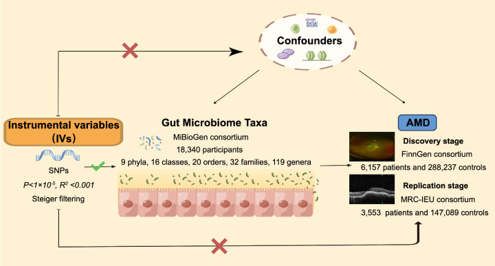
For the MR analysis, three assumptions had to be satisfied19 (Fig. 1) (terms appearing in the manuscript are explained in the Supplementary Materials): (1) independent instrumental variables (IVs) were associated with each GM taxon and were not associated with AMD; (2) independent IVs associated with each GM taxon were not associated with confounders; and (3) effects of the IVs were associated with AMD only through each GM taxon without other factors.
Figure 1.
Directed acyclic graph composed of the IVs (each GM taxon-related SNP from five levels), exposure (196 GM taxa from five levels), and outcome (AMD in two stages).
All of the genetic variables were quality controlled to obtain valid IVs. As with the current criteria for GM studies, we chose P < 1 × 10⁻⁵ screening IVs.20 The presence of a weak IV was assessed using F-values . For F < 10, the IV was defined as weak and was removed. To make the SNPs independent from each other, a linkage disequilibrium was carried out based on the European-based 1000 Genomes Project.21 The threshold R2 for identifying independent SNPs was set to 0.001, and the clumping distance was 10,000 kb. Finally, we performed Steiger filtering22 to remove the IVs that might cause inversion of causality.
Statistical Analysis
R 4.1.1 (R Foundation for Statistical Computing, Vienna, Austria) and the R package TwoSampleMR were used for the data analysis. For causal effect assessment using a single IV, the Wald ratio test was used to estimate the association between a single IV and each GM taxon. The inverse variance weighted (IVW) test was the main method used for calculations without horizontal pleiotropy,23 which occurs when the IV has an effect on AMD outside of its effect on the GM in MR. If the number of SNPs with pleiotropy was less than half, the weighted median (WM) estimator24 was used as an additional method. In addition, MR-Egger regression25 could provide valid results even when all IVs were invalid.
Sensitivity analysis was necessary for the MR results. Cochran's Q method was used to examine the heterogeneity of the results. MR-Egger regression tested the pleiotropy of the results. If P > 0.05, heterogeneity and pleiotropy were assumed not to exist. In addition, for the GM taxa with causality, we performed further pleiotropy tests using MR-PRESSO (Mendelian Randomization Pleiotropy RESidual Sum and Outlier) and removed the outliers.
Results
In this study, we first obtained effective IVs through quality control. Then, MR was conducted using IVs to evaluate the causal relationship between 196 GM taxa and AMD in the discovery stage (based on the FinnGen consortium). Finally, the causal relationship was further verified in the replication stage (based on the MRC-IEU consortium). For all MR results, we conducted sensitivity analyses to evaluate heterogeneity (Cochran's Q) and pleiotropy (MR-Egger regression and MR-PRESSO) (Fig. 1).
Gut Microbiota and AMD IVs
We conducted quality control and identified 2075 SNPs in the discovery stage (Supplementary Table S1) and 1580 SNPs in the replication stage (Supplementary Table S2) as IVs, which were associated with 196 GM taxa (including 119 genera, 32 families, 20 orders, 16 classes, and nine phyla) for AMD. These IVs were effective and independent (P < 1 × 10⁻⁵; R2 < 0.001). In the discovery stage (FinnGen consortium), the number of IVs for each GM taxon ranged from three to 19, and the F statistics varied from 14.59 to 88.43 (Supplementary Table S1). In the duplicated stage, the number of IVs for each GM taxon ranged from two to 15, and the F statistics varied from 17.68 to 88.43 (Supplementary Table S2). In the two stages, all of the F statistics were greater than 10, indicating that there was no weak IV. Additional information about the GM taxon genetic IVs in the AMD GWAS dataset can be found in Supplementary Tables S1 and S2.
Causal Association Based on MR Results in the Discovery Stage
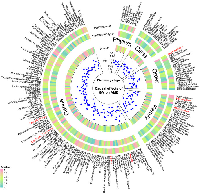
Based on IVW results, six GM taxa were suggestively associated with AMD (Table 1). At the order level, the IVW results confirmed that the order Rhodospirillales was an influential factor in AMD risk (odds ratio [OR] = 1.16; P = 3.38 × 10⁻²) (Fig. 2, Table 1). At the family level, the family Victivallaceae (OR = 1.12; P = 3.14 × 10⁻²) and family Rikenellaceae (OR = 0.82; P = 3.58 × 10⁻²) had a significant impact on AMD risk (Fig. 2, Table 1). At the genus level, it can be genetically expected that Slackia (OR = 0.81; P = 3.15 × 10⁻²), Faecalibacterium (OR = 1.31; P = 3.01 × 10⁻²), Bilophila (OR = 1.27; P = 1.11 × 10⁻²), and Candidatus Soleaferrea (OR = 0.80; P = 2.45 × 10⁻²) could significantly affect AMD risk (Fig. 2, Table 1). At the phylum and class levels, no statistically significant association was found between the GM taxa and the AMD risk (Fig. 2, Supplementary Tables S3 and S4). The MR results of 196 GM taxa based on WM and MR-Egger are presented in Supplementary Tables S3 and S4.
Table 1.
IVW Results for GM Taxa and AMD in the Discovery Stage
| Level | GM Taxa | OR | 95% CI | P |
|---|---|---|---|---|
| Order | Rhodospirillales | 1.16 | 1.01–1.32 | 3.38 × 10⁻² |
| Family | Victivallaceae | 1.12 | 1.01–1.25 | 3.14 × 10⁻² |
| Family | Rikenellaceae | 0.82 | 0.68–0.99 | 3.58 × 10⁻² |
| Genus | Slackia | 0.81 | 0.67–0.98 | 3.15 × 10⁻² |
| Genus | Faecalibacterium | 1.31 | 1.10–1.57 | 3.01 × 10⁻³ |
| Genus | Bilophila | 1.27 | 1.06–1.53 | 1.11 × 10⁻² |
| Genus | Candidatus Soleaferrea | 0.80 | 0.69–0.92 | 2.45 × 10⁻³ |
Figure 2.
Causal analyses and sensitivity analyses of each gut microbiome taxon from five levels and AMD based on IVW results (P < 1 × 10⁻⁵) in the discovery stage (FinnGen consortium). From the outside to the inside, they are, respectively, the GM taxa name, the P value of the pleiotropy test (MR-Egger regression), the P value of the heterogeneity test (Cochran's Q), the P value based on the IVW results, and the OR based on the IVW results. The color corresponding to the P value is based on the RGB color (P = 0, #66CCCC; P = 0.5, #CCFF66; P = 1, #FF99CC).
Sensitivity Analyses in the Discovery Stage
The results of the sensitivity analyses in the discovery stage for 196 GM taxa are displayed in Figure 2 and Supplementary Tables S3 and S4. No heterogeneity (IVW: P ≥ 0.30; MR-Egger: P ≥ 0.23) or horizontal pleiotropy (P ≥ 0.13) were observed in Rhodospirillales, Victivallaceae, Rikenellaceae, Slackia, Faecalibacterium, Bilophila, or Candidatus Soleaferrea (Fig. 2, Table 2). The MR-PRESSO results further confirmed the absence of horizontal pleiotropy in six GM taxa (P ≥ 0.32) (Fig. 2, Table 2). Among those sensitivity analyses, the IVW results were more reliable and further validated the accuracy of the Rhodospirillales, Victivallaceae, Rikenellaceae, Slackia, Faecalibacterium, Bilophila, and Candidatus Soleaferrea causal effects on AMD.
Table 2.
Sensitivity Analyses of MR Results Between GM Taxa and AMD in the Discovery Stage
| Heterogeneity (Cochran's Q), P | Pleiotropy, P | ||||
|---|---|---|---|---|---|
| Level | GM Taxa | IVW | MR-Egger | MR-Egger Regression | MR-PRESSO |
| Order | Rhodospirillales | 0.95 | 0.94 | 0.52 | 0.32 |
| Family | Victivallaceae | 0.30 | 0.23 | 0.93 | 0.38 |
| Family | Rikenellaceae | 0.41 | 0.52 | 0.13 | 0.98 |
| Genus | Slackia | 0.96 | 0.90 | 0.88 | 0.97 |
| Genus | Faecalibacterium | 0.59 | 0.56 | 0.44 | 0.63 |
| Genus | Bilophila | 0.89 | 0.93 | 0.26 | 0.91 |
| Genus | Candidatus Soleaferrea | 0.99 | 0.99 | 0.50 | 0.76 |
Causal Association Based on MR Results in the Replication Stage
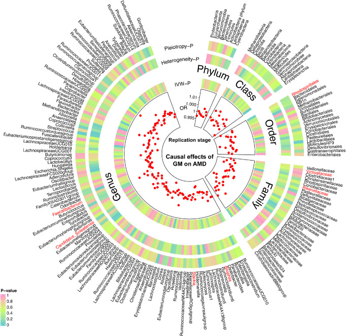
Based on MRC-IEU, we investigated causal relationships between 196 GM taxa and AMD (Fig. 3, Supplementary Tables S5 and S6). Note that only the effect of Rhodospirillales on the risk of AMD was further validated (P = 0.03) (Fig. 3, Supplementary Table S5). However, the effects of Victivallaceae, Rikenellaceae, Slackia, Faecalibacterium, Bilophila, and Candidatus Soleaferrea on the AMD risk were not significant at the replication stage (P > 0.05) (Fig. 3, Supplementary Tables S5 and S6). The results of WM and MR-Egger analysis of the GM taxa and AMD can be found in Supplementary Tables S5 and S6.
Figure 3.
Causal analyses and sensitivity analyses of each gut microbiome taxon from five levels and AMD based on IVW results (P < 1 × 10⁻⁵) in the replication stage (MRC-IEU consortium). From the outside to the inside, they are, respectively, the GM taxa name, the P value of the pleiotropy test (MR-Egger regression), the P value of the heterogeneity test (Cochran's Q), the P value based on the IVW results, and the OR based on the IVW results. The color corresponding to the P value is based on the RGB color (P = 0, #66CCCC; P = 0.5, #CCFF66; P = 1, #FF99CC).
Sensitivity Analyses in the Replication Stage
To ensure the accuracy of the causality at the replication stage, we performed a sensitivity analysis on the results for 196 GM taxa (Fig. 3, Supplementary Tables S5 and S6). If horizontal pleiotropy was detected, the results of these GM taxa were corrected through MR-PRESSO and PhenoScanner. For Rhodospirillales, Victivallaceae, Rikenellaceae, Slackia, Faecalibacterium, Bilophila, and Candidatus Soleaferrea, the final results confirmed that there was no heterogeneity (P > 0.05) or horizontal pleiotropy (P > 0.05) (Table 3). Overall, the combined results of the two stages suggest that Rhodospirillales impacts AMD risk.
Table 3.
Sensitivity Analyses of MR Results Between GM Taxa and AMD in the Replication Stage
| Heterogeneity (Cochran's Q), P | Pleiotropy, P | ||||
|---|---|---|---|---|---|
| Level | GM Taxa | IVW | MR-Egger | MR-Egger Regression | MR-PRESSO |
| Order | Rhodospirillales | 0.66 | 0.71 | 0.27 | 0.69 |
| Family | Victivallaceae | 0.53 | 0.43 | 0.85 | 0.70 |
| Family | Rikenellaceae | 0.60 | 0.66 | 0.23 | 0.72 |
| Genus | Slackia | 0.55 | 0.43 | 0.58 | 0.47 |
| Genus | Faecalibacterium | 0.25 | 0.27 | 0.36 | 0.06 |
| Genus | Bilophila | 0.45 | 0.52 | 0.23 | 0.33 |
| Genus | Candidatus Soleaferrea | 0.16 | 0.57 | 0.09 | 0.20 |
Discussion
Studies continue to reveal the mechanisms of nutrition and other environmental factors in the pathogenesis of AMD.26 As a factor that affects nutritional intake, the association between the GM and AMD is not yet clear. Compared with the current treatment methods, the relative accessibility of the GM taxa opens up new opportunities for interventions to modify the risk of AMD. In clinical research, many confounders interfere with exploring causal relationships between GM taxa and retinal diseases. For this reason, we studied GM taxa and identified associations with ICD codes. We choose to use MR for causal association analysis, minimized the interference of confounding factors through quality control, and obtained more reliable causal associations using IVs from large sample sources. Based on the largest GWAS of human microbiology, which was conducted by Kurilshikovet al.,14 we confirmed that the GM taxa of one order (Rhodospirillales), two families (Victivallaceae and Rikenellaceae), and four genera (Slackia, Faecalibacterium, Bilophila, and Candidatus Soleaferrea) had an impact on AMD risk. During the replication stage, only the effect of Rhodospirillales passed verification. The causal effect between other GM taxa and AMD still must be further investigated. Based on the principle of MR, the direction of these causal relationships is determined—that is, not reversed. This means that our results confirm that GM taxa are involved in the pathogenesis of AMD, which provides theoretical support for the study of the mechanism of the gut–retina axis.
Zhang et al.27 used a laser-induced mouse model that presented some features of AMD (neovascularization and inflammation) and compared the differences between germ-free (GF) mice (the gold standard for microbiome studies) and specific pathogen-free (SPF) mice under a normal diet. They found that, compared with SPF mice, the GF mice model had reduced neovascularization and peripheral microglia infiltration, which confirmed that some feature changes of AMD are regulated by the GM, indicating a connection between the gut–retina axis. Rowan et al.28 compared the GM composition of mice with and without the AMD phenotype through 16S rDNA sequencing and found significant differences in the GM taxa mediated by diet. In addition, they also found that Clostridia and Bacilli were risk factors for AMD. In our MR study, we explored the existence of a causal relationship between Rhodospirillales and AMD. Although different taxa were found, this may be due to the fact that the subjects of the study by Rowan et al.28 were mice (the subjects of the outcomes in our study were humans). It is undeniable that our study supports the idea of an association within the gut–retina axis. In addition to animal studies, the control study of Kiang et al.29 revealed that 89 patients with AMD and 49 healthy subjects had significant differences in their GMs. In that study, the GM taxa related to AMD were found to be enriched in an immunoglobulin A (IgA)-bound fraction and participate in immune regulation.29 Zinkernagel et al.12 found that Anaerotruncus, Ruminococcus torques, and Eubacterium ventriosum were enriched in AMD patients. These three bacterial genera belong to the Firmicutes phylum, Clostridia class, Clostridiales order, and Ruminococcaceae family, which had the same classification as Faecalibacterium and Candidatus Soleaferrea in our results. However, these results have not been verified in the replication phase, and a larger sample is required for further exploration.
Gastaldello et al.7 reported that a Mediterranean diet lowers the odds of developing AMD and also decreases the risk of progression to more advanced stages of the disease. Dietary factors and GMs can also interact. Rowan et al.28 showed that high fat intake can exacerbate AMD in a manner dependent on the GM. The Age-Related Eye Disease Study 2 found that nutritional supplements can delay the progression of AMD, and these supplements may play a role by regulating GM homeostasis.30 The GM is rich in genes involved in amino acid metabolism pathways such as arginine and glutamic acid. Studies in patients with AMD have found a decrease in the genes involved in these amino acid metabolic pathways.12,31 Therefore, GM involvement in the development of AMD is partially influenced by genetic and nutritional factors. This means that the intestinal microbiome may become a potential target for the prevention and treatment of AMD. In addition, clarifying specific taxa of the GM is of great value for the identification of AMD targets.
To the best of our knowledge, our study is the first to identify the role of Rhodospirillales in eye diseases, although the specific pathogenic mechanism for AMD has not been reported. Luo et al.32 tested the GM of hypoxia-induced pulmonary hypertension (PH) mice and found changes in Rhodospirillales, including reversal of the increase in disease-associated Rhodospirillales and a decrease in antiinflammatory and immunomodulatory functional Bacteroidaceae in the mesenchymal stem cell–treated group. The mRNA expression of interleukin-1β (IL-1β) and IL-6 was increased in the PH mice. Rhodospirillales was considered a biomarker of PH mice. The abundance change of Rhodospirillales also revealed the same trend in the study by Ma et al.33 In the colon cancer mice model, the abundance of Bacteroidetes, Rhodospirillales, and Muribaculateae was reduced. After treatment with Lactiplantibacillus plantarum-12, the change in GMs in colon cancer mice was reversed, and the levels of proinflammatory factors (tumor necrosis factor-alpha [TNF-α] and IL-1β) were reduced. Based on the role of Rhodospirillales in pulmonary hypertension and colon cancer, it can be found that red Rhodospirillales is involved in the regulation of inflammatory responses. It is well known that inflammation plays an integral role in the mechanisms of AMD. Some AMD-related studies have found significant changes in the levels of proinflammatory factors (IL-1β and TNF-α) in patients with AMD.34–36 Studies have confirmed that changes in diet affect the composition of the GM and increase local inflammation, thus increasing the risk of AMD.37 These inflammatory mediators have similar effects in the pathogenesis of PH, colon cancer, and AMD. Therefore, we speculate that the effect of Rhodospirillales on AMD might be similar to the mechanisms of pulmonary hypertension and colon cancer, and this connection can be extended to the retina through proinflammatory factors. Furthermore, Rhodospirillales can be considered an AMD biomarker.
The order of Rhodospirillales belongs to the Proteobacteria phylum and the Alphaproteobacteria class. Proteobacteria is a Gram-negative bacterium, and its metabolites, such as lipopolysaccharide and ethanol production, are involved in inflammation, immune escape, and other mechanisms.38 Sookoian et al.39 suggested that Proteobacteria mainly come from the intestine and can affect the risk of vascular disease through inflammatory injury. Sun et al.40 found that a deficiency of angiogenin will increase the abundance of Alphaproteobacteria, leading to the disruption of homeostasis and thus inducing colitis. The impact of Proteobacteria on disease risk depends on the combined effects of all the orders belonging to this phylum in the intestine. Rhodospirillales, as an order of the Proteobacteria phylum, is bound to participate in the occurrence and development of the Proteobacteria phylum in diseases. According to the research by Andriessen et al.,37 when the GM is altered or even dysregulated in homeostasis, it will alter pathogen-associated molecular pattern molecules (PAMPs) and subsequently make the intestinal permeability increase, which elevates ocular and systemic inflammation and enhances pathological neovascularization. Intestinal permeability is influenced by a combination of the GM and the mucosal immune system.41 Increased intestinal permeability enhances PAMP translocation.42 PAMPs affect proinflammatory signaling through pattern recognition receptors and induce inflammation, thus allowing intestinal-derived PAMPs to induce retinal inflammatory disease. Inflammation is considered to play a significant role in AMD pathogenesis.43 Singh et al.44 compared the difference in the frequency of immune cells between patients with AMD and normal subjects and found that the frequency of Th1 cells and CXCR3 CD4 T cells in patients with AMD was significantly reduced. Interestingly, studies have found that the GM can affect the homeostasis of microglia through metabolites45 and activate retinal-specific T cells.46 Horai et al.46 demonstrated that T-cell receptors can receive GM-derived activation signals and regulate autopathogenic T cells that cause diseases in distal tissues (such as the retina). These systemic inflammatory factors enhance the local inflammatory response in the eye, enhancing the secretion of vascular endothelial growth factor (VEGF) and triggering neovascularization.47 Due to the stimulation of local inflammatory factors, the retinal pigment epithelium becomes degenerated and the photoreceptor cells are gradually destroyed, forming irregular pigmentation.48 Therefore, GM taxa are associated with the pathogenesis of AMD through inflammation-related immune mechanisms, a relationship that to some extent reveals the intrinsic correlation of the gut–retina axis. This implies not only that AMD may be a local ophthalmic disease but also that its pathological mechanism may involve systemic factors, whether immune responses or the GM. It is undeniable that our research has confirmed the role of Rhodospirillales in AMD risk. Therefore, we believe that inflammation, as an important link in the pathogenesis of AMD, may connect Rhodospirillales and AMD through the gut–retina axis.
This MR study provides evidence for the direct causal effects of Rhodospirillales on the development of AMD. The limitation of the study is that the sample size in the replication stage was small, and the statistical power of the IVs may affect the validation effect. GWAS research based on large-sample sources would provide a greater theoretical basis for researchers to evaluate relationships between GM taxa and AMD. In future research, further analysis of AMD and GWASs based on optical coherence tomography images is needed to obtain more accurate conclusions from as much cohort information as possible while ensuring patient privacy. In addition, GM taxa are affected by diet, body shape, and other confounders, and it is difficult to completely balance these factors. The European population was selected for the two stages of the study. Considering that there may be racial differences in the GM taxa, we should be alert to this problem when evaluating our conclusions. The study of the gut–retina axis complements studies of retinal nutrition. Many studies have explored the effects of nutrition on the retina,8 but the specific GM taxa impact on the retina is not yet clear. We did not use overly conservative multiple corrections in order not to omit intestinal GM taxa with potential causal relationships.
In conclusion, our findings reinforced the impact of the GM on AMD, particularly the effects of specific taxa. Our study provides new impetus for development of the GM as an intervention to prevent the occurrence and development of AMD. New interventions may be able to reduce the risk of AMD.
Supplementary Material
Acknowledgments
The authors thank the participants and investigators of the MRC-IEU consortium, the FinnGen study, and the MiBioGen consortium for sharing the genetic data. We thank Figdraw (www.figdraw.com) for expert assistance in creating Figure 1 and Supplementary Figure S1. We also thank the Home for Researchers editorial team (www.home-for-researchers.com) for their language editing service.
Supported by grants from the National Natural Science Foundation of China (82260212) and Science, Science and Technology Planning Project of Jiangxi Provincial Health Commission (202210714) and Technology Innovation Base Construction–Clinical Medicine Research Centre Project (20221ZDG02012) to ZY; by the Jiangxi Province Postgraduate Innovation Special Fund Project (YC2022–B051); and by a grant from the Talent Development project of the Affiliated Eye Hospital of Nanchang University (No. 2022X05) to KL. The funders had no role in the study design, data collection, analysis, interpretation, or report writing.
Disclosure: K. Liu, None; J. Zou, None; R. Yuan, None; H. Fan, None; H. Hu, None; Y. Cheng, None; J. Liu, None; H. Zou, None; Z. You, None
References
- 1. Mitchell P, Liew G, Gopinath B, Wong TY.. Age-related macular degeneration. Lancet. 2018; 392: 1147–1159. [DOI] [PubMed] [Google Scholar]
- 2. Wong WL, Su X, Li X, et al.. Global prevalence of age-related macular degeneration and disease burden projection for 2020 and 2040: a systematic review and meta-analysis. Lancet Glob Health 2014; 2: e106–e116. [DOI] [PubMed] [Google Scholar]
- 3. Ebeling MC, Fisher CR, Kapphahn RJ, et al.. Inflammasome activation in retinal pigment epithelium from human donors with age-related macular degeneration. Cells. 2022; 11: 2075. [DOI] [PMC free article] [PubMed] [Google Scholar]
- 4. Curcio CA. Soft drusen in age-related macular degeneration: biology and targeting via the oil spill strategies. Invest Ophthalmol Vis Sci. 2018; 59: Amd160–Amd181. [DOI] [PMC free article] [PubMed] [Google Scholar]
- 5. Curcio CA. Antecedents of soft drusen, the specific deposits of age-related macular degeneration, in the biology of human macula. Invest Ophthalmol Vis Sci. 2018; 59: Amd182–Amd194. [DOI] [PMC free article] [PubMed] [Google Scholar]
- 6. Chen L, Yang P, Curcio CA.. Visualizing lipid behind the retina in aging and age-related macular degeneration, via indocyanine green angiography (ASHS-LIA). Eye (Lond). 2022; 36: 1735–1746. [DOI] [PMC free article] [PubMed] [Google Scholar]
- 7. Gastaldello A, Giampieri F, Quiles JL, et al.. Adherence to the Mediterranean-style eating pattern and macular degeneration: a systematic review of observational studies. Nutrients. 2022; 14: 2028. [DOI] [PMC free article] [PubMed] [Google Scholar]
- 8. Grant MB, Bernstein PS, Boesze-Battaglia K, et al.. Inside out: relations between the microbiome, nutrition, and eye health. Exp Eye Res. 2022; 224: 109216. [DOI] [PubMed] [Google Scholar]
- 9. Singh RK, Chang HW, Yan D, et al.. Influence of diet on the gut microbiome and implications for human health. J Transl Med. 2017; 15: 73. [DOI] [PMC free article] [PubMed] [Google Scholar]
- 10. Xu Q, Ni JJ, Han BX, et al.. Causal relationship between gut microbiota and autoimmune diseases: a two-sample Mendelian randomization study. Front Immunol. 2021; 12: 746998. [DOI] [PMC free article] [PubMed] [Google Scholar]
- 11. Rowan S, Jiang S, Korem T, et al.. Involvement of a gut–retina axis in protection against dietary glycemia-induced age-related macular degeneration. Proc Natl Acad Sci USA. 2017; 114: E4472–E4481. [DOI] [PMC free article] [PubMed] [Google Scholar]
- 12. Zinkernagel MS, Zysset-Burri DC, Keller I, et al.. Association of the intestinal microbiome with the development of neovascular age-related macular degeneration. Sci Rep. 2017; 7: 40826. [DOI] [PMC free article] [PubMed] [Google Scholar]
- 13. Haycock PC, Burgess S, Nounu A, et al.. Association between telomere length and risk of cancer and non-neoplastic diseases: a Mendelian randomization study. JAMA Oncol. 2017; 3: 636–651. [DOI] [PMC free article] [PubMed] [Google Scholar]
- 14. Kurilshikov A, Medina-Gomez C, Bacigalupe R, et al.. Large-scale association analyses identify host factors influencing human gut microbiome composition. Nat Genet. 2021; 53: 156–165. [DOI] [PMC free article] [PubMed] [Google Scholar]
- 15. Kurki MI, Karjalainen J, Palta P, et al.. FinnGen: Unique genetic insights from combining isolated population and national health register data. Available at: https://www.medrxiv.org/content/10.1101/2022.03.03.22271360v1. Accessed May 30, 2023.
- 16. Hemani G, Zheng J, Elsworth B, et al.. The MR-Base platform supports systematic causal inference across the human phenome. eLife. 2018; 7: e34408. [DOI] [PMC free article] [PubMed] [Google Scholar]
- 17. Mitchell RE, Elsworth BL, Mitchell R, et al.. MRC IEU UK Biobank GWAS pipeline version 2. Available at: https://research-information.bris.ac.uk/en/datasets/mrc-ieu-uk-biobank-gwas-pipeline-version-2. Accessed May 30, 2023.
- 18. Sudlow C, Gallacher J, Allen N, et al.. UK Biobank: an open access resource for identifying the causes of a wide range of complex diseases of middle and old age. PLoS Med. 2015; 12: e1001779. [DOI] [PMC free article] [PubMed] [Google Scholar]
- 19. Davey Smith G, Hemani G. Mendelian randomization: genetic anchors for causal inference in epidemiological studies. Hum Mol Genet. 2014; 23: R89–R98. [DOI] [PMC free article] [PubMed] [Google Scholar]
- 20. Sanna S, van Zuydam NR, Mahajan A, et al.. Causal relationships among the gut microbiome, short-chain fatty acids and metabolic diseases. Nat Genet. 2019; 51: 600–605. [DOI] [PMC free article] [PubMed] [Google Scholar]
- 21. Abecasis GR, Altshuler D, Auton A, et al.. A map of human genome variation from population-scale sequencing. Nature. 2010; 467: 1061–1073. [DOI] [PMC free article] [PubMed] [Google Scholar]
- 22. Hemani G, Tilling K, Davey Smith G. Orienting the causal relationship between imprecisely measured traits using GWAS summary data. PLoS Genet. 2017; 13: e1007081. [DOI] [PMC free article] [PubMed] [Google Scholar]
- 23. Verbanck M, Chen CY, Neale B, Do R. Detection of widespread horizontal pleiotropy in causal relationships inferred from Mendelian randomization between complex traits and diseases. Nat Genet. 2018; 50: 693–698. [DOI] [PMC free article] [PubMed] [Google Scholar]
- 24. Bowden J, Davey Smith G, Haycock PC, Burgess S. Consistent estimation in Mendelian randomization with some invalid instruments using a weighted median estimator. Genet Epidemiol. 2016; 40: 304–314. [DOI] [PMC free article] [PubMed] [Google Scholar]
- 25. Bowden J, Del Greco MF, Minelli C, Davey Smith G, Sheehan NA, Thompson JR. Assessing the suitability of summary data for two-sample Mendelian randomization analyses using MR-Egger regression: the role of the I2 statistic. Int J Epidemiol. 2016; 45: 1961–1974. [DOI] [PMC free article] [PubMed] [Google Scholar]
- 26. Rinninella E, Mele MC, Merendino N, et al.. The role of diet, micronutrients and the gut microbiota in age-related macular degeneration: new perspectives from the gut–retina axis. Nutrients. 2018; 10: 1677. [DOI] [PMC free article] [PubMed] [Google Scholar]
- 27. Zhang JY, Xie B, Barba H, et al.. Absence of gut microbiota is associated with RPE/choroid transcriptomic changes related to age-related macular degeneration pathobiology and decreased choroidal neovascularization. Int J Mol Sci. 2022; 23: 9676. [DOI] [PMC free article] [PubMed] [Google Scholar]
- 28. Rowan S, Taylor A.. Gut microbiota modify risk for dietary glycemia-induced age-related macular degeneration. Gut Microbes. 2018; 9: 452–457. [DOI] [PMC free article] [PubMed] [Google Scholar]
- 29. Kiang. L, McClintic. S, Saleh. M, et al.. The gut microbiome in advanced age-related macular degeneration. Invest Ophthalmol Vis Sci. 2017:58: 5739. [Google Scholar]
- 30. Lin P. The role of the intestinal microbiome in ocular inflammatory disease. Curr Opin Ophthalmol. 2018; 29: 261–266. [DOI] [PubMed] [Google Scholar]
- 31. Lin P. Importance of the intestinal microbiota in ocular inflammatory diseases: a review. Clin Exp Ophthalmol. 2019; 47: 418–422. [DOI] [PubMed] [Google Scholar]
- 32. Luo L, Chen Q, Yang L, Zhang Z, Xu J, Gou D.. MSCs therapy reverse the gut microbiota in hypoxia-induced pulmonary hypertension mice. Front Physiol. 2021; 12: 712139. [DOI] [PMC free article] [PubMed] [Google Scholar]
- 33. Ma F, Song Y, Sun M, et al.. Exopolysaccharide produced by Lactiplantibacillus plantarum-12 alleviates intestinal inflammation and colon cancer symptoms by modulating the gut microbiome and metabolites of C57BL/6 mice treated by azoxymethane/dextran sulfate sodium salt. Foods. 2021; 10: 3060. [DOI] [PMC free article] [PubMed] [Google Scholar]
- 34. Ten Berge JC, Fazil Z, van den Born I, et al.. Intraocular cytokine profile and autoimmune reactions in retinitis pigmentosa, age-related macular degeneration, glaucoma and cataract. Acta Ophthalmol. 2019; 97: 185–192. [DOI] [PMC free article] [PubMed] [Google Scholar]
- 35. Hagbi-Levi S, Tiosano L, Rinsky B, et al.. Anti-tumor necrosis factor alpha reduces the proangiogenic effects of activated macrophages derived from patients with age-related macular degeneration. Mol Vis. 2021; 27: 622–631. [PMC free article] [PubMed] [Google Scholar]
- 36. Khan AH, Pierce CO, De Salvo G, et al.. The effect of systemic levels of TNF-alpha and complement pathway activity on outcomes of VEGF inhibition in neovascular AMD. Eye (Lond). 2022; 36: 2192–2199. [DOI] [PMC free article] [PubMed] [Google Scholar]
- 37. Andriessen EM, Wilson AM, Mawambo G, et al.. Gut microbiota influences pathological angiogenesis in obesity-driven choroidal neovascularization. EMBO Mol Med. 2016; 8: 1366–1379. [DOI] [PMC free article] [PubMed] [Google Scholar]
- 38. Michail S, Lin M, Frey MR, et al.. Altered gut microbial energy and metabolism in children with non-alcoholic fatty liver disease. FEMS Microbiol Ecol. 2015; 91: 1–9. [DOI] [PMC free article] [PubMed] [Google Scholar]
- 39. Sookoian S, Salatino A, Castaño GO, et al.. Intrahepatic bacterial metataxonomic signature in non-alcoholic fatty liver disease. Gut. 2020; 69: 1483–1491. [DOI] [PubMed] [Google Scholar]
- 40. Sun D, Bai R, Zhou W, et al.. Angiogenin maintains gut microbe homeostasis by balancing α-Proteobacteria and Lachnospiraceae. Gut. 2021; 70: 666–676. [DOI] [PMC free article] [PubMed] [Google Scholar]
- 41. Bischoff SC, Barbara G, Buurman W, et al.. Intestinal permeability–a new target for disease prevention and therapy. BMC Gastroenterol. 2014; 14: 189. [DOI] [PMC free article] [PubMed] [Google Scholar]
- 42. Mehal WZ. The Gordian Knot of dysbiosis, obesity and NAFLD. Nat Rev Gastroenterol Hepatol. 2013; 10: 637–644. [DOI] [PubMed] [Google Scholar]
- 43. Kauppinen A, Paterno JJ, Blasiak J, Salminen A, Kaarniranta K.. Inflammation and its role in age-related macular degeneration. Cell Mol Life Sci. 2016; 73: 1765–1786. [DOI] [PMC free article] [PubMed] [Google Scholar]
- 44. Singh A, Subhi Y, Krogh Nielsen M, et al.. Systemic frequencies of T helper 1 and T helper 17 cells in patients with age-related macular degeneration: a case-control study. Sci Rep. 2017; 7: 605. [DOI] [PMC free article] [PubMed] [Google Scholar]
- 45. Erny D, Hrabě de Angelis AL, Jaitin D, et al.. Host microbiota constantly control maturation and function of microglia in the CNS. Nat Neurosci. 2015; 18: 965–977. [DOI] [PMC free article] [PubMed] [Google Scholar]
- 46. Horai R, Zárate-Bladés CR, Dillenburg-Pilla P, et al.. Microbiota-dependent activation of an autoreactive T cell receptor provokes autoimmunity in an immunologically privileged site. Immunity. 2015; 43: 343–353. [DOI] [PMC free article] [PubMed] [Google Scholar]
- 47. Tan W, Zou J, Yoshida S, Jiang B, Zhou Y.. The role of inflammation in age-related macular degeneration. Int J Biol Sci. 2020; 16: 2989–3001. [DOI] [PMC free article] [PubMed] [Google Scholar]
- 48. Ao J, Wood JP, Chidlow G, Gillies MC, Casson RJ.. Retinal pigment epithelium in the pathogenesis of age-related macular degeneration and photobiomodulation as a potential therapy? Clin Exp Ophthalmol. 2018; 46: 670–686. [DOI] [PubMed] [Google Scholar]
Associated Data
This section collects any data citations, data availability statements, or supplementary materials included in this article.


