

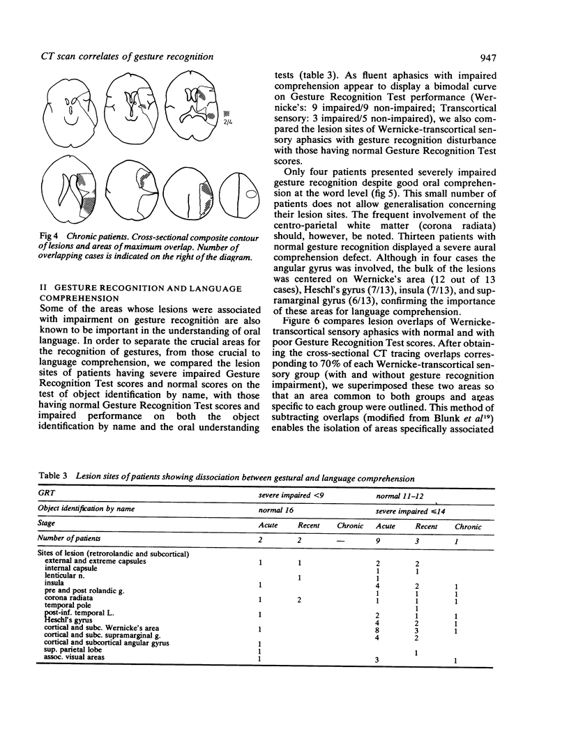
Abstract
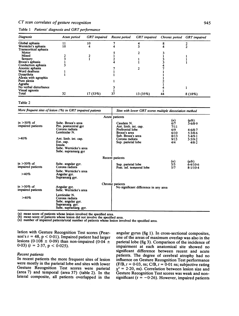
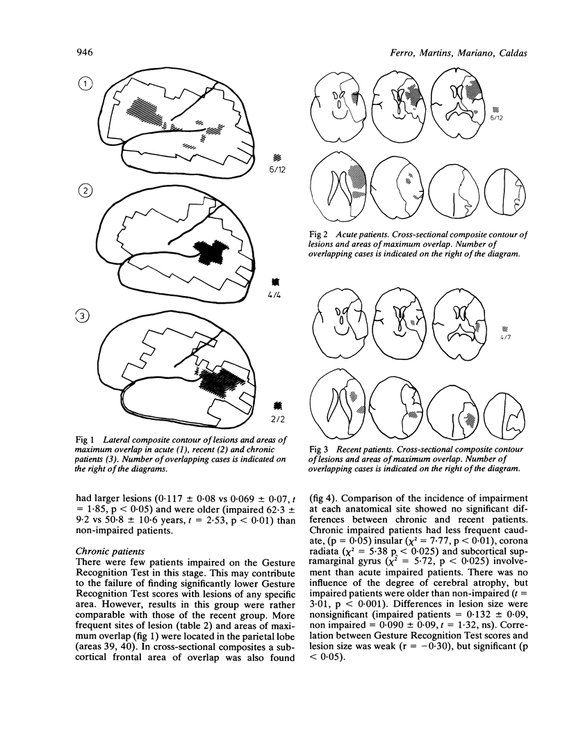
The ability to recognise gestures was studied in 65 left-hemispheric stroke patients whose lesions were located by CT scan. In the acute stage (first month) frontal lobe and basal ganglia were frequently involved in patients showing inability to recognise gestures. In the later (third to fourth month) and chronic stages (greater than 6 months) parietal lobe involvement was important; lesions causing gesture recognition impairment were larger, had more extensive and frequent parietal involvement and produced less temporal lobe damage than those causing aural comprehension defects. These findings are discussed in the light of recent models of cerebral localisation of complex functions.
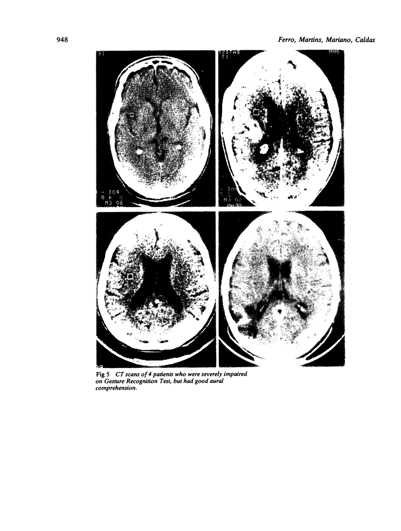
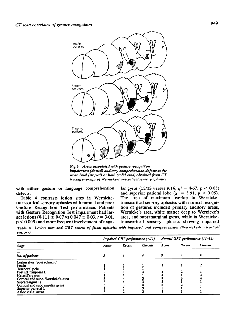
Full text
PDF
Images in this article
Selected References
These references are in PubMed. This may not be the complete list of references from this article.
- Assal G., Regli F. Syndrome de disconnexion visuo-verbale et visuo-gestuelle. Aphasie optique et apraxie optique. Rev Neurol (Paris) 1980;136(5):365–376. [PubMed] [Google Scholar]
- Blunk R., De Bleser R., Willmes K., Zeumer H. A refined method to relate morphological and functional aspects of aphasia. Eur Neurol. 1981;20(2):69–79. doi: 10.1159/000115210. [DOI] [PubMed] [Google Scholar]
- Cappa S., Cavallotti G., Vignolo L. A. Phonemic and lexical errors in fluent aphasia: correlation with lesion site. Neuropsychologia. 1981;19(2):171–177. doi: 10.1016/0028-3932(81)90102-0. [DOI] [PubMed] [Google Scholar]
- Duffy R. J., Duffy J. R. Pantomime recognition in aphasics. J Speech Hear Res. 1975 Mar;18(1):115–132. doi: 10.1044/jshr.1801.115. [DOI] [PubMed] [Google Scholar]
- Duffy R. J., Duffy J. R. Three studies of deficits in pantomimic expression and pantomimic recognition in aphasia. J Speech Hear Res. 1981 Mar;24(1):70–84. doi: 10.1044/jshr.2401.70. [DOI] [PubMed] [Google Scholar]
- Feyereisen P., Seron X., de Macar M. L'interprétation de différentes catégories de gestes chez des sujets aphasiques. Neuropsychologia. 1981;19(4):515–521. doi: 10.1016/0028-3932(81)90018-x. [DOI] [PubMed] [Google Scholar]
- Gado M., Hanaway J., Frank R. Functional anatomy of the cerebral cortex by computed tomography. J Comput Assist Tomogr. 1979 Feb;3(1):1–19. doi: 10.1097/00004728-197902000-00001. [DOI] [PubMed] [Google Scholar]
- Gainotti G., Lemmo M. S. Comprehension of symbolic gestures in aphasia. Brain Lang. 1976 Jul;3(3):451–460. doi: 10.1016/0093-934x(76)90039-0. [DOI] [PubMed] [Google Scholar]
- Hayward R. W., Naeser M. A., Zatz L. M. Cranial computed tomography in aphasia. Correlation of anatomical lesions with functional deficits. Radiology. 1977 Jun;123(3):653–660. doi: 10.1148/123.3.653. [DOI] [PubMed] [Google Scholar]
- Heilman K. M., Rothi L. J., Valenstein E. Two forms of ideomotor apraxia. Neurology. 1982 Apr;32(4):342–346. doi: 10.1212/wnl.32.4.342. [DOI] [PubMed] [Google Scholar]
- Huckman M. S., Fox J., Topel J. The validity of criteria for the evaluation of cerebral atrophy by computed tomography. Radiology. 1975 Jul;116(1):85–92. doi: 10.1148/116.1.85. [DOI] [PubMed] [Google Scholar]
- Kertesz A., Harlock W., Coates R. Computer tomographic localization, lesion size, and prognosis in aphasia and nonverbal impairment. Brain Lang. 1979 Jul;8(1):34–50. doi: 10.1016/0093-934x(79)90038-5. [DOI] [PubMed] [Google Scholar]
- Kievit J., Kuypers H. G. Organization of the thalamo-cortical connexions to the frontal lobe in the rhesus monkey. Exp Brain Res. 1977 Sep 28;29(3-4):299–322. doi: 10.1007/BF00236173. [DOI] [PubMed] [Google Scholar]
- Loizou L. A., Kendall B. E., Marshall J. Subcortical arteriosclerotic encephalopathy: a clinical and radiological investigation. J Neurol Neurosurg Psychiatry. 1981 Apr;44(4):294–304. doi: 10.1136/jnnp.44.4.294. [DOI] [PMC free article] [PubMed] [Google Scholar]
- Mazzocchi F., Vignolo L. A. Computer assisted tomography in neuropsychological research: a simple procedure for lesion mapping. Cortex. 1978 Mar;14(1):136–144. doi: 10.1016/s0010-9452(78)80018-5. [DOI] [PubMed] [Google Scholar]
- Mazzocchi F., Vignolo L. A. Localisation of lesions in aphasia: clinical-CT scan correlations in stroke patients. Cortex. 1979 Dec;15(4):627–653. doi: 10.1016/s0010-9452(79)80051-9. [DOI] [PubMed] [Google Scholar]
- Mesulam M. M. A cortical network for directed attention and unilateral neglect. Ann Neurol. 1981 Oct;10(4):309–325. doi: 10.1002/ana.410100402. [DOI] [PubMed] [Google Scholar]
- Mountcastle V. B., Lynch J. C., Georgopoulos A., Sakata H., Acuna C. Posterior parietal association cortex of the monkey: command functions for operations within extrapersonal space. J Neurophysiol. 1975 Jul;38(4):871–908. doi: 10.1152/jn.1975.38.4.871. [DOI] [PubMed] [Google Scholar]
- Seron X., van der Kaa M. A., Remitz A., van der Linden M. Pantomime interpretation and aphasia. Neuropsychologia. 1979;17(6):661–668. doi: 10.1016/0028-3932(79)90041-1. [DOI] [PubMed] [Google Scholar]
- Varney N. R. Linguistic correlates of pantomime recognition in aphasic patients. J Neurol Neurosurg Psychiatry. 1978 Jun;41(6):564–568. doi: 10.1136/jnnp.41.6.564. [DOI] [PMC free article] [PubMed] [Google Scholar]