Abstract
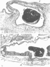
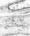
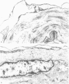
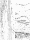
Electron microscopic data on human bridging veins show thin walls of variable thickness, circumferential arrangement of collagen fibres and a lack of outer reinforcement by arachnoid trabecules, all contributory to the subdural portion of the vein being more fragile than its subarachnoid portion. These features explain the laceration of veins and the subdural location of resultant haematomas.
Full text
PDF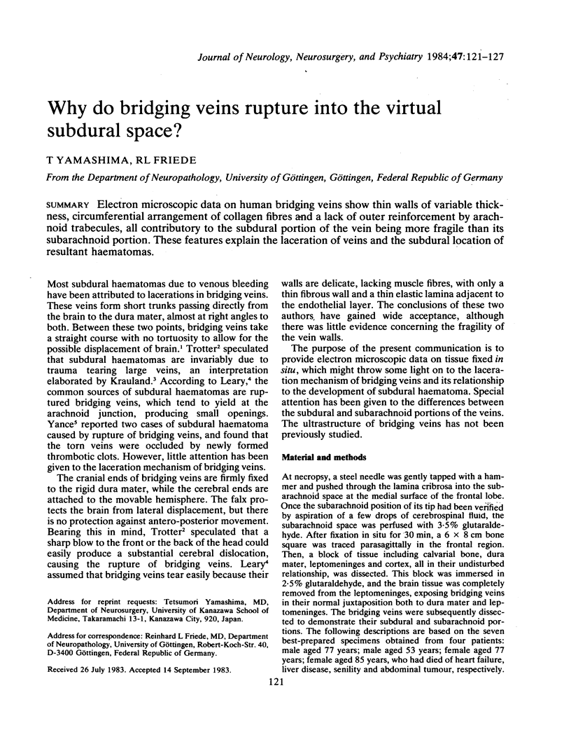
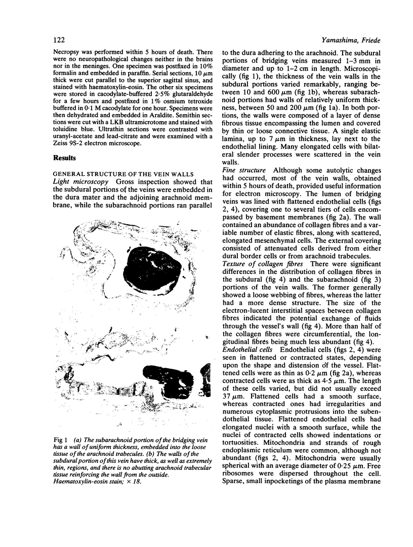
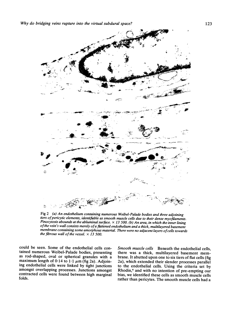
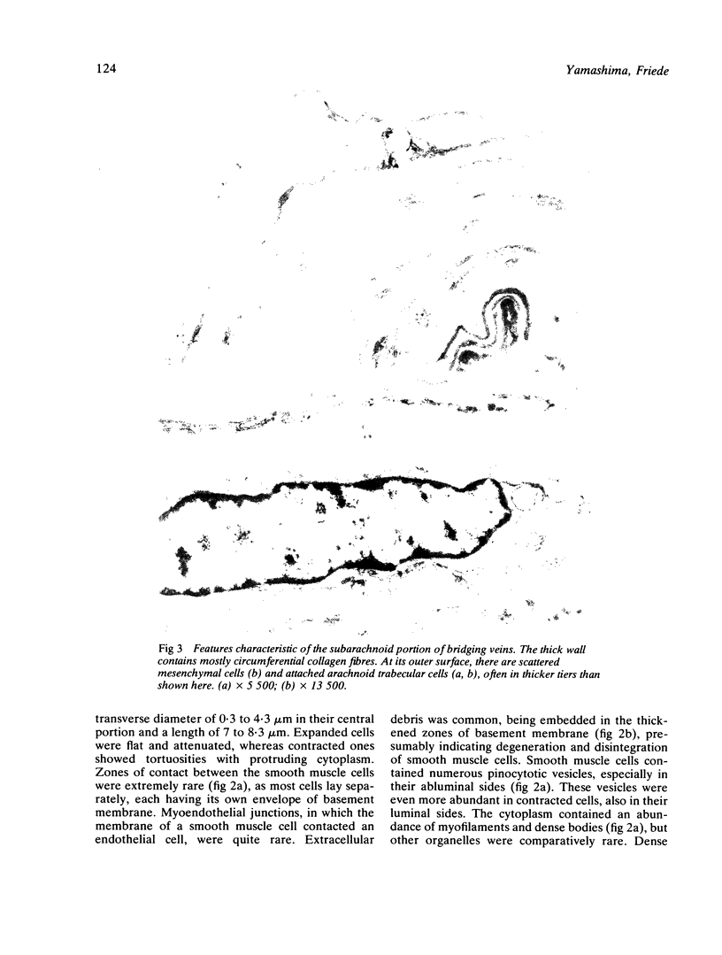

Images in this article
Selected References
These references are in PubMed. This may not be the complete list of references from this article.
- Friede R. L. Incidence and distribution of neomembranes of dura mater. J Neurol Neurosurg Psychiatry. 1971 Aug;34(4):439–446. doi: 10.1136/jnnp.34.4.439. [DOI] [PMC free article] [PubMed] [Google Scholar]
- Rhodin J. A. Ultrastructure of mammalian venous capillaries, venules, and small collecting veins. J Ultrastruct Res. 1968 Dec;25(5):452–500. doi: 10.1016/s0022-5320(68)80098-x. [DOI] [PubMed] [Google Scholar]
- Schachenmayr W., Friede R. L. The origin of subdural neomembranes. I. Fine structure of the dura-arachnoid interface in man. Am J Pathol. 1978 Jul;92(1):53–68. [PMC free article] [PubMed] [Google Scholar]
- Shenkin H. A. Acute subdural hematoma. Review of 39 consecutive cases with high incidence of cortical artery rupture. J Neurosurg. 1982 Aug;57(2):254–257. doi: 10.3171/jns.1982.57.2.0254. [DOI] [PubMed] [Google Scholar]
- VANCE B. M. Ruptures of surface blood vessels on cerebral hemispheres as a cause of subdural hemorrhage. AMA Arch Surg. 1950 Dec;61(6):992–1006. doi: 10.1001/archsurg.1950.01250021002002. [DOI] [PubMed] [Google Scholar]


