Abstract
OBJECTIVES:
The study aims to measure the mandibular condylar height (CH), ramus height (RH), total height (CH+RH), asymmetry index, and condylar volume (Cvol) in individuals with different anteroposterior and vertical skeletal discrepancies.
MATERIALS AND METHODS:
The study sample consisted of 131 subjects (60 females and 71 males) with a mean age of 35.06 ± 12.79 years. Pre-existing CBCT images were divided into groups according to the anteroposterior and vertical skeletal discrepancies. The investigator analyzed the data using t-tests to assess the mandibular bilateral sides of the individuals and gender differences. The mean difference between groups was determined using a one-way analysis of variance (ANOVA). The Chi-square test was used to study the association between the asymmetry index and groups.
RESULTS:
Each individual's bilateral sides exhibited statistically significant differences in CH, RH, and Cvol (P = 0.033, P = 0.039, P = 0.005, respectively), but not in CH+RH (P = 0.458). There were, however, statistically significant gender differences in CH+RH (P < 0.001). Skeletal Class III and hypodivergent groups revealed the highest linear and volumetric values compared to other groups. The asymmetry index was increased in CH (P = 0.006) and Cvol (P = 0.002) in skeletal Class II subjects.
CONCLUSIONS:
Significant differences in CH, RH, and Cvol were found on the right and left sides of the same individual. This study found increased linear and volumetric values in males, skeletal Class III, and hypodivergent subjects. Class II individuals had an increased CH and Cvol asymmetry index. This study highlights in-depth knowledge of mandibular asymmetry, which is extremely important to achieve an accurate diagnosis and provide the best treatment outcome.
Keywords: Asymmetry index, condylar height, condylar volume, ramus height
Introduction
Achieving a balanced and harmonious facial appearance is one of the main objectives of orthodontic treatment.[1] Craniofacial symmetry is one aspect of harmony that determines attractiveness regardless of varying cultural norms,[1,2] although minor variations in facial symmetry may still be perceived as esthetically pleasing with no esthetic or functional significance.[1,3] Exactly how to distinguish between pathological and normal asymmetry remains a matter of debate.[4] A lack of human facial symmetry should be addressed when it affects the function and esthetics of an individual.[1]
Various etiological factors can cause facial asymmetries, such as age, gender, growth pattern, dental occlusal changes, muscular activities, and pathological factors.[3,5,6,7,8,9] Furthermore, genetic factors can influence the development of facial asymmetries, such as PITX2 and ACTIN3 gene mutations.[10]
Facial asymmetry primarily affects the lower third of the face.[11] A systematic review presented a high prevalence of mandibular asymmetry in the overall sample, ranging from 17.43 to 72.95%.[12] In addition, Gribel et al.[13] reported a prevalence of 44% of mild-to-severe mandibular transverse discrepancy using cone-beam computed tomography (CBCT) in 250 subjects. This is possibly explained by the longer mandibular growth duration compared to other craniofacial bone structures.[14] Variations in the mandibular ramus height (RH) or condylar process can influence the development of unilateral mandibular hypoplasia; this kind of mandibular asymmetry can present dental surgeons and orthodontists with significant challenges.[10]
The anteroposterior relationship between the upper and lower jawbones can also affect craniofacial asymmetry.[13] A systematic review reported a greater prevalence of mandibular asymmetry in skeletal Class III cases.[12] In addition, vertical growth patterns can influence mandibular asymmetry, and a study has reported mandibular vertical asymmetry in patients with high-angle growth patterns.[15]
Mandibular asymmetry can present maxillofacial surgeons and orthodontists with significant challenges,[10] in terms of diagnosis and treatment approach, yet the assessment of mandibular asymmetry remains inconsistent. A recent systematic review stated that previous studies examined mandibular asymmetries without considering vertical growth patterns.[12] In addition, the asymmetry index was considered in only one study, which analyzed the prevalence of mandibular asymmetry in sagittal skeletal malocclusions.[12] To address these weaknesses, the present study sought to analyze the asymmetry index and mandibular vertical asymmetry in different sagittal and vertical skeletal patterns. Although it is recognized that investigators have reported information about asymmetry in different skeletal patterns in other ethnicities,[16,17] this is the first orthodontic paper to investigate mandibular asymmetry in the Saudi Arabian population. In addition, mandibular asymmetry analysis has not previously been investigated using modern 3D segmentation analysis.[16] Therefore, the study aims to assess mandibular condylar height (CH), RH, total height (CH+RH), asymmetry index, and condylar volume (Cvol) in individuals with different anteroposterior and vertical skeletal relationships.
There are several diagnostic tools for measuring mandibular asymmetry, such as panoramic, posterior-anterior, and submentovertex radiographs.[18] Currently, CBCT imaging has allowed us to overcome the limitations of conventional radiographs by providing high-resolution imaging of craniofacial structures without magnification or distortion.[4,18] Hence, this study aims to utilize CBCT imaging to achieve the aims discussed above. The present study highlights the prevalence of skeletal deformities with asymmetry which is extremely important to orthodontists to achieve an accurate diagnosis of skeletal asymmetry that affect the treatment time and outcomes. In addition, early identification of individuals with potential skeletal asymmetry will allow the implementation of orthopedic/orthodontic approaches to solve facial disharmony.[18]
Materials and Methods
This study included CBCT records from a retrospective screening of CBCT images at the dental department of the Ministry of National Guard Health Affairs and the College of Dentistry at King Saud bin Abdulaziz University for Health Sciences, acquired between January 2017 and April 2022. The ethical committee of the King Abdullah International Medical Research Center (KAIMRC) approved this study (NRC21R/207/06) on June 27 2021. All CBCTs were taken for dental and surgical treatment purposes, and subjects were not exposed to additional radiation for research reasons. The exclusion criteria included subjects who exhibited one or more of the following: previous orthodontic treatment, previous orthognathic surgery, temporomandibular disorder, condylar degenerative diseases, previous trauma, cleft lip and palate, craniofacial anomalies, CBCT images of poor quality, and non-Saudi ethnicity. In addition, the investigator excluded subjects under the age of 18 years old. The study included CBCT images obtained from a Planmeca Promax 3D Proface (Planmeca OY, Helsinki, Finland, 2015) with a voxel size of 0.4 mm and a field of view (20 × 19 cm). The subjects who met the inclusion criteria and were included in this study were 131 Saudi Arabian subjects (60 females and 71 males) with a mean age of 35.06 ± 12.79.
The investigator used CBCT Romexis software (version 6.1, Planmeca OY, Helsinki, Finland) in this study. The CBCT records were classified based on the anteroposterior skeletal relationship of Steiner's A point, nasion, and B point (ANB) angle into three groups[19]:
Skeletal Class I group was defined as having values of the ANB angle of 1° to 4°, indicating no anteroposterior skeletal discrepancy.
Skeletal Class II group was defined when the ANB angle was greater than 4°, indicating either a protrusive maxilla or retrognathic mandible.
Skeletal Class III group was defined when the ANB angle was less than or equal to 0°, indicating that the maxilla was positioned posteriorly relative to the mandible.
In addition, CBCT scans were classified based on the vertical pattern according to Tweed's Frankfort mandibular plane (FMA) angle into three groups[20]:
Hyperdivergent group was defined when the FMA angle was 30° or more, indicating an excessive vertical growth pattern.
Hypodivergent group was defined when the FMA angle was 20° or less, indicating a deficient vertical growth pattern.
Normodivergent group (normal) was defined as values of the FMA angle were 20° to 30°, indicating a skeletal pattern with normal growth direction.
Linear measurements
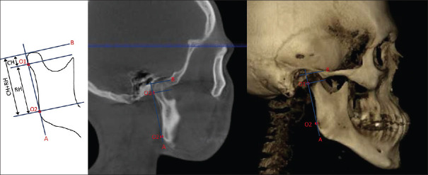
The investigator performed CH, RH, and CH+RH measurements as explained in the previous literature [Figure 1].[21] The most lateral point of the condyle was marked O1, while O2 marked the most posterior end of the mandibular ramus. The tangent line (A-line) was drawn to connect the two points (O1, O2). Another line (B-line) was drawn from the most superior point of the condyle (B-line) and perpendicular to the A-line. The CH was measured from the B-line to O1. The A-line connecting O1 and O2, meanwhile, represents the RH. CH+RH is the distance calculated from the B-line to the line that passes through O2 and is perpendicular to the A-line. All linear values were measured in millimeters.
Figure 1.
Definitions of linear landmarks (CH, RH, and CH+RH) on CBCT, sagittal view, and schematic drawing. Landmarks: O1: the most lateral point of the condyle, O2: the most posterior end of the mandibular ramus. (A) line: a tangent line connecting O1 and O2. (B) line: a line drawn from the most superior point of the condyle and perpendicular to the A-line. Abbreviations: CH: mandibular condylar height; RH: ramus height, total height (CH+RH)
The asymmetry index was calculated using the formula below[21]:

Habets et al.[21] suggested that an asymmetry index of less than 6% might be considered a technical error. If it is greater than 6%, it can be considered a true mandibular asymmetry.
Volumetric measurements
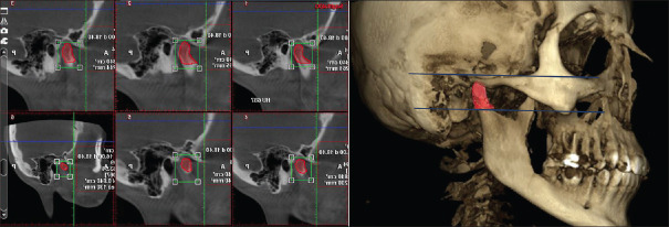
The investigator (N. A.) utilized the manual segmentation tool to measure Cvol on the sagittal view of the CBCT image using Romexis software (version 6.1, Planmeca OY, Helsinki, Finland). First, the sagittal slice of the CBCT image was adjusted to include the region of interest (ROI), which is the mandibular condyle measured from the deepest point of the sigmoid notch parallel to the Frankfort plane. Then, the number of slices in the sagittal view was adjusted using the viewport settings on the top-right corner of the cross-section slice to include nine slices of 1.2 mm thickness with a spacing of 5.0 mm between slices. After that, the manual segmentation tool was selected to define and mark the boundaries of the ROI on each slice. The inferior limit was the line parallel to the Frankfort plane that passes through the deepest point of the sigmoid notch. The superior limit was marked manually to include the boundaries of the mandibular condyle. The anterior and posterior limits were marked manually to involve the outline of the mandibular condyle. Finally, the volume of ROI was composed automatically in the Romex software after combining all the defined outlines from each slice.[22] The unit used to measure Cvol was cubic centimeters [Figure 2].
Figure 2.
Demonstration of condylar volume manual segmentation
Statistical analysis
The present study utilized the Number Cruncher Statistical (NCSS) Software (version 2021) (NCSS LLC company, Kaysville, Utah, USA) and the Power Analysis and Sample Size statistical software (version 15) (NCSS LLC company, Kaysville, Utah, USA) for sample size calculations, based on the effect size reported by Mendoza et al.[16] Based on that calculation, a minimum sample size of 19 subjects from each skeletal sagittal group was necessary to obtain a type I error of 5% and 90% power. In addition, a minimum sample size of 26 subjects per skeletal vertical group was adequate to get a type I error of 5% and 90% power. Data analysis was done using SAS (SAS Institute Inc., Cary, North Carolina, USA) statistical software. The paired t-test was used to assess the differences between the bilateral sides of the subjects. Independent t-tests were utilized to assess gender discrepancies regarding linear and volumetric measurements. ANOVA was performed to determine the CH, RH, CH+RH, and Cvol mean values between the independent skeletal groups. Following that, a post-hoc analysis was utilized for individual differentiation. The differences in the asymmetry index were assessed using ANOVA. The prevalence of an asymmetry index of more than 6% was estimated by performing the Chi-square analysis.
A logistic regression test was conducted to analyze the relationship between the asymmetry index and different cut-off points (1%, 3%, 6%, and 10%) according to gender, anteroposterior, and vertical skeletal patterns. The significance level in the study is set at 5% (P-value ≤ 0.05).
CBCT radiographs of 20 subjects were randomly selected, retraced, and re-digitized eight weeks after the initial analysis by the same examiner (N.A.). The Intraclass Correlation Coefficient (ICC) was calculated to assess intra-examiner reliability.
Results
Histograms revealed that the data were distributed homogeneously, and therefore, parametric tests were used in this study. Paired t-tests demonstrated a statistically significant difference in the means of CH, RH, and Cvol measurements between the mandibular bilateral sides of the same individual. The means of the CH+RH measurements on the bilateral sides showed no significant differences, however [Table 1 and Supplemental Figure 1 (688.4KB, tif) ].
Table 1.
Linear and volumetric measurements of the right and left sides of the same individual
| 95% CI | n | Right side | Left side | Mean difference | Paired T-test (P) | |
|---|---|---|---|---|---|---|
| Linear measurements | CH (mm) | 131.00 | 7.21 | 7.59 | -0.3831 | P=0.033* |
| RH (mm) | 131.00 | 40.54 | 39.91 | 0.6335 | P=0.039* | |
| CH+RH (mm) | 131.00 | 47.76 | 47.50 | 0.2538 | P=0.458 | |
| Volumetric measurements | Cvol (cm3) | 131.00 | 1.11 | 1.00 | 0.0709 | P=0.005** |
CI=confidence interval. *P<0.05. **P<0.01
Table 2 shows the linear and volumetric values concerning gender, sagittal, and vertical skeletal patterns. This reveals a statistically significant difference in CH+RH between genders, with male subjects having higher CH+RH values than female subjects (P < 0.001). It is also evident that the skeletal Class III group had significantly increased CH, RH, and CH+RH values compared to other groups. The Cvol means were not statistically significantly different between the skeletal Classes, however [Supplemental Figure 2 (939.1KB, tif) ]. RH and CH+RH means were significantly higher in the hypodivergent group, followed by the normal and hyperdivergent groups. Conversely, CH and Cvol means were significantly lower in the hyperdivergent group compared to the normal group (P = 0.009 and 0.029, respectively) [Supplemental Figure 3 (1.1MB, tif) ].
Table 2.
Linear and volumetric measurements according to gender, sagittal, and vertical skeletal patterns
| Total n=131 | Variables/statistical tests | n | Linear measurements (95% CI mean) | Volumetric measurements (95% CI mean) Cvol (cm3) | ||
|---|---|---|---|---|---|---|
|
| ||||||
| CH (mm) | RH (mm) | CH + RH (mm) | ||||
| Gender | Female | 60.00 | 7.15 | 38.17 | 44.76 | 0.97 |
| Male | 71.00 | 7.58 | 42.69 | 50.28 | 1.16 | |
| Student t-test P | 0.232 | 0.132 | <0.001** | 0.811 | ||
| Skeletal sagittal pattern | Class I | 52.00 | 7.45 | 41.08 | 48.53 | 1.08 |
| Class II | 57.00 | 6.89 | 38.77 | 44.97 | 1.00 | |
| Class III | 22.00 | 8.30 | 42.05 | 50.36 | 1.15 | |
| ANOVA P | P=0.006* | P=0.005* | P=0.001** | P=0.149 | ||
| Post-hoc | P=0.005 Class II vs. III | P=0.026* Class I vs. II | P=0.012* Class I vs. II | |||
| P=0.014* Class II vs. III | P=0.003* Class II vs. III | |||||
| Skeletal vertical pattern | Normal | 64.00 | 7.72 | 39.46 | 47.19 | 1.10 |
| Hyper | 35.00 | 6.60 | 38.79 | 45.39 | 0.91 | |
| Hypo | 32.00 | 7.42 | 43.22 | 49.42 | 1.12 | |
| ANOVA P | P=0.012* | P<0.0001 | P=0.046* | P=0.012* | ||
| Post-hoc | P=0.009* normal vs. hyper | P=0.0004 ** normal vs. hypo | P=0.036* normal vs. hypo | P=0.029* normal vs. hyper | ||
| P=0.0002** hyper vs. hypo | ||||||
CI=confidence interval. *P<0.05. **P<0.01
Table 3 shows the asymmetry index for linear and volumetric values regarding gender, anteroposterior, and vertical skeletal patterns. This study found a statistically significant difference in CH between genders (P = 0.016). Skeletal Class II had an increased CH and Cvol asymmetry index compared to Class I and Class III. The overall asymmetry index ranged from 12.1% to 64%. The highest prevalence of asymmetry was in respect to Cvol (64.1%), followed by CH (62.6%), RH (20.5%), and CH+RH (12.1%). The only statistically significant association, however, was found between CH asymmetry and anteroposterior skeletal pattern (P = 0.005) [Supplemental Table 1]. Regarding CH asymmetry, 69.2% of individuals were Class I, 68.4% were Class II, and 31.8% were Class I subjects.
Table 3.
Asymmetric index for the linear and volumetric values according to gender, sagittal, and vertical skeletal patterns
| Total n=131 | Variables/statistical tests | n | Linear measurements (95% CI mean) | Volumetric measurements (95% CI mean) Cvol (cm3) | ||
|---|---|---|---|---|---|---|
|
| ||||||
| CH (mm) | RH (mm) | CH + RH (mm) | ||||
| Gender | Female | 60.00 | 12.52 | 3.52 | 3.04 | 12.67 |
| Male | 71.00 | 11.27 | 3.53 | 3.61 | 11.03 | |
| t-test P | P=0.016* | P=0.22 | P=0.424 | P=0.142 | ||
| Skeletal sagittal pattern | Class I | 52.00 | 13.13 | 3.34 | 3.33 | 8.51 |
| Class II | 57.00 | 13.27 | 3.60 | 3.28 | 15.82 | |
| Class III | 22.00 | 5.70 | 3.73 | 3.30 | 9.87 | |
| ANOVA P | P=0.006* | P=0.835 | P=0.995 | P=0.002* | ||
| Post-hoc |
P=0.010* Class I vs. Class III |
P=0.002* Class I vs. II |
||||
|
P=0.007* Class II vs. Class III |
||||||
| Skeletal vertical pattern | Normal | 64.00 | 10.35 | 3.46 | 3.13 | 11.83 |
| Hyper | 35.00 | 13.57 | 4.26 | 4.04 | 14.12 | |
| Hypo | 32.00 | 13.37 | 2.87 | 2.84 | 9.69 | |
| ANOVA P | P=0.217 | P=0.133 | P=0.131 | P=0.287 | ||
| Post-hoc | ||||||
CI=confidence interval. *P<0.05
Supplemental Table 1.
Percentage of individuals with an asymmetric index of more than 6% according to gender, sagittal, and vertical patterns
| n | Linear measurements (95% CI) | Volumetric measurements (95% CI) Cvol (%) | ||||
|---|---|---|---|---|---|---|
|
| ||||||
| CH (%) | RH (%) | CH + RH (%) | ||||
| Gender | Female | 60.00 | 60.00 | 21.70 | 15.00 | 66.70 |
| Male | 71.00 | 64.80 | 19.40 | 9.70 | 62.00 | |
| Chi-square P | 0.573 | 0.753 | 0.355 | 0.577 | ||
| Skeletal sagittal pattern | Class I | 52.00 | 69.20 | 21.20 | 11.50 | 55.80 |
| Class II | 57.00 | 68.40 | 20.70 | 12.10 | 73.70 | |
| Class III | 22.00 | 31.80 | 18.20 | 13.60 | 59.10 | |
| Chi-square P | 0.005* | 0.957 | 0.968 | 0.13 | ||
| Skeletal vertical | Normal | 64.00 | 59.40 | 20.30 | 7.80 | 68.80 |
| pattern | Hyper | 35.00 | 62.90 | 22.90 | 22.90 | 65.70 |
| Hypo | 32.00 | 68.80 | 18.20 | 9.10 | 53.10 | |
| Chi-square P | 0.67 | 0.891 | 0.075 | 0.314 | ||
| Total | 131.00 | 62.60 | 20.50 | 12.10 | 64.10 | |
CI=confidence interval. *P<0.05
Logistic regression results for multivariate analysis are presented in Table 4. The investigator analyzed the prevalence of subjects with asymmetry at different cut-off points. An association was found between RH asymmetry (cut-off 3%) and a Class III pattern (odds ratio = 29.00 and 95% CI = 2.736, 307.448). In addition, Cvol asymmetry (cut-off 3%) was associated with hypodivergent patterns (odds ratio = 4.79 and 95% CI = 1.106, 20.784; Table 4). Based on ICC guidelines, the present study showed good to excellent reliability.[23] The values were between 0.75 and above 0.9.
Table 4.
Percentage of individuals with asymmetric index (cut-off points >1%, >3%, >6%, >10%)
| Linear measurements | Volumetric measurements Cvol (%) | |||
|---|---|---|---|---|
|
| ||||
| Asymmetry % | CH (%) | RH (%) | CH + RH (%) | |
| Cut-off 1% | 89.39 | 78.03 | 81.81 | 91.67 |
| Cut-off 3% | 80.30 | 49.24*a | 43.94 | 78.78*b |
| Cut-off 6% | 62.12 | 20.45 | 12.12 | 63.64 |
| Cut-off 10% | 46.21 | 1.52 | 3.79 | 42.42 |
Percentage of individuals with asymmetry (asymmetry index with cut-off points >1%, >3%, >6%, >10%). *a Associated with logistic regression by forward Selection to Class III. (OR=29.007; P=0.005). *b Associated with logistic regression by forward Selection to hypodivergent. Pattern (OR=4.79; P=0.036)
Discussion
Understanding mandibular asymmetry is essential if orthodontists are to offer proper diagnosis and treatment planning. Mandibular asymmetry compromises facial esthetics and can affect craniofacial function.[24] For example, a cohort study reported a high prevalence of temporomandibular disorder in subjects with mandibular asymmetry.[25] The present study's findings clearly show the assessment of the mandibular CH, RH, CH+RH, asymmetry index, and Cvol in adult subjects who presented with different anteroposterior and vertical skeletal relationships.
The study included subjects 18 years of age and older to eliminate the growth factor that could influence the skeletal dimensions.[26] Any subjects with previous orthodontic or surgical procedures were excluded since those procedures might change the maxillary and mandibular positions. Moreover, this study focused on subjects of the same race and ethnicity with the same genetic backgrounds.
To allow for reliable comparisons between studies, the subjects in this paper were classified according to their sagittal skeletal and vertical patterns.[15,16,27] Like previous studies, Steiner's ANB angle was used to determine the anteroposterior skeletal classificats[7,28] In addition, the vertical skeletal pattern was classified based on Tweed's FMA angle,[20] which is consistent with Nakawaki et al.'s study.[27] Moreover, the Cvol segmentation technique has proven to be a reliable and accurate method,[29] as is the assessment of vertical mandibular asymmetry according to Habets et al.'s[21] method.[30] Although Habets et al.[21] measured mandibular posterior asymmetry on a panoramic radiograph, a study found that CBCT images were slightly more reliable for measuring the asymmetry index than panoramic radiographs due to the higher image quality produced by the CBCT.[31] Accordingly, CBCT is considered the gold standard in detecting mandibular asymmetry.[31] In addition, Habets et al.[21] suggested a 6% cutoff to detect mandibular asymmetry, which was supported by Sadat-Khonsari et al.'s study.[32] Hence, the present study utilized the asymmetry index according to Habets et al.'s method on CBCT images using a 6% cutoff point to detect asymmetry.
Interestingly, although statistically significant differences were observed in CH and RH between the right and left sides of the same individual, there was no parallel significant difference in the CH+RH measurements, suggesting that the combination of CH+RH might act as a compensatory factor to reduce the level of CH and RH asymmetry in individuals. In agreement with the results of this study, Celik et al.[15] reported a slight difference in CH between both sides. Other studies, however, have reported no significant difference between the right and left sides of the same individual.[16,33] In regards to Cvol, the current study reported significant differences in Cvol between the right and left sides, a finding that concurs with the results of a study by Shetty et al.[34] On the other hand, other studies have reported different findings.[15,16] The lack of consensus in these results could be explained by the different ethnicities of the various studied populations.
While the linear measurements increased in males compared to females, this paper showed no significant differences between genders except for the considerable increase in CH+RH values in males compared to females. In contrast, Mendoza et al.[16] reported significant differences in all linear measurements. The strong sexual dimorphism in mandibular ramus measurements can be explained by the different growth rates, growth durations, and masticatory forces exerted between males and females.[35,36] Moreover, these data revealed significant differences in the CH asymmetry index between genders, whereas Saglam et al.[37] reported significant differences in the total asymmetry index. On the contrary, other studies revealed no significant differences between genders.[38,39,40]
Similar to the previous studies, this study reported increased linear measurements in skeletal Class III subjects compared to other Classes.[12,16] The explanation for this could be the prolonged active mandibular growth and vertical facial growth beyond the growth spurt in skeletal Class III individuals.[41] Class II showed greater CH and Cvol asymmetry compared to other Classes in regard to the asymmetry index. In agreement with the study results, Mendoza et al.[16] found higher Cvol asymmetry in the Class II group. On the other hand, other authors have reported no significant differences between the skeletal Classes.[13,27] The disagreement could be attributed to the different genetic backgrounds and ethnicities of the populations in these studies.
As for the vertical skeletal patterns, hypodivergent subjects showed significant increases in RH, CH+RH, and Cvol compared to the other vertical patterns. The study results agree with previous findings.[27,42] In addition, the results demonstrated reduced RH in the hyperdivergent group, which agrees with the findings of Lemes et al.'s study.[17] In contrast, other studies reported no significant differences between vertical skeletal groups, with increased values in hyperdivergent subjects.[15,16] Moreover, the results of the present study revealed that all vertical skeletal patterns exhibited increased CH and Cvol asymmetry indexes beyond 6%, however.
The prevalence of CH and Cvol asymmetry was high, a finding that agrees with Mendoza et al.'s[16] study. In addition, a large majority of the CH asymmetry was found in Class I, followed by Class II and Class III groups. Although Mendoza et al.[16] found no association between the prevalence of asymmetry and skeletal patterns, a relatively high prevalence of CH asymmetry was found in Class III (80.4%), Class II (71.70%), and Class I (68.98%) in the present study. The disagreement in findings could be due to different ethnic backgrounds.
Limitations of this study involve the absence of dental malocclusion and crossbite evaluation, which could contribute to mandibular asymmetry. Knowledge of the prevalence of mandibular asymmetry in different skeletal patterns is crucial to allow clinicians to minimize the risk of developing asymmetry and to plan patients’ treatment appropriately.
Conclusions
The results demonstrate significant differences in CH, RH, and Cvol on the mandibular bilateral sides of the same individual. In contrast, the values for total CH+RH revealed no significant difference between the mandibular bilateral sides of the same individual, suggesting that total CH+RH might act as a compensatory factor to reduce the level of CH and RH asymmetry.
The present study reported increased linear and volumetric measurement values in males, skeletal Class III, and hypodivergent subjects.
Financial support and sponsorship
The ethical committee of the King Abdullah International Medical Research Center (KAIMRC) approved this study (NRC21R/207/06) on June 27, 2021.
Conflicts of interest
There are no conflicts of interest.
Scatterplots showing the linear (CH, RH, and CH+RH) and volumetric (Cvol) according to side
Diffograms demonstrating the significant differences in linear and volumetric measurements between the sagittal skeletal Classes
Diffograms illustrating the significant differences in linear and volumetric measurements between hyperdivergent (hyper), hypodivergent (hypo), and normodivergent (normal) skeletal patterns
Acknowledgements
The author is thankful to Mr. Altaf Husain Khan for the statistical analysis of the manuscript.
References
- 1.Proffit WR, Fields HW, Larson B, Sarver DM. Contemporary Orthodontics-E-book. Elsevier Health Sciences. 2018 [Google Scholar]
- 2.Jackson Th, Clark K, Mitroff SR. Enhanced facial symmetry assessment in orthodontists. Vis Cogn. 2013;21:838–52. doi: 10.1080/13506285.2013.832450. [DOI] [PMC free article] [PubMed] [Google Scholar]
- 3.Andrade NN, Mathai P, Aggarwal N. Oral and Maxillofacial Surgery for the Clinician. Springer; 2021. Facial asymmetry; pp. 1549–76. [Google Scholar]
- 4.Srivastava D, Singh H, Mishra S, Sharma P, Kapoor P, Chandra L. Facial asymmetry revisited: Part I-diagnosis and treatment planning. J Oral Biol Craniofac Res. 2018;8(1):7–14. doi: 10.1016/j.jobcr.2017.04.010. [DOI] [PMC free article] [PubMed] [Google Scholar]
- 5.Arieta-Miranda JM, Silva-Valencia M, Flores-Mir C, Paredes-Sampen NA, Arriola-Guillen LE. Spatial analysis of condyle position according to sagittal skeletal relationship, assessed by cone beam computed tomography. Prog Orthod. 2013;14:1–9. doi: 10.1186/2196-1042-14-36. [DOI] [PMC free article] [PubMed] [Google Scholar]
- 6.Saccucci M, D’Attilio M, Rodolfino D, Festa F, Polimeni A, Tecco S. Condylar volume and condylar area in class I, class II and class III young adult subjects. Head Face Med. 2012;8:1–8. doi: 10.1186/1746-160X-8-34. [DOI] [PMC free article] [PubMed] [Google Scholar]
- 7.Sop B, Maricic M, Pavlic A, Legovic M, Spalj S. Biological predictors of mandibular asymmetries in children with mixed dentition. CRANIO®. 2016;34:303–8. doi: 10.1080/08869634.2015.1106809. [DOI] [PubMed] [Google Scholar]
- 8.Ueki K, Nakagawa K, Marukawa K, Takatsuka S, Yamamoto E. The relationship between temporomandibular joint disc morphology and stress angulation in skeletal Class III patients. Eur J Orthod. 2005;27:501–6. doi: 10.1093/ejo/cji029. [DOI] [PubMed] [Google Scholar]
- 9.Van Elslande DC, Russett SJ, Major PW, Flores-Mir C. Mandibular asymmetry diagnosis with panoramic imaging. Am J Orthod Dentofac Orthop. 2008;134:183–92. doi: 10.1016/j.ajodo.2007.07.021. [DOI] [PubMed] [Google Scholar]
- 10.Babczyńska A, Kawala B, Sarul M. Genetic factors that affect asymmetric mandibular growth—A systematic review. Symmetry. 2022;14:490. [Google Scholar]
- 11.Kim S-J, Baik H-S, Hwang C-J, Yu H-S. Diagnosis and evaluation of skeletal Class III patients with facial asymmetry for orthognathic surgery using three-dimensional computed tomography. Semin Orthod. 2015;21:274–82. [Google Scholar]
- 12.Evangelista K, Teodoro AB, Bianchi J, Cevidanes LHS, de Oliveira Ruellas AC, Silva MAG, et al. Prevalence of mandibular asymmetry in different skeletal sagittal patterns: A systematic review. Angl Orthod. 2022;92:118–26. doi: 10.2319/040921-292.1. [DOI] [PMC free article] [PubMed] [Google Scholar]
- 13.Thiesen G, Gribel BF, Kim KB, Pereira KCR, Freitas MPM. Prevalence and associated factors of mandibular asymmetry in an adult population. J Craniofac Surg. 2017;28:e199–203. doi: 10.1097/SCS.0000000000003371. [DOI] [PubMed] [Google Scholar]
- 14.Nahhas RW, Valiathan M, Sherwood RJ. Variation in timing, duration, intensity, and direction of adolescent growth in the mandible, maxilla, and cranial base: The Fels longitudinal study. Anat Record. 2014;297:1195–207. doi: 10.1002/ar.22918. [DOI] [PMC free article] [PubMed] [Google Scholar]
- 15.Celik S, Celikoglu M, Buyuk SK, Sekerci AE. Mandibular vertical asymmetry in adult orthodontic patients with different vertical growth patterns: A cone beam computed tomography study. Angl Orthod. 2016;86:271–7. doi: 10.2319/030515-135.1. [DOI] [PMC free article] [PubMed] [Google Scholar]
- 16.Mendoza LV, Bellot-Arcís C, Montiel-Company JM, García-Sanz V, Almerich-Silla JM, Paredes-Gallardo V. Linear and volumetric mandibular asymmetries in adult patients with different skeletal classes and vertical patterns: A cone-beam computed tomography study. Sci Rep. 2018;8:1–10. doi: 10.1038/s41598-018-30270-7. [DOI] [PMC free article] [PubMed] [Google Scholar]
- 17.Lemes CR, Tozzi CF, Gribel S, Gribel BF, Venezian GC, do Carmo Menezes C, et al. Mandibular ramus height and condyle distance asymmetries in individuals with different facial growth patterns: A cone-beam computed tomography study. Surg Radiol Anat. 2021;43:267–74. doi: 10.1007/s00276-020-02577-6. [DOI] [PubMed] [Google Scholar]
- 18.Thiesen G, Gribel BF, Freitas MPM. Facial asymmetry: A current review. Dent Press J Orthod. 2015;20:110–25. doi: 10.1590/2177-6709.20.6.110-125.sar. [DOI] [PMC free article] [PubMed] [Google Scholar]
- 19.Steiner CC. The use of cephalometrics as an aid to planning and assessing orthodontic treatment: Report of a case. Am J Orthod. 1960;46:721–35. [Google Scholar]
- 20.Tweed C. The frankfort-mandibular plant angle in orthodontic diagnosis, classification, treatment planning, and prognosis. Plast Reconstr Surg. 1947;2:513. doi: 10.1016/0096-6347(46)90001-4. [DOI] [PubMed] [Google Scholar]
- 21.Habets L, Bezuur J, Naeiji M, Hansson T. The orthopantomogram®, an aid in diagnosis of temporomandibular joint problems. II. The vertical symmetry. J Oral Rehabil. 1988;15:465–71. doi: 10.1111/j.1365-2842.1988.tb00182.x. [DOI] [PubMed] [Google Scholar]
- 22.Planmeca Romexis® User's Manual. Publication number 10029550, Revision 5, Helsinki, Finland. 2017 [Google Scholar]
- 23.Bobak CA, Barr PJ, O’Malley AJ. Estimation of an inter-rater intra-class correlation coefficient that overcomes common assumption violations in the assessment of health measurement scales. BMC Med Res Methodol. 2018;18:1–11. doi: 10.1186/s12874-018-0550-6. [DOI] [PMC free article] [PubMed] [Google Scholar]
- 24.Bishara SE, Burkey PS, Kharouf JG. Dental and facial asymmetries: A review. Angl Orthod. 1994;64:89–98. doi: 10.1043/0003-3219(1994)064<0089:DAFAAR>2.0.CO;2. [DOI] [PubMed] [Google Scholar]
- 25.Toh QJ, Chan JLH, Leung YY. Mandibular asymmetry as a possible etiopathologic factor in temporomandibular disorder: A prospective cohort of 134 patients. Clin Oral Investig. 2021;25:4445–50. doi: 10.1007/s00784-020-03756-w. [DOI] [PubMed] [Google Scholar]
- 26.Björk A, Helm S. Prediction of the age of maximum puberal growth in body height. Angle Orthod. 1967;37:134–43. doi: 10.1043/0003-3219(1967)037<0134:POTAOM>2.0.CO;2. [DOI] [PubMed] [Google Scholar]
- 27.Nakawaki T, Yamaguchi T, Tomita D, Hikita Y, Adel M, Katayama K, et al. Evaluation of mandibular volume classified by vertical skeletal dimensions with cone-beam computed tomography. Angle Orthod. 2016;86:949–54. doi: 10.2319/103015-732.1. [DOI] [PMC free article] [PubMed] [Google Scholar]
- 28.Steiner C. Cephalometrics for you and me. Am J Orthod. 1053;31:141–56. [Google Scholar]
- 29.Kim JJ, Lagravere MO, Kaipatur NR, Major PW, Romanyk DL. Reliability and accuracy of a method for measuring temporomandibular joint condylar volume. Oral Surg Oral Med Oral Pathol Oral Radiol. 2021;131:485–93. doi: 10.1016/j.oooo.2020.08.014. [DOI] [PubMed] [Google Scholar]
- 30.Paknahad M, Shahidi S, Bahrampour E, Beladi AS, Khojastepour L. Cone beam computed tomographic evaluation of mandibular asymmetry in patients with cleft lip and palate. Cleft Palate Craniofac J. 2018;55:919–24. doi: 10.1597/15-280. [DOI] [PubMed] [Google Scholar]
- 31.Lim Y-S, Chung D-H, Lee J-W, Lee S-M. Reliability and validity of mandibular posterior vertical asymmetry index in panoramic radiography compared with cone-beam computed tomography. Am J Orthod Dentofac Orthop. 2018;153:558–67. doi: 10.1016/j.ajodo.2017.08.019. [DOI] [PubMed] [Google Scholar]
- 32.Sadat-Khonsari R, Fenske C, Behfar L, Bauss O. Panoramic radiography: Effects of head alignment on the vertical dimension of the mandibular ramus and condyle region. Eur J Orthod. 2012;34:164–9. doi: 10.1093/ejo/cjq175. [DOI] [PubMed] [Google Scholar]
- 33.Türker G, Öztürk Yaşar M. Evaluation of associations between condylar morphology, ramus height, and mandibular plane angle in various vertical skeletal patterns: A digital radiographic study. BMC Oral Health. 2022;22:1–10. doi: 10.1186/s12903-022-02365-1. [DOI] [PMC free article] [PubMed] [Google Scholar]
- 34.Shetty SR, Al-Bayatti S, AlKawas S, Talaat W, Narasimhan S, Gaballah K, et al. Analysis of the volumetric asymmetry of the mandibular condyles using CBCT. Int Dent J. 2022;72:797–804. doi: 10.1016/j.identj.2022.06.019. [DOI] [PMC free article] [PubMed] [Google Scholar]
- 35.Loth SR, Henneberg M. Sexually dimorphic mandibular morphology in the first few years of life. Am J Physical Anthropol. 2001;115:179–86. doi: 10.1002/ajpa.1067. [DOI] [PubMed] [Google Scholar]
- 36.Loth SR, Henneberg M. Mandibular ramus flexure: A new morphologic indicator of sexual dimorphism in the human skeleton. Am J Physical Anthropol. 1996;99:473–85. doi: 10.1002/(SICI)1096-8644(199603)99:3<473::AID-AJPA8>3.0.CO;2-X. [DOI] [PubMed] [Google Scholar]
- 37.Sağlam ŞN. The condylar asymmetry measurements in different skeletal patterns. J Oral Rehabil. 2000;;30:738–42. doi: 10.1046/j.1365-2842.2003.01089.x. [DOI] [PubMed] [Google Scholar]
- 38.Uysal T, Sisman Y, Kurt G, Ramoglu SI. Condylar and ramal vertical asymmetry in unilateral and bilateral posterior crossbite patients and a normal occlusion sample. Am J Orthod Dentofac Orthop. 2009;136:37–43. doi: 10.1016/j.ajodo.2007.06.019. [DOI] [PubMed] [Google Scholar]
- 39.Sezgin OS, Celenk P, Arici S. Mandibular asymmetry in different occlusion patterns: A radiological evaluation. Angle Orthod. 2007;77:803–7. doi: 10.2319/092506-392. [DOI] [PubMed] [Google Scholar]
- 40.Kasimoglu Y, Tuna EB, Rahimi B, Marsan G, Gencay K. Condylar asymmetry in different occlusion types. CRANIO®. 2015;33:10–4. doi: 10.1179/0886963414Z.00000000039. [DOI] [PubMed] [Google Scholar]
- 41.Baccetti T, Franchi L, McNamara JA., Jr Growth in the untreated Class III subject. Semin Orthod. 2007;13:130–42. [Google Scholar]
- 42.Mangla R, Singh N, Dua V, Padmanabhan P, Khanna M. Evaluation of mandibular morphology in different facial types. Contemp Clin Dent. 2011;2:200–6. doi: 10.4103/0976-237X.86458. [DOI] [PMC free article] [PubMed] [Google Scholar]
Associated Data
This section collects any data citations, data availability statements, or supplementary materials included in this article.
Supplementary Materials
Scatterplots showing the linear (CH, RH, and CH+RH) and volumetric (Cvol) according to side
Diffograms demonstrating the significant differences in linear and volumetric measurements between the sagittal skeletal Classes
Diffograms illustrating the significant differences in linear and volumetric measurements between hyperdivergent (hyper), hypodivergent (hypo), and normodivergent (normal) skeletal patterns


