

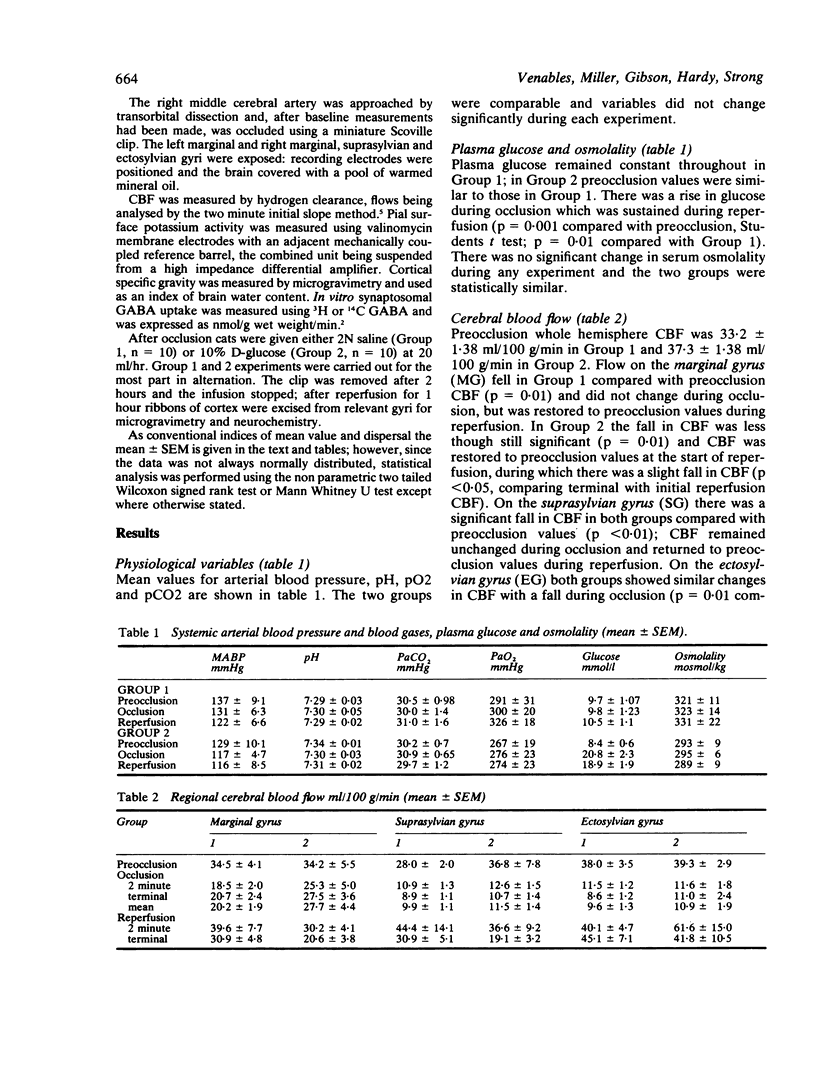
Abstract
The effects of isosmolar loads of glucose and saline after onset of focal cerebral ischaemia (middle cerebral artery occlusion) were compared in cats. In cats given saline cerebral blood flow (CBF) fell and then rose slightly on the marginal gyrus (infarct penumbra). There was a sustained fall in CBF on the suprasylvian and ectosylvian gyri (infarct core). Reperfusion restored blood flow to preocclusion levels with no overall postischaemic hypoperfusion. Below ischaemic flows of 14 ml/100 g/min brain specific gravity was reduced in a smaller proportion of gyri by contrast with non reperfused cortex, suggesting that in some gyri resolution of cerebral oedema had taken place. GABA uptake was normal in the infarct core, but was reduced within the ischaemic penumbra. In animals given glucose after occlusion, CBF fell on the marginal gyrus during reperfusion. The degree of resolution of cerebral oedema was less than in saline infused cats. GABA uptake showed a pattern of abnormality similar to that seen in saline infused cats, except that uptake values were lower in the infarct core. Pial surface potassium activity remained elevated in the penumbra following reperfusion in glucose infused cats, but returned to normal in saline infused cats. Implications for the management of cerebral ischaemia in man are discussed.
Full text
PDF


Selected References
These references are in PubMed. This may not be the complete list of references from this article.
- Bowen D. M., Goodhardt M. J., Strong A. J., Smith C. B., White P., Branston N. M., Symon L., Davison A. N. Biochemical indices of brain structure, function and "hypoxia" in cortex from baboons with middle cerebral artery occlusion. Brain Res. 1976 Dec 3;117(3):503–507. doi: 10.1016/0006-8993(76)90757-5. [DOI] [PubMed] [Google Scholar]
- Eklöf B., Siesjö B. K. The effect of bilateral carotid artery ligation upon acid-base parameters and substrate levels in the rat brain. Acta Physiol Scand. 1972 Dec;86(4):528–538. doi: 10.1111/j.1748-1716.1972.tb05354.x. [DOI] [PubMed] [Google Scholar]
- Ginsberg M. D., Budd M. W., Welsh F. A. Diffuse cerebral ischemia in the cat: I. Local blood flow during severe ischemia and recirculation. Ann Neurol. 1978 Jun;3(6):482–492. doi: 10.1002/ana.410030605. [DOI] [PubMed] [Google Scholar]
- Hansen A. J. The extracellular potassium concentration in brain cortex following ischemia in hypo- and hyperglycemic rats. Acta Physiol Scand. 1978 Mar;102(3):324–329. doi: 10.1111/j.1748-1716.1978.tb06079.x. [DOI] [PubMed] [Google Scholar]
- Ljunggren B., Norberg K., Siesjö B. K. Influence of tissue acidosis upon restitution of brain energy metabolism following total ischemia. Brain Res. 1974 Sep 6;77(2):173–186. doi: 10.1016/0006-8993(74)90782-3. [DOI] [PubMed] [Google Scholar]
- Marmarou A., Tanaka K., Shulman K. An improved gravimetric measure of cerebral edema. J Neurosurg. 1982 Feb;56(2):246–253. doi: 10.3171/jns.1982.56.2.0246. [DOI] [PubMed] [Google Scholar]
- Myers R. E., Yamaguchi S. Nervous system effects of cardiac arrest in monkeys. Preservation of vision. Arch Neurol. 1977 Feb;34(2):65–74. doi: 10.1001/archneur.1977.00500140019003. [DOI] [PubMed] [Google Scholar]
- Palumbo P. J., Elveback L. R., Whisnant J. P. Neurologic complications of diabetes mellitus: transient ischemic attack, stroke, and peripheral neuropathy. Adv Neurol. 1978;19:593–601. [PubMed] [Google Scholar]
- Rehncrona S., Rosén I., Siesjö B. K. Excessive cellular acidosis: an important mechanism of neuronal damage in the brain? Acta Physiol Scand. 1980 Dec;110(4):435–437. doi: 10.1111/j.1748-1716.1980.tb06692.x. [DOI] [PubMed] [Google Scholar]
- Riddle M. C., Hart J. Hyperglycemia, recognized and unrecognized, as a risk factor for stroke and transient ischemic attacks. Stroke. 1982 May-Jun;13(3):356–359. doi: 10.1161/01.str.13.3.356. [DOI] [PubMed] [Google Scholar]
- Salford L. G., Siesjö B. K. The influence of arterial hypoxia and unilateral carotid artery occlusion upon regional blood flow and metabolism in the rat brain. Acta Physiol Scand. 1974 Sep;92(1):130–141. doi: 10.1111/j.1748-1716.1974.tb05729.x. [DOI] [PubMed] [Google Scholar]
- Schuier F. J., Hossmann K. A. Experimental brain infarcts in cats. II. Ischemic brain edema. Stroke. 1980 Nov-Dec;11(6):593–601. doi: 10.1161/01.str.11.6.593. [DOI] [PubMed] [Google Scholar]
- Siemkowicz E., Hansen A. J. Brain extracellular ion composition and EEG activity following 10 minutes ischemia in normo- and hyperglycemic rats. Stroke. 1981 Mar-Apr;12(2):236–240. doi: 10.1161/01.str.12.2.236. [DOI] [PubMed] [Google Scholar]
- Siesjö B. K. Cerebral circulation and metabolism. J Neurosurg. 1984 May;60(5):883–908. doi: 10.3171/jns.1984.60.5.0883. [DOI] [PubMed] [Google Scholar]
- Steen P. A., Michenfelder J. D., Milde J. H. Incomplete versus complete cerebral ischemia: improved outcome with a minimal blood flow. Ann Neurol. 1979 Nov;6(5):389–398. doi: 10.1002/ana.410060503. [DOI] [PubMed] [Google Scholar]
- Strong A. J., Tomlinson B. E., Venables G. S., Gibson G., Hardy J. A. The cortical ischaemic penumbra associated with occlusion of the middle cerebral artery in the cat: 2. Studies of histopathology, water content, and in vitro neurotransmitter uptake. J Cereb Blood Flow Metab. 1983 Mar;3(1):97–108. doi: 10.1038/jcbfm.1983.12. [DOI] [PubMed] [Google Scholar]
- Strong A. J., Venables G. S., Gibson G. The cortical ischaemic penumbra associated with occlusion of the middle cerebral artery in the cat: 1. Topography of changes in blood flow, potassium ion activity, and EEG. J Cereb Blood Flow Metab. 1983 Mar;3(1):86–96. doi: 10.1038/jcbfm.1983.11. [DOI] [PubMed] [Google Scholar]
- Symon L., Branston N. M., Chikovani O. Ischemic brain edema following middle cerebral artery occlusion in baboons: relationship between regional cerebral water content and blood flow at 1 to 2 hours. Stroke. 1979 Mar-Apr;10(2):184–191. doi: 10.1161/01.str.10.2.184. [DOI] [PubMed] [Google Scholar]
- Symon L., Pasztor E., Branston N. M. The distribution and density of reduced cerebral blood flow following acute middle cerebral artery occlusion: an experimental study by the technique of hydrogen clearance in baboons. Stroke. 1974 May-Jun;5(3):355–364. doi: 10.1161/01.str.5.3.355. [DOI] [PubMed] [Google Scholar]
- Traupe H., Kruse E., Heiss W. D. Reperfusion of focal ischemia of varying duration: postischemic hyper- and hypo-perfusion. Stroke. 1982 Sep-Oct;13(5):615–622. doi: 10.1161/01.str.13.5.615. [DOI] [PubMed] [Google Scholar]