Abstract
Fibroblast activation protein (FAP) is expressed as a pro-inflammatory agent from fibrous tissue in liver cirrhosis and in the tumor microenvironment. Cirrhosis is the last stage of any chronic liver disease, and the natural course of cirrhosis is the progression from the asymptomatic phase to the symptomatic decompensated phase with the development of ascites. Although various clinical features suggest cirrhosis in patients with chronic liver disease, non-invasive methods should follow the clinical approach before a definitive diagnosis. Herein, we present three cases of liver cirrhosis with fibroblast activation protein inhibitor (FAPI) uptake to demonstrate the usefulness of 68Ga-FAPI-04 positron emission tomography/computed tomography (PET/CT) scan in cirrhosis.
Keywords: PET/CT, 68Ga FAPI-04, cirrhosis, liver, molecular imaging
Abstract
Fibroblast aktivasyon proteini (FAP), karaciğer sirozunda ve tümör mikroçevresinde fibröz dokudan proenflamatuvar bir ajan olarak eksprese edilir. Siroz, herhangi bir kronik karaciğer hastalığının son aşamasını gösterir ve sirozun doğal seyri, asit gelişimi ile asemptomatik fazdan semptomatik dekompanse faza ilerlemedir. Kronik karaciğer hastalığı olan hastalarda çeşitli klinik özellikler sirozu düşündürse de, kesin tanıdan önce invaziv olmayan yöntemler klinik yaklaşımı takip etmelidir. Burada, sirozda 68Ga-fibroblast aktivasyon protein inhibitörü (FAPI)-04 pozitron emisyon tomografisi/bilgisayarlı tomografi taramasının yararlılığını göstermek için FAPI tutulumu olan üç karaciğer sirozu olgusunu sunuyoruz.
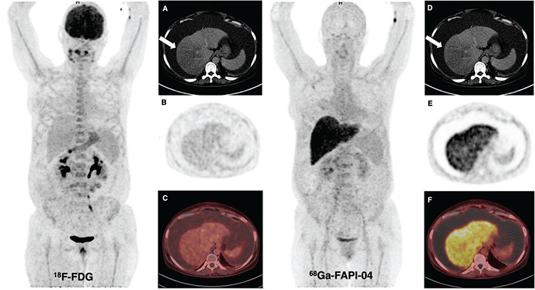
Figure 1.

A 50-year-old woman patient presented with abdominal pain and was found to have massive ascites and had undergone18F-fluorodeoxyglucose (FDG) and 68Ga-fibroblast activation protein inhibitor (FAPI)-04 positron emission tomography/computed tomography (PET/CT) imaging for a suspected gastric tumor. Abnormal liver activity was not observed on 18F-FDG PET/CT, but diffuse intense FAPI uptake [maximum standardized uptake value (SUVmax): 11.2] was seen in the liver on 68Ga-FAPI-04 PET/CT. The hypodense area detected in CT sections [(A, D) arrow] at the right lobe of the liver did not show prominent 18F-FDG [(B) PET, (C) fusion images] or FAPI uptake [(E) PET, (F) fusion images] in the parenchyma. Also, no malignant cells were found in the peritoneal aspiration fluid. The patient is being followed up with the diagnosis of decompensated alcoholic cirrhosis.
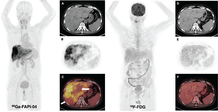
Figure 2.

18F-FDG and 68Ga-FAPI-04 PET/CT imaging were performed on a 55-year-old patient who was investigated for a liver malignancy that may occur on the basis of cirrhosis. Heterogeneous intense FAPI uptake was detected in the liver, especially in the right lobe, whereas there was no significant pathological uptake apart from parenchymal areas protected from cirrhosis [(C) arrows] [(A) CT, (B) PET, (C) fusion images] and no prominent liver activity on 18F-FDG PET/CT [(D) CT, (E) PET, (F) fusion images]. In addition, mild to moderately increased thyroidal uptake suggestive of thyroiditis was revealed on the maximum intensity projection image of 68Ga-FAPI-04 PET/CT.
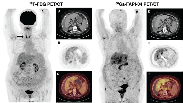
Figure 3.

18F-FDG and 68Ga-FAPI-04 PET/CT scans were obtained on a 70-year-old woman patient with free fluid in the peritoneum for the evaluation of liver cirrhosis [(A, D) CT)]. Slightly increased tracer activity in the liver [(B) PET, (C) fusion images] and diffusely increased thyroid uptake (arrow) indicated thyroiditis were seen on 18F-FDG PET/CT. The axial view of PET (E) and fusion images (F) showed intense heterogeneous FAPI uptake (SUVmax: 9.7) in the liver, but no FAPI uptake in the thyroid gland on 68Ga-FAPI-04 PET/CT.
The histological diagnosis of cirrhosis indicates the last stage of any chronic liver disease, and the natural course of cirrhosis is the progression from the asymptomatic phase to the symptomatic decompensated phase with the development of ascites (1,2). FAPI was superior to 18F-FDG in detecting liver tumors and many types of cancer (3,4,5). In a few cases published in the literature, diffuse intense FAPI uptake in the liver has been defined in patients with cirrhosis and 68Ga-FAPI-04 PET/CT has been shown to have a place in the differentiation of liver lesions due to its preferred image contrast (6). In a study evaluating liver lesions comparatively both with FAPI and 18F-FDG PET/CT on twenty patients, 68Ga-FAPI-04 PET/CT was found superior to 18F-FDG in detecting liver lesions and Shi et al. (7) also detected significantly higher hepatic FAPI uptake in nine patients with cirrhosis than the non-cirrhotic study group. Low background activity is among the important advantages of imaging with FAPI, but high uptake on the liver in patients with cirrhosis may limit the evaluation of hepatic lesions in terms of malignant nature. Therefore, in patients without cirrhosis, some liver lesions may be better differentiated in the absence of intense FAPI uptake. However, Zhao et al. (8) reported a case of cirrhosis with multiple FAPI-negative benign hepatic nodules with increased liver FAPI uptake. According to this patient example, they stated that 68Ga-FAPI-04 PET/CT might be an appropriate tool for differentiating benign nodular lesions from primary malignant tumors. Our results, together with the published cases, suggest that 68Ga- FAPI-04 PET/CT may have a place in the treatment plan and management of liver cirrhosis in addition to tumor imaging in the future molecular era.
Footnotes
Ethics
Informed Consent: Informed consent was obtained from the patients included in the study.
Peer-review: Externally and internally peer-reviewed.
Authorship Contributions
Surgical and Medical Practices: G.T., E.B., Ö.E.F., İ.S., N.E., T.F.Ç., Concept: G.T., E.B., Ö.E.F., İ.S., N.E., T.F.Ç., Design: G.T., E.B., Ö.E.F., İ.S., N.E., T.F.Ç., Data Collection or Processing: G.T., E.B., Ö.E.F., İ.S., N.E., T.F.Ç., Analysis or Interpretation: G.T., E.B., Ö.E.F., İ.S., N.E., T.F.Ç., Literature Search: G.T., E.B., Ö.E.F., İ.S., Writing: G.T., T.F.Ç.
Conflict of Interest: No conflict of interest was declared by the authors.
Financial Disclosure: The authors declared that this study received no financial support.
References
- 1.Ginès P, Krag A, Abraldes JG, Solà E, Fabrellas N, Kamath PS. Liver cirrhosis. Lancet. 2021;398:1359–1376. doi: 10.1016/S0140-6736(21)01374-X. [DOI] [PubMed] [Google Scholar]
- 2.Zhou WC, Zhang QB, Qiao L. Pathogenesis of liver cirrhosis. World J Gastroenterol. 2014;20:7312–7324. doi: 10.3748/wjg.v20.i23.7312. [DOI] [PMC free article] [PubMed] [Google Scholar]
- 3.Çermik TF, Ergül N, Yılmaz B, Mercanoğlu G. Tumor imaging with 68Ga-DOTA-FAPI-04 PET/CT: comparison with 18F-FDG PET/CT in 22 different cancer types. 2022;47:333–339. doi: 10.1097/RLU.0000000000004073. [DOI] [PubMed] [Google Scholar]
- 4.Chen H, Zhao L, Ruan D, Pang Y, Hao B, Dai Y, Wu X, Guo W, Fan C, Wu J, Huang W, Lin Q, Sun L, Wu H. Usefulness of [68Ga]Ga-DOTA-FAPI-04 PET/CT in patients presenting with inconclusive [18F]FDG PET/CT findings. Eur J Nucl Med Mol Imaging. 2021;48:73–86. doi: 10.1007/s00259-020-04940-6. [DOI] [PubMed] [Google Scholar]
- 5.Ballal S, Yadav MP, Moon ES, Kramer VS, Roesch F, Kumari S, Tripathi M, ArunRaj ST, Sarswat S, Bal C. Biodistribution, pharmacokinetics, dosimetry of [68Ga]Ga-DOTA.SA.FAPi, and the head-to-head comparison with [18F]F-FDG PET/CT in patients with various cancers. Eur J Nucl Med Mol Imaging. 2021;48:1915–1931. doi: 10.1007/s00259-020-05132-y. [DOI] [PubMed] [Google Scholar]
- 6.Shi X, Xing H, Yang X, Li F, Yao S, Congwei J, Zhao H, Hacker M, Huo L, Li X. Comparison of PET imaging of activated fibroblasts and 18F-FDG for diagnosis of primary hepatic tumours: a prospective pilot study. Eur J Nucl Med Mol Imaging. 2021;48:1593–1603. doi: 10.1007/s00259-020-05070-9. [DOI] [PubMed] [Google Scholar]
- 7.Shi X, Xing H, Yang X, Li F, Yao S, Zhang H, Zhao H, Hacker M, Huo L, Li X. Fibroblast imaging of hepatic carcinoma with 68Ga-FAPI-04 PET/CT: a pilot study in patients with suspected hepatic nodules. Eur J Nucl Med Mol Imaging. 2021;48:196–203. doi: 10.1007/s00259-020-04882-z. [DOI] [PubMed] [Google Scholar]
- 8.Zhao L, Gu J, Fu K, Lin Q, Chen H. 68Ga-FAPI PET/CT in assessment of liver nodules in a cirrhotic patient. Clin Nucl Med. 2020;45:430–432. doi: 10.1097/RLU.0000000000003015. [DOI] [PubMed] [Google Scholar]


