F1000Research logoLink to F1000Research
. 2023 Mar 13;11:933. Originally published 2022 Aug 15. [Version 3] doi: 10.12688/f1000research.123044.3

Enhancement of the activity of the antimicrobial peptides HNP1 and LL-37 by bovine pancreatic ribonuclease A

Bryan Ericksen 1,a
PMCID: PMC10285323  PMID: 37360940

Version Changes

Revised. Amendments from Version 2

Biofilms are no longer mentioned in the Abstract. Figure 9, a composite mean of Figures 2, 5 and 6, has been added with error bars indicating standard error of the mean. Various other changes have been made to the Results and Discussion sections as recommended by the second and third reviewers.

Abstract

Background: HNP1, LL-37, and HBD1 are antimicrobial against Escherichia coli ATCC 25922 at the standard inoculum but less active at higher inocula.  

Methods: The virtual colony count (VCC) microbiological assay was adapted for high inocula and the addition of yeast tRNA and bovine pancreatic ribonuclease A (RNase).  96-well plates were read for 12 hours in a Tecan Infinite M1000 plate reader and photographed under 10x magnification.   

Results: Adding tRNA 1:1 wt/wt to HNP1 at the standard inoculum almost completely abrogated activity.  Adding RNase 1:1 to HNP1 at the standard inoculum of 5x10 5 CFU/mL did not enhance activity.  Increasing the inoculum to 6.25x10 7 CFU/mL almost abrogated HNP1 activity.  However, adding RNase 25:1 to HNP1 enhanced activity at the highest tested concentration of HNP1.  Adding both tRNA and RNase resulted in enhanced activity, indicating that the enhancement effect of RNase overwhelms the inhibiting effect of tRNA when both are present.  HBD1 activity at the standard inoculum was almost completely abrogated by the addition of tRNA, but LL-37 activity was only slightly inhibited by tRNA.  At the high inoculum, LL-37 activity was enhanced by RNase.  HBD1 activity was not enhanced by RNase.  RNase was not antimicrobial in the absence of antimicrobial peptides.  Cell clumps were observed at the high inoculum in the presence of all three antimicrobial peptides and at the standard inoculum in the presence of HNP1+tRNA and HBD1+tRNA.   

Conclusions: Antimicrobial peptide-ribonuclease combinations have the potential to be active against high cell concentrations, conditions where the antimicrobial agent alone is relatively ineffective.

Keywords: antimicrobial peptide, ribonuclease

Introduction

Although cationic antimicrobial peptides (CAPs) have been studied as possible therapeutic agents for many years, few have survived clinical trials to become useful antibiotics ( Mishra et al. 2017). Three CAPs are representative of three different structural classes that contribute to the human innate immune system: human neutrophil peptide 1 (HNP1), an alpha defensin; human beta defensin 1 (HBD1); and the human cathelicidin LL-37 ( De Smet 2005). One reason why CAP drug candidates have failed to gain approval is a lack of efficacy ( Magana 2020). I demonstrated a pronounced inoculum effect when the defensin HNP1 was assayed against high inocula of Escherichia coli ATCC 25922, such that the antimicrobial peptide almost completely lost activity under those conditions ( Ericksen 2020). A pronounced inoculum effect was also observed when HNP1 was assayed against Staphylococcus aureus ATCC 29213 and Bacillus cereus ATCC 10876. What might cause this decrease in efficacy at high cell concentrations? The molecular basis of the inoculum effect is unclear. However, one possibility is that bacterial cells produce defensin inhibitors that are at a higher concentration when there are more cells present.

One possible type of inhibition is that polyanions might bind and inhibit CAPs by electrostatic attraction. Here I hypothesize that the polyanion tRNA might inhibit CAPs, that inhibition by RNA (quite possibly a general property of RNA, not specific to tRNA) is partially responsible for the inoculum effect, and that the addition of ribonuclease could enhance antimicrobial peptide activity, restoring some of the efficacy lost at high cell concentrations.

Methods

The VCC assay was adapted for high inocula as previously described ( Ericksen 2020), and volumes were adjusted to allow for the addition of yeast tRNA (Sigma from Saccharomyces cerevisiae) and/or bovine pancreatic ribonuclease (Roche or Macherey-Nagel (MN)). HNP1, LL-37 and HBD1 were synthesized with an ABI 433A synthesizer using an optimized HBTU activation/DIEA in situ neutralization protocol developed by Kent and coworkers for Boc chemistry solid phase peptide synthesis as previously described ( Zhao 2013; Pazgier 2013; Bharucha 2021). Two inocula of E. coli ATCC 25922 were studied: the standard inoculum of 5×10 5 CFU/mL, with cells from a seed culture diluted in 10 mM sodium phosphate pH 7.4, and a high inoculum of 6.25×10 7 CFU/mL, equivalent to adding undiluted seed culture. Antimicrobial peptides were incubated in 10 mM sodium phosphate pH 7.4 plus 1% tryptic soy broth (TSB) for two hours at 37°C shaking every 5 minutes for 3 seconds in a Tecan Infinite M1000 plate reader. An equal volume of twice-concentrated Mueller Hinton Broth was then added and 96-well plates were read for 12 hours in the plate reader and then some wells containing cell clumps were photographed under 10x magnification. In one experiment, the concentration of TSB present in phosphate buffer was adjusted.

Results

Adding tRNA 1:1 to HNP1 at the standard inoculum almost completely abrogated activity ( Figure 1). Adding Roche RNase 1:1 to HNP1 at the standard inoculum of 5×10 5 CFU/mL did not enhance activity. Increasing the inoculum to 6.25×10 7 CFU/mL almost abrogated HNP1 activity ( Figure 2). However, adding RNase 25:1 to HNP1 enhanced activity abruptly at the high inoculum. Adding both tRNA and RNase resulted in enhanced activity, indicating that the enhancement effect of RNase overwhelms the inhibiting effect of tRNA when both are present. HBD1 activity at the standard inoculum was almost completely abrogated by the addition of tRNA, but LL-37 activity was only slightly inhibited by tRNA ( Figure 3). At the high inoculum, LL-37 activity was enhanced, but LL-37 showed greater activity than HNP1 in the absence of RNase ( Figure 4). HBD1 activity was not enhanced by RNase. RNase was not antimicrobial in the absence of antimicrobial peptides. The observations with HNP1 at the high inolculum were repeated using a second RNase manufacturer, Macherey-Nagel ( Figure 5). The experiment with MN RNase was repeated ( Figure 6). 1% TSB was used in most assays, but the %TSB was varied in one experiment, resulting in maximum activity at 4% TSB with either 5× or 25× MN RNase added ( Figure 7). Cell clumps similar to those previously described ( Ericksen 2020) were observed at the high inoculum in the presence of all three antimicrobial peptides with or without RNase and at the standard inoculum in the presence of HNP1+tRNA and HBD1+tRNA ( Figure 8). The VCC assays were conducted with TSB added to the 10 mM sodium phosphate incubation buffer. Although biofilm formation was not directly assayed, it is assumed that the cell clumps photographed at 10x magnification are biofilms. Ribonuclease did not enhance HBD1 activity at the 6.25×10 7 CFU/mL inoculum, demonstrating a strong inoculum effect with HBD1 vs. E. coli. LL-37 had a much lesser inoculum effect against E. coli. The effect of ribonuclease on HNP1 is strongest with lowest amounts of TSB present in the phosphate buffer during the 2 hour incubation. The ability of tRNA to abrogate HNP1 and HBD1 activity, and the failure of tRNA to affect LL-37 activity, at the standard inoculum cannot be explained by net charge. Possibly, hydrophobic interactions play a role in tRNA binding and inhibition. It is also possible that tRNA inducing biofilm formation impacts HNP1 and HBD1 more than LL-37. The results shown in Figures 2, 5, and 6 can be regarded as triplicate experiments, assuming the two RNase preparations are equivalent. The mean virtual survival of HNP1 + 5× RNase was 0.75, and the standard deviation was 0.21. The mean virtual survival of HNP1 + 25× RNase was 0.70, and the standard deviation was 0.10. The mean virtual survival of HNP1 alone was 0.88, and the standard deviation was 0.02. Based on these values, the two-tailed p-value for HNP1 + 5× RNase compared to HNP1 alone was 0.38, whereas the two-tailed p-value for HNP1 + 25× RNase compared to HNP1 alone was 0.10. Therefore, the slight differences in activity observed were not statistically significant (p>0.05). The mean composite virtual survival from the experiments shown in Figures 2, 5 and 6 of HNP1 + 25× RNase is plotted in Figure 9. Error bars represent the standard error of the mean (SEM).

Figure 1. Activity of HNP1 with or without tRNA and RNase at the standard inoculum.
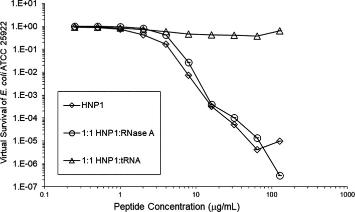
Figure 1.

Figure 2. Activity of HNP1 at the high inoculum with or without tRNA and three concentrations of RNase.
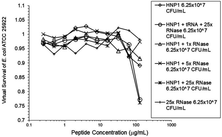
Figure 2.

Activity with HNP1 and both tRNA and the highest concentration of RNase was essentially the same as HNP1 plus RNase alone, indicating the enhancement of activity overcomes inhibition by tRNA. RNase in the absence of antimicrobial peptides was not antimicrobial.

Figure 3. LL-37 was assayed at the standard inoculum with or without tRNA, and HBD1 was assayed with tRNA.
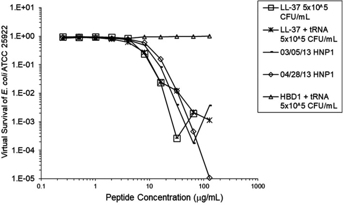
Figure 3.

HBD1 was assayed at the standard inoculum in the presence of 1:1 tRNA. Two preparations of HNP1 were assayed in the absence of tRNA as positive controls.

Figure 4. LL-37 was assayed at the high inoculum with or without RNase.
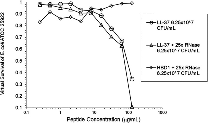
Figure 4.

HBD1 was assayed at the high inoculum with RNase.

Figure 5. HNP1 was assayed at the high inoculum in the presence and absence of RNase from a second manufacturer, and in the presence of both tRNA and RNase.
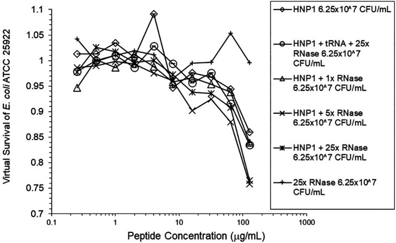
Figure 5.

Figure 6. The assay shown in Figure 5 was repeated.
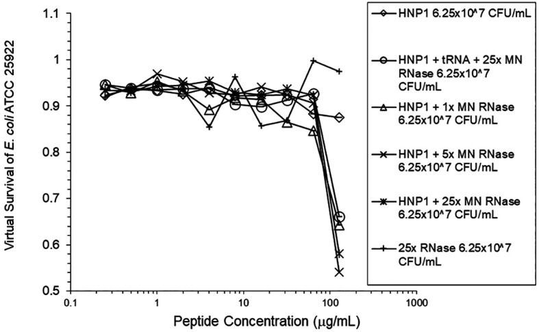
Figure 6.

Figure 7. HNP1 was assayed at the high inoculum with variation in the amount of TSB present during the two-hour incubation in 10 mM sodium phosphate buffer.
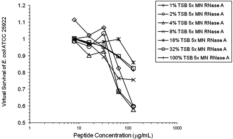
Figure 7.

Figure 8. Cell clumps photographed at 10× magnification.
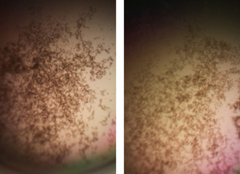
Figure 8.

Left panel: 128 μg/mL HNP1 at the high inoculum. Right panel: 128 μg/mL HNP1 + 1:5 RNase at the high inoculum.

Figure 9. Composite mean of data in Figures 2, 5 and 6 ± standard error of the mean.
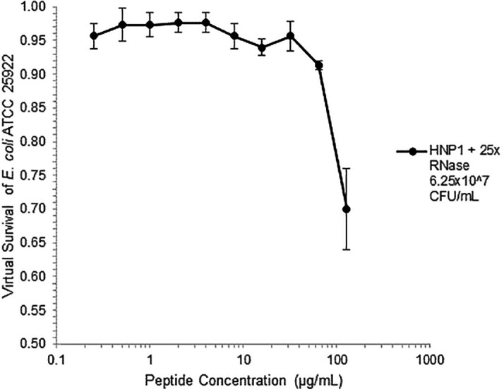
Figure 9.

Conclusions

Antimicrobial assays are ordinarily conducted using a single antimicrobial agent, studying its effect in isolation. However, the experiments presented here may offer a glimpse into a more realistic in vivo scenario, in which multiple antimicrobial agents work in concert against infection. Eight RNases are encoded by the human genome, many of which have potent antimicrobial activity, such as RNase 7 expressed in epithelial cells ( Sorrentino 2010). Bovine pancreatic RNase A, on the other hand, has a digestive function degrading RNA and an antimicrobial function has not normally been ascribed to it. RNase A is a basic protein (pI = 9.63). It is unknown whether the RNA-degrading activity of RNase or its cationicity is responsible for the enhancement of HNP1 and LL-37 activity. Product literature suggests assaying RNase A using 100 mM Tris buffer, pH 7.4. Enzymatic activity in 10 mM sodium phosphate buffer was not tested, but RNase A is very stable with four disulfide bonds.

The variation in the amount of TSB present in 10 mM phosphate buffer revealed that the increase in activity caused by a small amount of nutrients present, allowing some growth during the two-hour incubation, is counterbalanced by the inhibition of defensin activity at higher TSB concentrations, presumably by the salt content of TSB. This same effect is probably partially responsible for the almost complete abrogation of activity of HNP1 when undiluted seed culture is added to the 96-well plate at the high inoculum in the absence of RNase, since the salt concentration is much higher than in assays at the standard inoculum where the seed culture is diluted in 10 mM sodium phosphate buffer before adding to the 96-well plate.

The vast majority of published VCC assays were conducted at the standard inoculum, reflecting a general reliance on the standard inoculum in a wide range of published antimicrobial assays. Under these conditions, cells are predominantly planktonic. However, a high inoculum may be more medically relevant, since high cell concentrations and biofilms can accompany acute infections. It should be emphasized that these experiments do not prove that the observed cell clumps are biofilms. This study demonstrates the utility of conducting assays at a high inoculum, revealing details of antimicrobial activity that would be missed if the antimicrobial agents were studied only at the standard inoculum. Further studies using animal models are necessary to determine whether the enhancement of activity observed at the high inoculum is sufficient to enable the infected host to overcome bacterial infections.

It should be emphasized that both RNA and ribonucleases are ubiquitous in vivo. Therefore, these experiments may be more biologically relevant than VCC experiments lacking RNA or ribonuclease. However, the presence of tRNA in the medium at the high inoculum has not been demonstrated. There are several possible sources of bacterial RNA that might be present at the site of a bacterial infection. Firstly, bacteria normally secrete RNA during their growth, which may have a role in the extracellular matrix of biofilms ( Ozoline 2019). Any RNA, especially RNAs normally secreted as part of bacterial growth or incorporated into biofilms, could inhibit antimicrobial peptides. The results of the experiments presented here suggest that this secreted RNA may also be a bacterial defense mechanism against antimicrobial peptides. Secondly, once antimicrobial peptides are released at the infection site, cell lysis may result in the release of intracellular RNAs, including mRNA and tRNA. Thirdly, host RNA may be present. Therefore, inhibition by RNA must be regarded as a common obstacle to effective antimicrobial activity that frequently occurs in real world scenarios.

The combination of an antimicrobial peptide with a ribonuclease could be regarded as a novel invention that could possibly be used as a therapy to treat bacterial infections. LL-37 and RNase 1 have been shown to act synergistically to kill E. coli ( Eller 2020). RNases have been tested in clinical trials as chemotherapeutics for the treatment of cancer ( Ardelt 2009).

Further studies are warranted to determine whether these results are biologically relevant or could be generalized to antimicrobial peptide-nuclease combinations, as might be suggested by the presence of DNA in biofilms. A combination of an antimicrobial peptide with both deoxyribonuclease (DNase) and RNase might be expected to be more potent than the combination of the antimicrobial peptide and RNase in the absence of DNase, because DNA is considered a more prevalent structural component of biofilms than RNA ( Gilan 2013). DNase is an approved drug, dornase alfa (Pulmozyme), which cuts apart extracellular DNA in the lungs of cystic fibrosis patients, making the mucus thinner and easier to expel ( Wagener 2012). It is possible that DNase in combination with an antimicrobial peptide and RNase would form an effective treatment against acute bacterial infections. Although the effect of HNP1+RNase was not statistically significant compared to HNP1 alone, it is possible that the effect of an antimicrobial peptide+DNase+RNase would be significant compared to the antimicrobial peptide alone. A new generation of antimicrobial peptide-nuclease combinations would offer a new hope that peptides that are sometimes defeated by the resistance mechanism of biofilm formation can be repurposed to degrade biofilms instead, with increased activity to fight infections.

Data availability

Underlying data

Figshare: Enhancement of Antimicrobial Peptide Activity by Ribonuclease (virtual colony count data), https://doi.org/10.6084/m9.figshare.20352996.v1 ( Ericksen 2022).

Data are available under the terms of the Creative Commons Zero “No rights reserved” data waiver (CC0 1.0 Public domain dedication).

Acknowledgments

I thank Wuyuan Lu for providing antimicrobial peptides and helpful discussions.

Funding Statement

Peprotech, Inc. provided funding.

The funders had no role in study design, data collection and analysis, decision to publish, or preparation of the manuscript.

[version 3; peer review: 1 approved

References

  1. Ardelt W, Ardelt B, Darzynkiewicz Z: Ribonucleases as potential modalities in anticancer therapy. Eur. J. Pharmacol. 2009;625(1-3):181–189. 10.1016/j.ejphar.2009.06.067 [DOI] [PMC free article] [PubMed] [Google Scholar]
  2. Bharucha JP, Sun L, Lu W, et al. : Human beta-defensin 2 and 3 Inhibit HIV-1 replication in macrophages. Front. Cell. Infect. Microbiol. 2021;11:535352. 10.3389/fcimb.2021.535352 [DOI] [PMC free article] [PubMed] [Google Scholar]
  3. De Smet K, Contreras R: Human antimicrobial peptides: defensins, cathelicidins and histatins. Biotechnol. Lett. 2005;27(18):1337–1347. 10.1007/s10529-005-0936-5 [DOI] [PubMed] [Google Scholar]
  4. Eller CH, Raines RT: Antimicrobial synergy of a ribonuclease and a peptide secreted by human cells. ACS Infectious Diseases. 2020;6(11):3083–3088. 10.1021/acsinfecdis.0c00594 [DOI] [PMC free article] [PubMed] [Google Scholar]
  5. Ericksen B: Virtual colony count. Wiklj. Sci. 2020;3(1):3. 10.15347/WJS/2020.003 [DOI] [Google Scholar]
  6. Ericksen B: Enhancement of Antimicrobial Peptide Activity by Ribonuclease. Figshare. [Dataset]. 2022. 10.6084/m9.figshare.20352996.v1 [DOI]
  7. Gilan I, Sivan A: Extracellular DNA plays an important structural role in the biofilm of the plastic degrading actinomycete Rhodococcus ruber. Adv. Microbiol. 2013;3(8):40674. [DOI] [PubMed] [Google Scholar]
  8. Magana M, Pushpanathan M, Santos AL, et al. : The value of antimicrobial peptides in the age of resistance. Lancet Infect. Dis. 2020;20(9):e216–e230. 10.1016/S1473-3099(20)30327-3 [DOI] [PubMed] [Google Scholar]
  9. Mishra B, Reiling S, Zarena D, et al. : Host defense antimicrobial peptides as antibiotics: design and application strategies. Curr. Opin. Chem. Biol. 2017;38:87–96. 10.1016/j.cbpa.2017.03.014 [DOI] [PMC free article] [PubMed] [Google Scholar]
  10. Ozoline O, Jass J: Editorial: secretion and signalling of bacterial RNAs. FEMS Microbiol. Lett. 2019;366(1):fny281. 10.1093/femsle/fny281 [DOI] [PubMed] [Google Scholar]
  11. Pazgier M, Ericksen B, Ling M, et al. : Structural and functional analysis of the pro-domain of human cathelicidin, LL-37. Biochemistry. 2013;52(9):1547–1558. 10.1021/bi301008r [DOI] [PMC free article] [PubMed] [Google Scholar]
  12. Sorrentino S: The eight human “canonical” ribonucleases: Molecular diversity, catalytic properties, and special biological actions of the enzyme proteins. FEBS Lett. 2010;584:2194–2200. 10.1016/j.febslet.2010.04.018 [DOI] [PubMed] [Google Scholar]
  13. Wagener JS, Kupfer O: Dornase alfa (Pulmozyme). Curr. Opin. Pulm. Med. 2012;18(6):609–614. 10.1097/MCP.0b013e328358d51f [DOI] [PubMed] [Google Scholar]
  14. Zhao L, Tolbert WD, Ericksen B, et al. : Single, double and quadruple alanine substitutions at oligomeric interfaces identify hydrophobicity as the key determinant of human neutrophil alpha defensin HNP1 function. PLoS One. 2013;8(11):e78937. 10.1371/journal.pone.0078937 [DOI] [PMC free article] [PubMed] [Google Scholar]
F1000Res. 2023 Jun 6. doi: 10.5256/f1000research.145427.r174366

Reviewer response for version 3

Paweł M Krzysciak 1

The provided text discusses the challenges in developing cationic antimicrobial peptides (CAPs) as effective antibiotics and proposes a hypothesis regarding the inoculum effect observed when testing the defensin HNP1 against high bacterial cell concentrations. The text suggests that the decrease in efficacy at high cell concentrations could be attributed to the presence of defensin inhibitors, particularly polyanions like tRNA, which may bind and inhibit CAPs through electrostatic attraction.

Overall, the text presents an interesting hypothesis that adds to our understanding of the factors influencing the efficacy of CAPs. The identification of potential inhibitors, such as polyanions, and their impact on antimicrobial peptide activity is a valuable contribution to the field. The proposed role of RNA inhibition, potentially not limited to tRNA, in the inoculum effect adds depth to the discussion.

I suggest that  to address the all reviewers' concerns and strengthen the conclusions, it is recommended to conduct additional analyses using standard microbiological methods, such as colony-forming unit (CFU) counting or metabolic tests, such as the MTT assay. These analyses would provide complementary data to the Virtual Colony Count (VCC) assay and enhance the reliability and comprehensiveness of the findings.

It would be beneficial to discuss the potential limitations associated with using TSB in the experimental setup and acknowledge the need for further investigations to validate the observed effects in a more controlled environment.

The assumption made regarding the presence of biofilms based solely on the observation of cell clumps at 10x magnification should be addressed more cautiously. Biofilm formation is a complex process and typically requires specific conditions and timeframes. Without conducting specific biofilm assays or providing further evidence, it is uncertain whether the observed cell clumps can be definitively classified as biofilms. Acknowledging the limitations and potential alternative explanations for the observed clumps would strengthen the validity of the conclusions drawn from this observation.

One area of improvement could be the inclusion of gene expression analysis related to biofilm formation. Since biofilm formation is a complex process involving the regulation of specific genes, it would be valuable to assess the expression levels of key biofilm-related genes. This could provide insights into the potential role of the observed cell clumps in biofilm formation and their relationship to the inoculum effect. Including gene expression analysis would strengthen the study by providing a molecular understanding of the observed phenomenon.

It would be beneficial to provide more comprehensive statistical information, such as the t statistic value (or other suitable statistical test), degrees of freedom (df), power of the test, and the sample size (n) for each comparison made. These details are essential for the readers to evaluate the strength and reliability of the statistical analysis performed. By including the t statistic value and degrees of freedom, the readers can assess the magnitude of the observed differences and the precision of the estimates. Additionally, reporting the power of the test would provide insights into the sensitivity of the statistical analysis to detect true differences if they exist. Lastly, providing the sample size (n) would give readers an understanding of the reliability of the observed results.

In summary, by incorporating the suggested improvements, including additional microbiological analyses, addressing limitations, considering alternative explanations, and providing comprehensive statistical information, the text would make a valuable contribution to the field of antimicrobial peptide research. The proposed hypothesis and insights into the inoculum effect and potential inhibitory factors enhance our understanding of CAPs' efficacy as antibiotics and pave the way for future investigations in this area.

Is the work clearly and accurately presented and does it cite the current literature?

Yes

If applicable, is the statistical analysis and its interpretation appropriate?

Partly

Are all the source data underlying the results available to ensure full reproducibility?

Yes

Is the study design appropriate and is the work technically sound?

Yes

Are the conclusions drawn adequately supported by the results?

Yes

Are sufficient details of methods and analysis provided to allow replication by others?

Yes

Reviewer Expertise:

medical microbiology, medical mycology, antimicrobial susceptibility testing,

I confirm that I have read this submission and believe that I have an appropriate level of expertise to confirm that it is of an acceptable scientific standard, however I have significant reservations, as outlined above.

F1000Res. 2023 Mar 27. doi: 10.5256/f1000research.145427.r166245

Reviewer response for version 3

Ramakrishnan Nagaraj 1

The authors have adequately answered my recommendations

Is the work clearly and accurately presented and does it cite the current literature?

Partly

If applicable, is the statistical analysis and its interpretation appropriate?

Not applicable

Are all the source data underlying the results available to ensure full reproducibility?

Yes

Is the study design appropriate and is the work technically sound?

Partly

Are the conclusions drawn adequately supported by the results?

No

Are sufficient details of methods and analysis provided to allow replication by others?

Partly

Reviewer Expertise:

NA

I confirm that I have read this submission and believe that I have an appropriate level of expertise to confirm that it is of an acceptable scientific standard.

F1000Res. 2023 Mar 7. doi: 10.5256/f1000research.141606.r163610

Reviewer response for version 2

Ramakrishnan Nagaraj 1

The brief report titled “Enhancement of the activity of the antimicrobial peptides HNP1 and LL-37 by bovine pancreatic ribonuclease A” by Ericksen describes the effect of tRNA and RNase A on antibacterial activity of HNP1, HBD1 and LL37 at low and high inoculum by VCC assay. Activity of HNP1 is inhibited by tRNA at 1:1 but restored when RNase A is present at 25:1. Similar effects are not observed with LL37. RNase A does not modulate the activity of HBD1 at high inoculum. The fact that inhibition of activity was observed at high inoculums, but not when RNase is present, suggesting that peptide binds to tRNA, is interesting. It is interesting as it could provide insights into HNP1 inactivation. However, more definitive experiments to confirm this result are necessary and worth doing.

Points

  1. The author argues that RNase 25:1 to HNP1 enhanced activity at high inoculum. It appears that the antibacterial activity of HNP1 that is abrogated at high inoculum is restored not enhanced. Antibacterial activity is restored in the presence of tRNA and RNase A.

  2. Wherever the activity is restored, antibacterial activity is restored abruptly (Figs 2, 5,6) unlike in controls (Figs 1, 3, 4).

  3. The presence of tRNA in the medium under high inoculum has not been demonstrated.

  4. The concentrations should have been represented as molar concentrations.

  5. DNA could also inhibit antibacterial activity which has not been demonstrated.

  6. The enzyme activity of RNase A could have been determined in 10 mM sodium phosphate buffer. There is too much speculation in the Conclusion section. For example, “It is unknown whether the RNA-degrading activity of RNase or its cationicity is responsible for the enhancement of HNP1 and LL-37 activity”.

  7. As the author states “Further studies are warranted to determine whether these results are biologically relevant or could be generalized to antimicrobial peptide-nuclease combinations, as might be suggested by the presence of DNA in biofilms”.

The data are preliminary and more definitive experiments are necessary. There is no enhancement in activity in the presence of RNase A, only restoration of activity. The author should demonstrate the presence of bacterial tRNA in high inoculum and also should address more conclusively the varied effects on LL37 and HBD1. The author should provide details of synthetic HNP1 characterization to rule out any trivial explanation.

Is the work clearly and accurately presented and does it cite the current literature?

Partly

If applicable, is the statistical analysis and its interpretation appropriate?

Not applicable

Are all the source data underlying the results available to ensure full reproducibility?

Yes

Is the study design appropriate and is the work technically sound?

Partly

Are the conclusions drawn adequately supported by the results?

No

Are sufficient details of methods and analysis provided to allow replication by others?

Partly

Reviewer Expertise:

Biology of host-defence peptides such as defensins, peptide antibiotics, mechanisms of bacterial killing by defensins.

I confirm that I have read this submission and believe that I have an appropriate level of expertise to confirm that it is of an acceptable scientific standard, however I have significant reservations, as outlined above.

F1000Res. 2023 Mar 9.
Bryan Ericksen 1

Dear Dr. Nagaraj,

Thank you for your detailed review.  I will respond to each of your points.

  1.  The more general term “enhanced” is more appropriate than “restored”, because the activity of HNP1 at the standard inoculum is far greater than the activity of HNP1 + 25x RNase at the high inoculum. Therefore, we cannot say that activity has been fully “restored” but it has certainly been “enhanced” by RNase.

  2. I have added the word “abruptly” in the Results section, description of Figure 2.

  3. The sentence “However, the presence of tRNA in the medium at the high inoculum has not been demonstrated” has been added to the Discussion section.

  4. There is a long history of expressing concentrations as µg/mL instead of µM. Please refer to the seven Journal of Biological Chemistry publications referenced in the Ericksen 2020 reference.

  5. DNA could certainly inhibit antibacterial activity, as I speculated in the Discussion section. I hope that speculation will lead another researcher to test DNA+RNA+antimicrobial peptide combinations.

  6. I apologize for neglecting to demonstrate the enzyme activity of RNase in 10 mM sodium phosphate buffer pH 7.4, but I feel it Is reasonable to speculate that the enzyme would be active under these conditions, especially given its structural robustness.

  7. Once again, I hope my speculation encourages another researcher to test DNA in experiments like these.

F1000Res. 2023 Jan 25. doi: 10.5256/f1000research.141606.r157025

Reviewer response for version 2

Bouke K H L Boekema 1,2

Concerning Figure 2, 5 and 6: you state that standard deviations have been added but this is not the case. If I understand you correctly, the results shown in these figures are single values. These must be averaged in 1 figure. There is no added value of showing them separately. The excel file is missing, but there should be  raw data (or a back-up of the raw data), which can be used to generate a new figure?

"Evidence that tRNA induces biofilm is the observation of cell clumps", then you should mention clumping and not biofilm.

Is the work clearly and accurately presented and does it cite the current literature?

Partly

If applicable, is the statistical analysis and its interpretation appropriate?

No

Are all the source data underlying the results available to ensure full reproducibility?

Partly

Is the study design appropriate and is the work technically sound?

Partly

Are the conclusions drawn adequately supported by the results?

Yes

Are sufficient details of methods and analysis provided to allow replication by others?

Partly

Reviewer Expertise:

Microbiology, wound infection, antimicrobial peptides/therapies.

I confirm that I have read this submission and believe that I have an appropriate level of expertise to confirm that it is of an acceptable scientific standard, however I have significant reservations, as outlined above.

F1000Res. 2023 Mar 9.
Bryan Ericksen 1

Dear Dr. Boekema,

Thank you for suggesting adding a figure showing the mean of Figures 2, 5 and 6.  Figure 9 also shows the standard error of the mean. I do believe that there is added value showing the figures separately, as it allows the reader to grasp fully the variation between experiments.

Regarding mentioning clumping and not biofilm, the word biofilm has been removed from the abstract. In addition, the sentence, “It should be emphasized that these experiments do not prove that the observed cell clumps are biofilms” has been added to the discussion. I should mention that subsequent experiments in the laboratory of Dr. Wuyuan Lu strongly suggest that cell clumps observed in VCC assays are in fact biofilms. Those experiments have yet to be published.

F1000Res. 2022 Nov 18. doi: 10.5256/f1000research.135107.r155649

Reviewer response for version 1

Bouke K H L Boekema 1,2

The idea of enhancing antimicrobial activity (of AMPs) is appealing and might be extended to other molecules/antimicrobials. Although there is a clear effect of tRNA on bacterial survival, the current paper is lacking in depth and does not meet the standards of scientific reporting. The introduction, materials & methods and results sections are rather limited. By what mechanisms might tRNA and RNase influence the AMP activities? There are several options which need to be addressed and experimentally tested.

The low number of AMPs approved for clinical use is mainly due to i) unfavorable pharmacokinetic profile, ii) safety issues and iii) reduced activity in clinically relevant environments. In view of this, the limited activity of the AMP-RNase combination does not show high potential.

Minor Issues

When treating more bacteria, it seems only logical that more antibacterial is required. Is it  biologically relevant if there is only bactericidal activity in case of low level of bacteria?

An alternative for VCC would be the use of a microcalorimeter for real time measurements.

The bacterial species is not mentioned in MM.

Why was the percentage TSB varied? And how would TSB affect activity?

The ratio tRNA to HNP, is this in mol/mol?

Addition of RNase 25:1 to HNP1 enhanced activity (figure 2) but only at the highest concentration of HNP1.

‘LL-37 was enhanced’ (Figure 4)? Meant is that LL37 activity was enhanced? But in fig 4 more LL37 is needed for same effect as in fig3 (so it is not enhanced)? Please check/explain.

‘RNase was not active in the absence AMP’: this is probably not true. RNase will still be active (against RNA) but not detectable as antimicrobial.

For all Figures: how many replicates were used, where is standard deviation/statistics?

Figure 3: where are the results with HBD1 without tRNA?

There is mention of using washed cells but it is not clear how this was done (timing of washing).

The conditions for biofilm formation seem rather random, please elaborate.

‘possible that tRNA inducing biofilm’: is there evidence for this?

Furthermore, I fully agree with the comments by reviewer Jacobs.

In case bacteria secrete peptide inactivating substances this can be tested by using culture supernatants.

Restructuring the graphs would improve presentation of the results.

There is a clear effect of tRNA on bacterial killing, which might be demonstrated more clearly by using for example LIVE/DEAD stain, which does not involve dilution and culture and can be used on both low and high bacterial concentrations.

Is the work clearly and accurately presented and does it cite the current literature?

Partly

If applicable, is the statistical analysis and its interpretation appropriate?

No

Are all the source data underlying the results available to ensure full reproducibility?

Partly

Is the study design appropriate and is the work technically sound?

Partly

Are the conclusions drawn adequately supported by the results?

Yes

Are sufficient details of methods and analysis provided to allow replication by others?

Partly

Reviewer Expertise:

Microbiology, wound infection, antimicrobial peptides/therapies.

I confirm that I have read this submission and believe that I have an appropriate level of expertise to state that I do not consider it to be of an acceptable scientific standard, for reasons outlined above.

F1000Res. 2022 Nov 26.
Bryan Ericksen 1

Dear Dr. Boekema,

Thank you for your detailed comments. I will respond to each paragraph point by point in the same order as in your reviewer report.

First paragraph:

Although this article is lacking in depth and does not include mechanistic experiments, it is merely a Brief Report, intended as an initial study of antimicrobial activity only. Although the Introduction, Methods, and Results sections are limited, version 1 was very close to the 2500 word limit for a Brief Report. I cannot expand these sections without going over the word limit. I feel a Brief Report is appropriate because the results of only one method, VCC, are presented. 

Second paragraph:

Although the activity of antimicrobial peptides in combination with RNase is slight, I feel it is significant because any activity at the high inoculum could be the difference between a lethal infection and an infection that the immune system has time to control in vivo, especially given the lack of activity of HNP1 alone at the high inoculum. Furthermore, these results are a stepping stone to antimicrobial peptide+DNase+RNase combinations, which may have statistically significant activity. As an initial study of an interesting phenomenon these results deserve to be published.

Minor issues:

Further studies are required to determine whether the amount of activity reported here is biologically relevant. I added the words, “are biologically relevant or” to the first sentence in the last paragraph of the Conclusions section.

I agree microcalorimetry can be used to detect antibacterial activity. I would look forward to a report by another researcher using such a method to confirm these results. 

“Of E. coli ATCC 25922” was added to the Methods section.

The percentage of TSB was varied because it has two opposite effects. The nutrients increase activity, whereas the added salt decreases activity. I had been using 1% TSB for many years as instructed by Robert I. Lehrer, the original developer of the VCC assay, but I never tested other TSB concentrations until this article. I feel the results of the TSB variation are of interest to the study of defensins and so I included them in this article.

The ratio of tRNA to HNP1 is wt/wt, not molar. I added “wt/wt” to the Abstract.

I added “at the highest tested concentration of HNP1” to the Abstract.

The difference between Figure 3 and Figure 4 is that Figure 3 is at the standard inoculum, whereas Figure 4 is at the high inoculum. Therefore, Figure 4 does show an enhancement of activity due to the addition of RNase at the high inoculum. I added the word “activity” before enhanced in the Results section.

I replaced the word “active” with “antimicrobial” regarding RNase alone.

Figures 2, 5 and 6 can be regarded as triplicate experiments, as I mentioned in the response to Reviewer 1. Standard deviations and p-values have been added.

The results of HBD1 without RNA were not tested because there was insufficient room on the 96-well plate. I apologize for the mistreatment of HBD1 in this article. In general, the emphasis of the article is on HNP1 more than LL-37, and LL-37 more than HBD1. Further studies of HBD1 are warranted. I considered omitting the HBD1 results but I included them for completeness.

Because data are not shown, the mention of washed cells has been deleted.

Biofilms formed at high inocula under the standard conditions of VCC assays. See the Ericksen 2020 reference for a further discussion. I added “similar to those previously described (Ericksen 2020)” to the Results section.

The evidence that tRNA induces biofilm is the observation of cell clumps in the 96 well plate after the assay. I believe this evidence is clear from the article as is.

I have fully responded to the comments of Reviewer 1.

I agree testing culture supernatants would be interesting. I look forward to a future article presenting such results.

There is a long history of this style of graph in VCC articles. For example, see the seven Journal of Biological Chemistry articles cited in the Ericksen 2020 reference. In any case, the hard drive of the computer containing the Excel files failed, so these figures cannot be remade.

I agree that further experiments using LIVE/DEAD stain in conjunction with flow cytometry would be interesting, although well beyond the scope of this article, which is intended as an initial study using just the VCC method.

F1000Res. 2022 Sep 7. doi: 10.5256/f1000research.135107.r147551

Reviewer response for version 1

William R Jacobs 1

Hi Dr. Erickson,

            Thank you for sharing this research with us. The idea of using antimicrobial peptide-ribonuclease combinations could be beneficial for use when the microbial agent alone is ineffective, such as when used against high bacterial concentrations and biofilms. However, the virtual colony count has many caveats that would make the data not reflective of the true killing effect of the peptides. For this paper, why not simply use colony forming units? The author hypothesizes that higher densities of bacteria cause inactivation of the peptides by secretion of tRNAs, but provides no data substantiating this hypothesis. The author could have taken the supernatant fluid from the more densely populated culture and identified compounds that inactivated the peptides. An alternative hypothesis is that the bacterial cells are entering into a persistent state where they are expressing a genetic program that prevents killing by the peptides. Overall, the data is convincing that tRNA inhibits peptide activity, but it’s unclear that this has real world relevance. The author does show a modest effect of peptide-ribonuclease combinations on high inoculum cultures, but the VCC is an inadequate measure of killing. I believe that parts of this manuscript should be reworked and more data should be obtained to prove the effectiveness of these combinations.

Minor Comments:

  1. In the results section of the abstract I think the ordering could be more streamlined. Currently it goes through all the results for HNP1 and then goes through the HBD1 and LL-37 results together and I am not sure this is the most effective way to convey this information succinctly, especially seeing it is mostly pulled as written from the beginning of the results section. An alternative way would be to describe all three together.

  2. In methods “HNP1, LL-37 and HBD1 were synthesized ABI 433A synthesizer using an optimized” may read better as “were synthesized with an ABI 433A synthesizer.”

  3. In some figures, especially 2, 5, and 6, the different shapes are difficult to parse. There’s a bunch of small, similar looking, overlapping shapes, and the way the shapes look on the key do not always perfectly correspond to the graph. For example, in Figure 2 in the key 25x RNase looks like a single uptick but I’m pretty sure from looking at the graph section it’s actually a plus sign. I think that if you write the inoculum (with CFUs/mL not just high or standard) in the figure title you don't need it in the key. It just makes things more cluttered to look at and harder to read for the relevant information differentiating each line. If you disagree that’s fine, but it should be consistent: Figure 1 doesn’t have CFUs/mL in the key, Figure 2 does, Figure 3 does for some of the conditions, etc.

  4. Figure 3 is called “LL-37 was assayed at the standard inoculum with or without tRNA,” but this is also where you show your HBD1 tRNA data so the title should reflect that. Also you should show how effective HBD1 is before tRNA is used for inhibition especially since you say that tRNA is abrogating the HBD1 activity but I’m not seeing what the activity looked like originally. I have a similar issue with Figure 4 where I think the HBD1 data without RNase should be given to match LL-37.

  5. You write that a “pronounced inoculum effect was also observed when HNP1 was assayed against Staphylococcus aureus ATCC 29213 and Bacillus cereus ATCC 10876.” Do you believe that doing the RNase and tRNA experiments with these bacteria would show similar results?

  6. You write that “the enhancement of activity caused by RNase was observed with LL-37 but not HNP1 when washed cells were used, indicating that RNase operates by different mechanisms with the two antimicrobial peptides” I think you must mean HBD1 instead of HNP1 or what I am getting from your graphs is wildly different from what you are. Additionally, I don’t see a difference between LL-37 alone vs LL-37 with 25x RNase in Figure 4 in anything besides the highest peptide condition, and there’s nothing on any of your graphs that mark differences as statistically significant.

  7. What mechanism would you think the RNase improves LL-37 activity by since it is much less impacted by tRNA and high inoculum as the defensins so there’s less of a problem for the RNase to solve. Additionally, it would be good to show RNase either reversing or failing to reverse the effect of tRNA on HBD1 like you do for HNP1, especially if you want to claim that RNase is not enhancing HBD1 activity.

  8. Regarding mechanism, has the author isolated mutants of the bacterial strains that are resistant to peptide function? A rigorous genetic analysis could provide important insights into the peptides specificities and functions.

  9. Further, you use a log scale for experiments done at a standard inoculum, but not for those at a high inoculum, so it seems clear that even when using the RNase, these peptides are orders of magnitude less effective at high bacterial concentrations. Is a 30% decrease in virtual survival at the highest peptide concentration enough to consider these combinations effective when compared with non-CAP antibiotics?

Is the work clearly and accurately presented and does it cite the current literature?

Partly

If applicable, is the statistical analysis and its interpretation appropriate?

No

Are all the source data underlying the results available to ensure full reproducibility?

No

Is the study design appropriate and is the work technically sound?

No

Are the conclusions drawn adequately supported by the results?

No

Are sufficient details of methods and analysis provided to allow replication by others?

No

Reviewer Expertise:

Microbial genetics, tuberculosis, leprosy, herpes.

I confirm that I have read this submission and believe that I have an appropriate level of expertise to state that I do not consider it to be of an acceptable scientific standard, for reasons outlined above.

F1000Res. 2022 Sep 8.
Bryan Ericksen 1

Dear Dr. Jacobs,

You have not provided any rational reason for your prejudice against the virtual colony count (VCC) assay. You suggest using the colony count assay, but VCC has one major advantage: in the colony count assay, cells must be diluted after exposure to antimicrobial agents in order to result in 30-300 colonies per plate. This dilution is problematic when cells are assayed at the standard inoculum, but much more so at a high inoculum. If an inoculum of 6.25x10^7 CFU/mL is used, and survival is .5, then cells must be diluted 5 orders of magnitude to reach the range of 30-300 colonies per plate. This dilution series adds inaccuracy to the assay. In the VCC procedure, by contrast, cells are not pipetted after the exposure to antimicrobial agents. Instead, twice-concentrated Mueller-Hinton broth is added and the 96-well plate is read for 12 hours in a plate reader. The VCC assay is much more precise. Its published results since 2005 in journals such as Antimicrobial Agents and Chemotherapy and The Journal of Biological Chemistry have demonstrated that the assay is highly reproducible except for paradoxical points, as is once again demonstrated in the present work for the triplicate results of HNP1+RNase at the high inoculum, assuming both preparations of RNase are equivalent. I encourage you to read the Ericksen 2020 reference, which includes a review of VCC studies up to that point and provides a peer-reviewed precedent for the use of VCC at high inocula.

Sincerely,

Bryan Ericksen, Ph.D.

F1000Res. 2022 Sep 30.
William Jacobs 1

Dear Bryan,

Thank you for your thoughtful response. Our lab has done death curves on Mtb for the last 10 years to look at the presence of persisters when subjected to bacteriocidal antibiotics. Typically we see a biphasic death curve that either leads to sterilization or drug resistance. I am not understanding with your technique how you are distinguishing amongst the four population: 1. live cells from, 2. dead cells, 3. persisters, and 4. drug resistant cells. While I await your answer, I will review the paper you cited.

Furthermore, I did a search on Pubmed for the paper and cannot find it, can you provide the PMID number.

Best,

Bill

F1000Res. 2022 Nov 26.
Bryan Ericksen 1

Dear Dr. Jacobs,

Thank you for your detailed comments.  I will respond to each paragraph point by point.

Initial paragraph:

I am not sure what caveats you are referring to, but it is true that the VCC assay is sensitive not only to bactericidal killing but also to lag times.  Therefore, it can be regarded as an assay for general antibacterial activity, not killing, as is reflected in the title of the first VCC publication in 2005, “Antibacterial activity and specificity of the six human alpha defensins”.  It should be emphasized that the ability to cause bacterial cells to lag before growing could allow the rest of the innate and adaptive immune systems time to respond to the infection in vivo; therefore, both types of activity are significant.  VCC is an appropriate method for an initial study determining antibacterial activity, which can later be followed up by a study using the colony count assay determining bactericidal killing if a distinction is important between killing and causing lag times.  The advantages of VCC, such as using less media and the absence of a dilution series after exposure to the antimicrobial agent, make it ideal for a first study.  I feel the Ericksen 2020 reference adequately discusses all of these points and they do not need to be repeated in this publication.  As an initial study using only one method, I feel it is appropriate that this manuscript is a Brief Report, not a full-length article where one might see the results of multiple assay methodologies.  I would welcome a future publication by other researchers reporting colony forming units.  

You mischaracterized my hypothesis somewhat.  I had to choose a convenient source of RNA for the study, so I chose tRNA, but my hypothesis is that any RNA, especially RNA normally secreted as part of bacterial growth or incorporated into biofilms, could inhibit antimicrobial peptides.  I do not mean to imply that tRNA is actively secreted by bacterial cells in a manner similar to other RNA, although undoubtedly some tRNA is released upon exposure to antimicrobial peptides due to the lysis of cells.  To emphasize this distinction, I have added “quite possibly a general property of RNA, not specific to tRNA” to the sentence where the hypothesis is first described in the introduction. In addition, in the conclusions section I have added, “Any RNA, especially RNAs normally secreted as part of bacterial growth or incorporated into biofilms, could inhibit antimicrobial peptides.”  Your alternative hypothesis is certainly also occurring simultaneously: persister cells that do not grow are not killed by defensins, as was demonstrated in the 1980s by the original developer of the VCC assay, Robert I. Lehrer.  I consider a study of the phenomenon of persistence to be beyond the scope if this Brief Report.  Please note that I wanted to acknowledge Dr. Lehrer for sharing with me an unpublished manuscript hypothesizing that RNA inhibits defensins, but he has retired and did not respond to my request for permission to name him in the Acknowledgments section.

Minor Comments:

1.    I am not sure what you mean by “describe all three together”.  Do you mean include the results from all three in one long sentence?  I believe the abstract is ideal as is, because results are presented in the same order as in the figures.  I especially think it is appropriate to go through all the results for HNP1 first, since HNP1 was the primary focus of this study, with more emphasis and more experiments than LL-37 or HBD1.

2.    The words, “with an” were added to the methods section.

3.    You are correct that one of the Microsoft Excel shapes is a plus sign.  The legend shows both the shape and the line; therefore, the horizontal part of the plus sign cannot be distinguished from the line.  I was consistent in that I used “standard” and “high” to describe the inocula in the figure titles but used the CFU/mL in the legend keys.  Having both types of description in the same figure gives all relevant information succinctly.  I apologize for leaving the CFU/mL off of the legends for Figure 1 and Figure 6.  Unfortunately, the hard drive of the computer containing the Excel files failed, so these figures cannot be remade short of reading each value off the plots and manually reentering the data.  There is a long history of using this style of figure to present VCC results, as you can appreciate if you look at the seven Journal of Biological Chemistry publications cited in the Ericksen 2020 reference, for example.

4.    The title of Figure 3 has been edited to include “and HBD1 was assayed with tRNA.”  I apologize for the mistreatment of HBD1 in this study.  HBD1 was not assayed without tRNA in the experiment shown in Figure 3, nor was it assayed without RNase in the experiment shown in Figure 4, due to limited space available on the 96-well plate.  As such, the HBD1 data can be regarded as preliminary.  Further experiments using HBD1 would be of interest.  I considered omitting HBD1 from this manuscript but I decided to include it for completeness.  To improve the information presented on HBD1, I included “and HBD1+tRNA” in the Abstract sentence mentioning cell clumps and in the Results section regarding cell clumps.

5.    Yes I expect this phenomenon is not limited to E. coli.  I look forward to future publications by other researchers testing other strains.

6.    Because data were not shown, the mention of washed cells has been deleted.  Although statistics cannot be reported for the single experiments using LL-37, a standard deviation can be calculated for the triplicate experiments using HNP1 shown in Figures 2, 5, and 6, assuming the two RNase preparations are equivalent.   The following was appended to the Results section: “The results shown in Figures 2, 5, and 6 can be regarded as triplicate experiments, assuming the two RNase preparations are equivalent.  The mean virtual survival of HNP1 + 5x RNase was 0.75, and the standard deviation was 0.21.  The mean virtual survival of HNP1 + 25x RNase was 0.70, and the standard deviation was 0.10.  The mean virtual survival of HNP1 alone was 0.88, and the standard deviation was 0.02.  Based on these values, the two-tailed p-value for HNP1 + 5x RNase compared to HNP1 alone was 0.38, whereas the two-tailed p-value for HNP1 + 25x RNase compared to HNP1 alone was 0.10.  Therefore, the slight differences in activity observed were not statistically significant (p>0.05).”  Finally, the following was added to the Conclusions section: “Although the effect of HNP1+RNase was not statistically significant compared to HNP1 alone, it is possible that the effect of an antimicrobial peptide+DNase+RNase would be significant compared to the antimicrobial peptide alone.”

7.    I prefer not to speculate too much regarding the mechanism in the main text of the publication, but there are probably two mechanisms at work here: direct binding of the anionic RNA to the cationic antimicrobial peptide, and indirect induction of biofilm by RNA, in which case it is the biofilm that inhibits the antimicrobial peptide, not RNA itself.  It is possible that both mechanisms are important for HNP1, but only the indirect mechanism is important for LL-37.  This explanation highlights the assumption that hydrophobic interactions must be important for RNA binding to antimicrobial peptides, since electrostatic charge cannot explain this difference.

8.    I agree that a rigorous genetic analysis would be of interest and I hope the publication of this initial Brief Report encourages another researcher to undertake and publish those studies.

9.    I agree that the observed differences are slight.  I included the word “slight” in the changes made in response to comment number 6, above.  A comparison with non-CAP antibiotics is beyond the scope of this study, but I would speculate that a 30% difference might be important simply because achieving any activity against biofilms at the high inoculum could be the difference between a lethal infection and an infection delayed such that the host immune system can mount an effective defense.  Any activity against biofilms might be important, especially given the minimal activity of HNP1 at the high inoculum.

F1000Res. 2022 Nov 26.
Bryan Ericksen 1

Dear Dr. Jacobs,

The Ericksen 2020 reference is not indexed in Pubmed.  It can be found in the peer-reviewed journal WikiJournal of Science:  WikiJournal of Science/Virtual colony count - Wikiversity

Dr. Lehrer and I speculated in the initial VCC publication ( Antibacterial Activity and Specificity of the Six Human α-Defensins | Antimicrobial Agents and Chemotherapy (asm.org) that biphasic survival curves were due to the presence of persister cells.  They may also explain paradoxical data, some of which is presented in this publication.  However, optical density measurements in a plate reader cannot distinguish between actively growing cells and persisters.  The study of persisters is well beyond the scope of this Brief Report, which is intended as an initial study using VCC alone.


Articles from F1000Research are provided here courtesy of F1000 Research Ltd

RESOURCES