FIG 3.
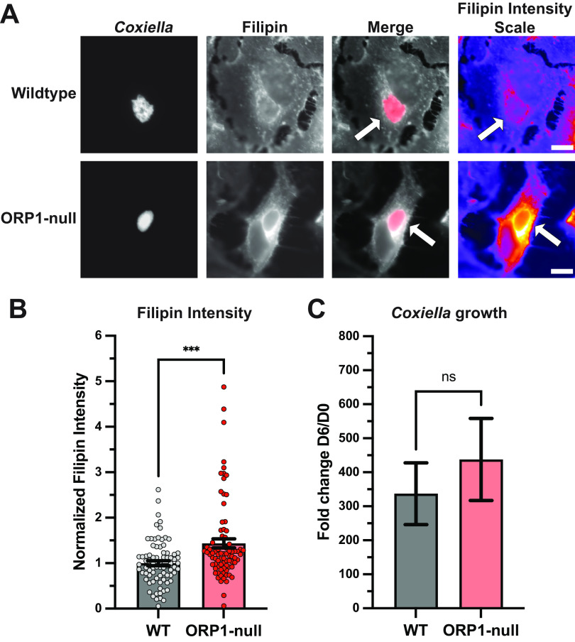
Absence of ORP1 alters CCV sterol content in HeLa cells. (A) Representative images of wild-type and ORP1-null HeLa cells infected with mCherry-expressing C. burnetii for 4 days, then stained with filipin, a fluorescent sterol stain. Filipin intensity scale images show strong filipin signal on the CCV in ORP1-null cells. White arrows indicate CCVs. Scale bars, 10 μm. (B) Filipin intensity in CCVs of HeLa and ORP1-null cells was measured using ImageJ. Images were acquired under identical settings. Intensity values were normalized to area, and background intensity was subtracted from CCV intensity. Data shown are mean ± SEM of at least 20 cells per condition in each of four independent experiments as determined by a nonparametric Mann-Whitney test. ***, P < 0.001. (C) C. burnetii growth was determined by CFU assay at 4 days postinfection in wild-type and ORP1-null HeLa cells, then normalized to day zero values to control for initial infection. Data shown are means ± SEM of eight independent experiments; in each independent experiment, each condition was performed at least in duplicate. No statistically significant differences were observed between wild-type HeLa cells and ORP1-null cells at any time point. Data were analyzed using an unpaired two-tailed t test.

