Abstract
The presence of coagulopathy as part of the systemic inflammatory response syndrome is a characteristic feature of severe coronavirus disease 2019 (COVID-19). Hematological changes (increased D-dimer [DD], prolonged activated partial thromboplastin clotting time [APTT] and prothrombin time [PT], high fibrinogen levels) have been observed in hospitalized patients with COVID-19, which characterize the risk of thrombotic events. Against the background of COVID-19 there is endothelial dysfunction, hypoxia and pulmonary congestion, mediated by thrombosis and microvascular occlusion. Up to 71.4% of patients who died from COVID-19 had disseminated intravascular coagulation syndrome, compared with only 0.6% of survivors. The main manifestation of COVID-19-associated coagulopathy is a significant increase in DD without a decrease in platelet count or prolongation of APTT and PT, indicating increased thrombin formation and the development of local fibrinolysis. An increase in DD levels of more than 3–4 times was associated with higher in-hospital mortality. Therefore, COVID-19 requires assessment of the severity of the disease for further tactics of thromboprophylaxis. The need for continued thromboprophylaxis, or therapeutic anticoagulation, in patients after inpatient treatment for two weeks using imaging techniques to assess of thrombosis assessment.
Keywords: coronavirus disease 2019 (COVID-19) infection, COVID-19-induced coagulopathy, SARS-CoV-2
Introduction
Since 2019, the outbreak of the then new coronavirus disease 2019 (COVID-19), which first originated at Wuhan, located in the Chinese province of Hubei, has spread around the world, and on 11 March 2020, the World Health Organization (WHO) announced the new COVID-19 as a pandemic [1, 2].
The number of confirmed cases and deaths from COVID-19 disease started to grow each day [3]. Mortality rates vary from country to country and depend on the capacity and effectiveness of the health care system. Since the beginning of the pandemic, Iran has been and is still considered a high-risk area with a much higher mortality rate than most cases in China [4–6].
The clinical spectrum of infection with the new severe acute respiratory syndrome coronavirus 2 (SARS-CoV-2) is different, ranging from the absence of any symptoms to lethal septic shock. 81% of all reported cases of COVID-19 by children and adults had a mild course of the disease, 14% — severe (eg, shortness of breath or oxygen saturation ≤ 93%) and 5% — critical condition (respiratory failure, septic shock, death) [7, 8].
The transition from mild to severe in patients with COVID-19 may be caused by cytokine storms and increased hypercoagulation with a significant risk of thromboembolic complications, affecting mainly venous, but are also registered in the arterial system [9–11].
The presence of comorbidities (cardiovascular, obesity, diabetes) that contribute to the development of coagulopathy, including those caused by sepsis, increased levels of D-dimer (DD), C-reactive protein (CRP), troponins and other markers of disseminated intravascular coagulation (DIC) in more than 6 times from the reference, is associated with a worse prognosis in hospitalized patients with severe coronavirus COVID-19, reaching 42% of in-hospital mortality [7].
A new term for COVID-induced coagulopathy (CIC) has been introduced to describe changes in blood clotting in patients with COVID-19 [12]. Characteristic laboratory data of CIC are observed, namely, elevated levels of DD and fibrin degradation products, which indicate an enhanced thrombotic state with high fibrin turnover. However, other CIC markers remain relatively unchanged.
Initial anticoagulant therapy, especially by direct injection, reduces mortality by 48% after 7 days and by 37% after 28 days and achieves a significant improvement in inhaled oxygen/O2 (PaO2/FiO2) by mitigating microthrombi and associated pulmonary coagulopathy [13].
Most protocols for the prevention of venous thromboembolism (VTE) with the use of therapeutic (full) doses of anticoagulants have been introduced in many countries [14–23], mainly for hospitalized adult patients. However, there are a limited number of publications on thromboprophylaxis in pediatric patients [23–26].
The present study offers a practical approach to CIC in patients with COVID-19, based on experience in the current literature and of our own experience in the treatment of patients hospitalized in local institutions.
Epidemiology
Since the outbreak of COVID-19 in December of 2019, which originated in the city of Wuhan, in the Chinese province of Hubei, this disease has spread around the world [7, 27]. On February 11, 2020, the International Coronavirus Taxonomy Study Group officially named the new coronavirus that causes COVID-19 “SARS-CoV-2”, and on March 11, 2020, the WHO announced a new COVID-19 pandemic [4–6, 8, 12, 14, 25, 28], which had its epicenter on the Asian continent (China), which later moved to the European (mainly to Italy and Spain), and then — to the Americas, first to the United States, and is now present in the United States and Latin America (Mexico and Brazil).
SARS-CoV-2 is a single-stranded RNA coronavirus that enters the cell mainly through binding to the type 2 angiotensin-converting enzyme (ACE-2) receptor, which is abundant in the alveolar clitoris, lungs, cardiac myocytes, vascular endothelium and other cells [29]. SARS-CoV-2 is transmitted mainly by inhalation of viral particles and entry into the respiratory tract. This virus can survive up to 24–72 hours on the surfaces of various objects indoors, which contributes to its spread [9, 19, 27, 30, 31].
Respiratory viral infection caused by COVID-19 is usually asymptomatic or with mild symptoms including fever, cough, fatigue, shortness of breath, diarrhea, headache and myalgia (up to 81.4% of patients). Severe cases are characterized by an increase in respiratory failure with a respiratory rate > 30 per minute, a decrease in saturation < 93% at rest, PaO2/FiO2 < 300 mmHg and infiltration > 50% of the lungs within 24–48 hours (up to 13.9% of patients) and may progress to the development of a critical condition (up to 4.7% of patients), demonstrating rapid deterioration and development of acute respiratory distress syndrome, septic shock, metabolic acidosis and coagulopathy, including cytokine storm [8, 12, 14–19, 32–36].
These clinical manifestations and paraclinical signs change with the further development of the pandemic worldwide, and also depend on the severity of the infection.
Coagulopathy
The most persistent hemostatic changes in COVID-19 are thrombocytopenia and increased DD [37], which are associated with the development of liver failure due to active inflammation in the lungs, intestines, myocardium, resulting in frequent use of ventilation requiring intensive care. In some cases this disease leads to the death of patients. Elderly patients with comorbidities with higher levels of DD had a higher risk of nosocomial mortality [20–22]. Given the clinical consequences of elevated DD levels, even in the absence of other serious symptoms, inpatient treatment should be considered, as this indicates an increase in thrombin levels and a higher risk of COVID-induced complications.
Blood clotting profile studies should be performed in hospitalized patients with suspected or confirmed COVID-19, including DD, activated partial thromboplastin clotting time (APTT), prothrombin time (PT), platelet count (PLT), and fibrinogen levels (Fib). Changes in these parameters may occur 7–11 days after the onset of symptoms or 4–10 days after hospitalization. It is important to repeat the study of these indicators to detect coagulopathy (DD, APTT, PT, Fib, PLT) in patients with severe COVID-19 at least every 2–3 days [9, 21, 38–44].
The combination of thrombocytopenia, prolonged prolongation of APTT and PT, increased DD indicates the development of DIC, the manifestations of which differ from those observed in septic processes of other etiologies, where thrombocytopenia is more pronounced and does not reach DD values observed in COVID-19. Recent evidence suggests that COVID-19-associated coagulopathy is a combination of low-grade DIC and pulmonary thrombotic microangiopathy, which has a significant effect on organ dysfunction in patients with the most severe condition [20, 45].
The presence of coagulopathy as part of the systemic inflammatory response syndrome is a common sign of severe COVID-19. Importantly, hematological changes in coagulation tests (elevated DD-dimer, prolonged APTT and PT, thrombocytopenia, and/or low fibrinogen levels) were observed in hospitalized patients with COVID-19, characterizing thrombotic events rather than hemorrhagic events. That is, COVID discoagulopathy causes thrombotic complications, in particular, VTE). Against the background of CIC, and hence endothelial dysfunction, there is hypoxia and pulmonary congestion, mediated by thrombosis and microvascular occlusion (including cerebrovascular events, limb ischemia, etc.) [9, 16, 21, 23–25, 46].
In patients who died of COVID-19, fibrin and thrombin deposition occurred predominantly in the pulmonary microcirculatory tract, which contributed to the development of acute respiratory distress syndrome and coagulopathy. In addition, hypoxia due to severe COVID-19 exacerbated thrombosis not only due to increased blood viscosity but also through a signaling pathway dependent on the transcription factor induced in the development of hypoxia [16, 23, 26, 47–52].
SARS-CoV-2 infection develops endothelial disease with profound microcirculatory changes, in which there is excessive thrombin formation and impaired fibrinolysis, similar to endothelial dysfunction in coagulopathy caused by sepsis (SIC). The receptor for viral adhesion is ACE-2 on endothelial cells, and virus replication causes inflammatory cell infiltration, endothelial apoptosis, and microvascular prothrombotic events. Viral inclusions in endothelial cells and infiltration by mononuclear and polymorphonuclear cells with evidence of endothelial apoptosis were observed in postmortem analysis of SARS-CoV-2 infection. Impaired microcirculation contributes to the development of severe clinical consequences in patients with COVID-19 [9, 16, 53, 54]. Other abnormalities that may be relevant in the context of coagulopathy are decreased fibrinogen levels, increased lactate dehydrogenase, and, in some patients, marked increases in serum ferritin levels [26, 55, 56].
Another important feature of COVID-19 infection is the development of an acute procoagulant response in elevated levels of FVIII, von Willebrand, and fibrinogen, which are associated with an increased risk of thrombosis. In severe stages of the disease there is an increase in inflammatory cytokines (tumor necrosis factor [FNO-α] and interleukins [IL]: 1 and 6). IL-6 induces the expression of tissue factor in macrophages, which initiates the activation of coagulation and thrombin formation [32]. FNO-α and IL-1 are major mediators of endogenous coagulation inhibition. Severely compromised patients with COVID-19 may experience a cytokine storm characterized by high concentrations of proinflammatory cytokines and chemokines [18, 20, 32].
The International Society for Thrombosis and Hemostasis (ISTH) has proposed a new category for the detection of early-stage DIC associated with sepsis, called SIC. This indicator can be used in patients with COVID-19, and in those who meet these criteria, it is optimal to use anticoagulants.
Up to 71.4% of patients who died from COVID-19 had an ICE, compared with only 0.6% of survivors. The main change in this coagulopathy is a marked increase in DD without a decrease in PLT, or a prolongation of APTT and PT, indicating the process of thrombin formation and local rather than systemic fibrinolysis. The highest level of hospital mortality was found in patients with DD values more than 3–4 times [24, 26, 28, 38, 39].
Given the occurrence of signs of coagulation system dysfunction in COVID-19, it is proposed to assess the severity of the disease (COVID-19-associated hemostatic abnormalities — CAHA-score) [28, 30, 41, 57–59]:
— Stage 1 CAHA: The patient has mild symptoms and can be cared for at home or in hospital. At this stage, pulmonary microthrombi may be missed on computed tomography;
— Stage 2 CAHA: The patient may develop more severe symptoms and need resuscitation. Computed tomography scans of these patients may reveal lung filling defects due to blood clots or embolism. They may also have asymptomatic deep vein thrombosis in the lower extremities, which means significant activation of coagulation;
— Stage 3 CAHA: The patient’s clinical condition is deteriorating, which requires the maintenance of a higher level of intensive care. Extensive pulmonary thrombi and systemic thrombosis, including DIC, have been reported.
Thus, there is a need to identify an increased risk of thrombotic events at an early stage and to prevent thrombotic events and organ damage as much as possible.
Deterioration of laboratory parameters associated with blood coagulation indicates the progression of the severity of COVID-19 infection and suggests the need for greater and more aggressive intensive care, while improvement in these parameters along with improvement or clinical stability indicates an adequate evolution [24].
Thromboprophylaxis
Hospitalized patients with COVID-19 have both internal and external risk factors for thrombosis, including old age, obesity, immobilization, neurological disease, cancer, intensive care, previous cases of thromboembolism or thrombophilia [42].
Pharmacological thromboprophylaxis should be considered in all hospitalized patients with COVID-19 if there are no contraindications (active bleeding or severe thrombocytopenia). Different scales (Padua, Caprini, IMPROVE) can be used to assess risk. The dose should be adjusted according to renal function. Although existing protocols should be followed when selecting the drug, the WHO recommends the use of unfractionated or low molecular weight heparins (LMWH) and, if contraindicated, mechanical thromboprophylaxis should be considered. Pharmacological thromboprophylaxis is recommended once a day, as it reduces the risk of skipping additional doses and is associated with less impact on medical personnel during its use. If LMWH is not available, unfractionated heparin may be considered, keeping in mind that this requires more frequent injections. Patients with more severe infections may require higher doses of thromboprophylaxis due to their hypercoagulable state. The use of direct anticoagulants for thromboprophylaxis is not recommended in this context due to the possible interactions that may occur with the various drugs and treatments available and studied for the treatment of COVID-19 [5, 20, 21, 42].
Some of the non-anticoagulant properties of LMWH include the potential for binding to inflammatory cytokines, inhibition of neutrophil chemotaxis and leukocyte migration, neutralization of positively charged complement factor C5a, and sequestration of acute phase proteins [18, 44].
In view of the above it is suggested that LMWH, administered in the early stages of SARS-CoV-2 infection may have a positive effect not only in preventing thrombosis, but also in reducing systemic and pulmonary inflammation and limiting viral invasion [12, 19], among others. Non-anticoagulant properties of heparin: antiviral action (experimental models), reduction of collagen deposits and antiarrhythmic action (animal models), modulation of endothelial dysfunction, improvement of microvascular dysfunction and alleviation of pulmonary coagulopathy [33, 42].
Patients who remain fully immobilized may benefit from drug thromboprophylaxis due to periodic pneumatic compression. This therapy should also be considered if there is severe thrombocytopenia (platelets from 25 G/L to 50 G/L) [8, 34, 35, 42].
According to the authors [8, 27, 42], long-term outpatient thromboprophylaxis (14 to 45 days) should be considered in patients at high risk of deep vein thrombosis, regardless of COVID-19 infection, including reduced mobility, previous thromboembolic events, comorbidities (eg. active cancer) and elevated DD levels of more than 2 times). Thromboprophylaxis is not recommended for patients in quarantine due to a mild form of COVID-19 but with significant comorbidities, or patients without COVID-19 but functionally limited in quarantine. These patients should be advised to stay active at home.
Pharmacological thromboprophylaxis in adult patients with COVID-19
The results of studies on the prevention of deep vein thrombosis in adult patients in critical condition with COVID-19-associated pneumonia have been published [10, 35, 36].
Histopathological examinations revealed diffuse lesions of the alveoli with deep inflammation, thrombosis and thrombotic microangiopathy of small vessels and capillaries of the lungs. In extrapulmonary organs, endothelial cell damage and diffuse microvascular thrombosis have also been reported, indicating thrombotic microangiopathy, which may explain the acute onset of multiorgan failure [10, 35, 36].
According to current guidelines, patients with stage 1 CAHA should receive LMWH in the absence of contraindications [35, 41]. Several trials are currently underway for patients with treatment disorders corresponding to stage 2 CAHA to determine whether a full dose of anticoagulants compared to prophylactic can help prevent the development of coagulopathy and ischemia in the extrapulmonary circulation. After the detection of blood clots, the standard practice is to treat such patients with anticoagulants in a therapeutic dose [35, 41, 42]. Intensification of antithrombotic therapy (prevention of double-dose LMWH) in stage 2 CAHA can be performed in combination with several other experimental measures, such as thrombolysis, which may be effective in patients in stage 3 CAHA [41].
The developed guidelines provide guidance to physicians caring for both COVID-19 patients and patients with chronic thrombotic conditions requiring ongoing treatment. Recently, there has been published work focusing on anticoagulant strategies for the prevention and treatment of deep vein thrombosis in patients with proven SARS-CoV-2 infection who are in outpatient treatment and hospitalized for the prevention or treatment of thrombosis [35, 42].
Pharmacological thromboprophylaxis in pediatric and adolescent patients with COVID-19
Rare deaths of children from COVID-19 infection have been reported in worldwide [60]. Most often diagnosed with asymptomatic disease, or with mild or moderate severity [46, 61]. As in adults, the risk of serious illness and death in children is higher in people with comorbidities [47, 62]. In general, the experience of monitoring infants and children with COVID-19 in hospitals is limited [63].
From the standpoint that a significant number of clinical issues deserve further study and clarification, we have prepared general recommendations for thromboprophylaxis childrens regimens.
Outpatients with mild COVID-19 should not be prescribed pharmacological thromboprophylaxis, but it is important to encourage increased mobility of the child and to control adequate hydration in the presence of fever or vomiting. Given the rapid deterioration reported in many adult patients with mild symptoms, regular monitoring of DD, Fib, PLT, PT and APTT every 48 hours for 5–7 days may be recommended. Patients with DD levels above 300 ng/mL have a high risk of thrombosis and should receive LMWH prophylaxis with continuous assessment of deep vein thrombosis.
Patients with moderate COVID-19 who require hospitalization should receive anticoagulant therapy [64] using prophylactic doses of LMWH.
Intensification of anticoagulant therapy is recommended in case of severe COVID-19 (DD > > 500 ng/mL, serum ferritin > 500 ng/mL, worsening of clinical signs of disease). Especially when the patient is in critical condition (DD > 2500 ng/mL, PLT > 450 G/L and CRP > 100 mg/dL; Table 1).
Table 1.
Anticoagulant prophylaxis with Enoxaparin for patients with moderate, severe and critical form of COVID-19, which require hospitalization.
| Profile dose | Prophylactic intensified dose | Treatment dose | |
|---|---|---|---|
| Target anti-Xa | 0.2–0,4 U/mL | 0.4–0.8 U/mL | 0.6–1.1 U/mL |
| ≤ 2 months | 0.75 mg/kg, every 12 h | 1.0 mg/kg, every 12 h | 1.5 mg/kg, every 12 h |
| From 2 months till ≤ 18 years: | |||
| < 40 kg | 0.5 mg/kg, every 12 h | 0.75 mg/kg, every 12 h | 1.0 mg/kg, every 12 h |
| > 40 kg | 40 mg, 1 time a day | 60 mg, every 12 h | 40 mg, every 12 h |
Coagulation should be closely monitored during treatment to prevent excessive anticoagulant therapy and bleeding [65, 66]. Anti-Xa levels and ARTT ratios are good ways to determine the effectiveness of LMWH in patients with confirmed deep vein thrombosis. After reaching the desired level, the test can be repeated every 6–7 days. If an invasive procedure is required, it is recommended to skip two doses of LMWH before the procedure [26].
In patients with renal insufficiency, unfractionated heparins of 75 IU/kg intravenous infusion over 10 minutes (maximum 5000 IU) as the loading dose are the anticoagulants of choice. This should be followed by a continuous maintenance intravenous infusion (maximum initial rate of 1300 IU/h). The recommended infusion rate is 28 IU/kg/h in infants and children under 12 months of age, 20 IU/kg/h in children 1 to 15 years of age, and 18 IU/kg/h in children 16 years of age and older.
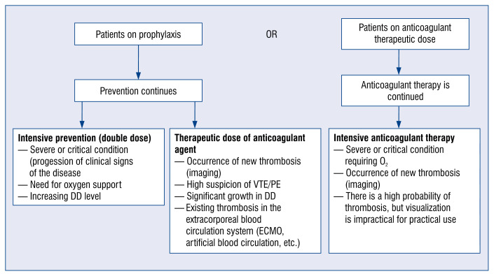
For patients already receiving anticoagulant therapy, the proposed algorithm approach is illustrated in Figure 1.
Figure 1.
Algorithm for the use of anticoagulants in coronavirus disease 2019 (COVID-19) patients with ≤ 18 years who have already taken an anticoagulant. Heparin resistance in COVID-19: if the heparin dose is > 25 IU/kg/day and there is no activated partial thromboplastin clotting time prolongation anti-Xa monitoring at the level of 0.3–0.7 IU/mL; DD — D-dimer; ECMO — extracorporeal membrane oxygenation; PE — pulmonary embolism; VTE — venous thromboembolism.
Transferring a patient from an inpatient setting to an outpatient setting is the next step, including reassessing anticoagulant therapy and ensuring adequate communication between physicians, the patient, family members, or caregivers. The use of thromboprophylaxis in patients with COVID-19 after inpatient treatment should be considered if the stratification of deep vein thrombolism risk indicates a persistently increased risk of co-morbidity due to comorbidities, as well as a concentration twice or more above the upper control range. This is evidenced by studies suggesting thromboprophylaxis or therapeutic anticoagulation in patients after inpatient treatment. At least 2 weeks of anticoagulant therapy and imaging studies to assess thrombosis are recommended, as well as extended anticoagulant therapy [49].
Conclusions
Severe complications and a high number of deaths due to COVID-19 have once again drawn the attention of experts to the prevention and treatment of thrombotic and thromboembolic diseases.
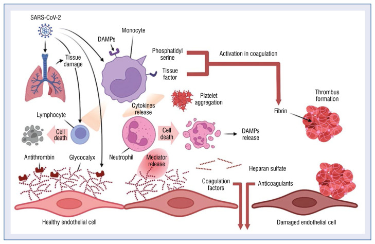
The direct and indirect effects of COVID-19 are associated with the development of a cytokine storm, which accelerates the onset of systemic inflammatory response syndrome and promotes thrombotic events (Central illustration).
Central illustration.
Mechanism of thrombus formation during COVID-19 infection; DAMP — danger/damage associated molecular patterns; SARS-CoV-2 — severe acute respiratory syndrome coronavirus 2.
Antiviral and pathogenetic agents of COVID-19 (lopinavir/ritonavir, remdesivir, tocilizumab, sarylumab, fingolimod, chloroquine/hydroxychloroquine, interferon, azithromycin) may have drug interactions with antiplatelet agents and/or anticoagulants.
At the same time, recommendations for social distancing may adversely affect the treatment of patients without COVID-19 who have thrombotic events, and fear of developing COVID-19 or complications may result in some patients not receiving or discontinuing anticoagulant therapy.
Some clinical issues deserve further research and refinement to better understand the specific features, needs and concerns of critically ill children and adults with COVID-19 infection, especially those who already have comorbidities.
Protocols for thromboprophylaxis, in particular the use of anticoagulants or additional considerations for the treatment of covid-induced coagulopathy, should be used in accordance with the latest national and international guidelines.
Footnotes
Conflict of interest: None declared
References
- 1.Ruetzler K, Szarpak L, Filipiak K, et al. The COVID-19 pandemic — a view of the current state of the problem. Disaster Emerg Med J. 2020;5(2):106–107. doi: 10.5603/demj.a2020.0015.. [DOI] [Google Scholar]
- 2.Dzieciatkowski T, Szarpak L, Filipiak KJ, et al. COVID-19 challenge for modern medicine. Cardiol J. 2020;27(2):175–183. doi: 10.5603/CJ.a2020.0055. [DOI] [PMC free article] [PubMed] [Google Scholar]
- 3.Merajikhah A, Beigi-khoozani A, Soleimani M. Risk of spreading delta coronavirus to operating room personnel after COVID-19 vaccination. Disaster Emerg Med J. 2021;6(4):206–207. doi: 10.5603/demj.a2021.0026.. [DOI] [Google Scholar]
- 4.Aguiar D, Lobrinus JA, Schibler M, et al. Inside the lungs of COVID-19 disease. Int J Legal Med. 2020;134(4):1271–1274. doi: 10.1007/s00414-020-02318-9. [DOI] [PMC free article] [PubMed] [Google Scholar]
- 5.American Society of Hematology. [Accessed May 5, 2020]. https://www.hematology.org/covid-19/covid-19-and-coagulopathy .
- 6.Branchford BR, Betensky M, Goldenberg NA. Pediatric issues in thrombosis and hemostasis: The how and why of venous thromboembolism risk stratification in hospitalized children. Thromb Res. 2018;172:190–193. doi: 10.1016/j.thromres.2018.02.010. [DOI] [PubMed] [Google Scholar]
- 7.Smereka J, Szarpak L, Filipiak K. Modern medicine in COVID-19 era. Disaster Emerg Med J. 2020 doi: 10.5603/demj.a2020.0012.. [DOI] [Google Scholar]
- 8.Ackermann M, Verleden SE, Kuehnel M, et al. Pulmonary vascular endothelialitis, thrombosis, and angiogenesis in COVID-19. N Engl J Med. 2020;383(2):120–128. doi: 10.1056/NEJMoa2015432. [DOI] [PMC free article] [PubMed] [Google Scholar]
- 9.Branchford BR, Mourani P, Bajaj L, et al. Risk factors for inhospital venous thromboembolism in children: a case-control study employing diagnostic validation. Haematologica. 2012;97(4):509–515. doi: 10.3324/haematol.2011.054775. [DOI] [PMC free article] [PubMed] [Google Scholar]
- 10.Thachil J, Tang N, Gando S, et al. ISTH interim guidance on recognition and management of coagulopathy in COVID-19. J Thromb Haemost. 2020;18(5):1023–1026. doi: 10.1111/jth.14810. [DOI] [PMC free article] [PubMed] [Google Scholar]
- 11.Zhou Y, Hou Y, Shen J, et al. Network-based drug repurposing for novel coronavirus 2019-nCoV/SARS-CoV-2. Cell Discov. 2020;6:14. doi: 10.1038/s41421-020-0153-3. [DOI] [PMC free article] [PubMed] [Google Scholar]
- 12.BSH Haemostatis and Thrombosis Task Force. [Accessed May 5, 2020]. https://b-s-h.org.uk/media/18206/dic-score-in-covid-19-pneumonia_01-04-2020.pdf .
- 13.Gómez-Mesa JE, Galindo-Coral S, Montes MC, et al. Thrombosis and Coagulopathy in COVID-19. Curr Probl Cardiol. 2021;46(3):100742. doi: 10.1016/j.cpcardiol.2020.100742. [DOI] [PMC free article] [PubMed] [Google Scholar]
- 14.Cao X. COVID-19: immunopathology and its implications for therapy. Nat Rev Immunol. 2020;20(5):269–270. doi: 10.1038/s41577-020-0308-3. [DOI] [PMC free article] [PubMed] [Google Scholar]
- 15.Fan E, Beitler J, Brochard L, et al. COVID-19-associated acute respiratory distress syndrome: is a different approach to management warranted? Lancet Respir Med. 2020;8(8):816–821. doi: 10.1016/s2213-260020303040. [DOI] [PMC free article] [PubMed] [Google Scholar]
- 16.Faustino EV, Raffini LJ. Prevention of hospital-acquired venous thromboembolism in children: a review of published guidelines. Front Pediatr. 2017;5:9. doi: 10.3389/fped.2017.00009. [DOI] [PMC free article] [PubMed] [Google Scholar]
- 17.Hoffmann M, Kleine-Weber H, Schroeder S, et al. SARS-CoV-2 cell entry depends on ACE2 and TMPRSS2 and is blocked by a clinically proven protease inhibitor. Cell. 2020;181(2):271–280e8. doi: 10.1016/j.cell.2020.02.052. [DOI] [PMC free article] [PubMed] [Google Scholar]
- 18.Hou YJ, Okuda K, Edwards CE, et al. SARS-CoV-2 reverse genetics reveals a variable infection gradient in the respiratory tract. Cell. 2020;182(2):429–446e14. doi: 10.1016/j.cell.2020.05.042. [DOI] [PMC free article] [PubMed] [Google Scholar]
- 19.Huang C, Wang Y, Li X, et al. Clinical features of patients infected with 2019 novel coronavirus in Wuhan, China. Lancet. 2020;395(10223):497–506. doi: 10.1016/s0140-6736(20)30183-5. [DOI] [PMC free article] [PubMed] [Google Scholar]
- 20.Jaffray J, Witmer C, O’Brien SH, et al. Peripherally inserted central catheters lead to a high risk of venous thromboembolism in children. Blood. 2020;135(3):220–226. doi: 10.1182/blood.2019002260. [DOI] [PubMed] [Google Scholar]
- 21.Jaimes JA, Millet JK, Whittaker GR. Proteolytic cleavage of the SARS-CoV-2 spike protein and the role of the novel S1/S2 site. iScience. 2020;23(6):101212. doi: 10.1016/j.isci.2020.101212. [DOI] [PMC free article] [PubMed] [Google Scholar]
- 22.Kirchdoerfer RN, Ward AB. Structure of the SARS-CoV nsp12 polymerase bound to nsp7 and nsp8 co-factors. Nat Commun. 2019;10(1):2342. doi: 10.1038/s41467-019-10280-3. [DOI] [PMC free article] [PubMed] [Google Scholar]
- 23.Li Q, Guan X, Wu P, et al. Early transmission dynamics in Wuhan, china, of novel coronavirus-infected pneumonia. N Engl J Med. 2020;382(13):1199–1207. doi: 10.1056/NEJMoa2001316. [DOI] [PMC free article] [PubMed] [Google Scholar]
- 24.Yu Li, Zhang Z, Yang Li, et al. The MERS-CoV receptor DPP4 as a candidate binding target of the SARS-CoV-2 spike. iScience. 2020;23(6):101160. doi: 10.1016/j.isci.2020.101160. [DOI] [PMC free article] [PubMed] [Google Scholar]
- 25.Mahajerin A, Branchford BR, Amankwah EK, et al. Hospital-associated venous thromboembolism in pediatrics: a systematic review and meta-analysis of risk factors and risk-assessment models. Haematologica. 2015;100(8):1045–1050. doi: 10.3324/haematol.2015.123455. [DOI] [PMC free article] [PubMed] [Google Scholar]
- 26.Marini JJ, Gattinoni L. Management of COVID-19 respiratory distress. JAMA. 2020;323(22):2329–2330. doi: 10.1001/jama.2020.6825. [DOI] [PubMed] [Google Scholar]
- 27.Andersen KG, Rambaut A, Lipkin WI, et al. The proximal origin of SARS-CoV-2. Nat Med. 2020;26(4):450–452. doi: 10.1038/s41591-020-0820-9. [DOI] [PMC free article] [PubMed] [Google Scholar]
- 28.Xu Z, Shi L, Wang Y, et al. Pathological findings of COVID-19 associated with acute respiratory distress syndrome. Lancet Respir Med. 2020;8(4):420–422. doi: 10.1016/s2213-2600(20)30076-x. [DOI] [PMC free article] [PubMed] [Google Scholar]
- 29.Szarpak L, Pruc M, Filipiak KJ, et al. Myocarditis: A complication of COVID-19 and long-COVID-19 syndrome as a serious threat in modern cardiology. Cardiol J. 2022;29(1):178–179. doi: 10.5603/CJ.a2021.0155. [DOI] [PMC free article] [PubMed] [Google Scholar]
- 30.Zhang YZ, Holmes EC. A genomic perspective on the origin and emergence of SARS-CoV-2. Cell. 2020;181(2):223–227. doi: 10.1016/j.cell.2020.03.035. [DOI] [PMC free article] [PubMed] [Google Scholar]
- 31.Nucera G, Chirico F, Rafique Z, et al. Need to update cardiological guidelines to prevent COVID-19 related myocardial infarction and ischemic stroke. Cardiol J. 2022;29(1):174–175. doi: 10.5603/CJ.a2021.0120. [DOI] [PMC free article] [PubMed] [Google Scholar]
- 32.Szarpak Ł, Nowak B, Kosior D, et al. Cytokines as predictors of COVID-19 severity: evidence from a meta-analysis. Pol Arch Intern Med. 2021;131(1):98–99. doi: 10.20452/pamw.15685. [DOI] [PubMed] [Google Scholar]
- 33.Song JC, Wang G, Zhang W, et al. Chinese expert consensus on diagnosis and treatment of coagulation dysfunction in COVID-19. Mil Med Res. 2020;7(1):19. doi: 10.1186/s40779-020-00247-7. [DOI] [PMC free article] [PubMed] [Google Scholar]
- 34.Sungnak W, Huang Ni, Bécavin C, et al. SARS-CoV-2 entry factors are highly expressed in nasal epithelial cells together with innate immune genes. Nat Med. 2020;26(5):681–687. doi: 10.1038/s41591-020-0868-6. [DOI] [PMC free article] [PubMed] [Google Scholar]
- 35.Tang N, Li D, Wang X, et al. Abnormal coagulation parameters are associated with poor prognosis in patients with novel coronavirus pneumonia. J Thromb Haemost. 2020;18(4):844–847. doi: 10.1111/jth.14768. [DOI] [PMC free article] [PubMed] [Google Scholar]
- 36.Taylor F, Toh CH, Hoots K, et al. Towards definition, clinical and laboratory criteria, and a scoring system for disseminated intravascular coagulation. J Thromb Haemost. 2017;86(11):1327–1330. doi: 10.1055/s-0037-1616068.. [DOI] [PubMed] [Google Scholar]
- 37.Ruetzler K, Szarpak Ł, Ładny JR, et al. D-dimer levels predict COVID-19 severity and mortality. Kardiol Pol. 2021;79(2):217–218. doi: 10.33963/KP.15830. [DOI] [PubMed] [Google Scholar]
- 38.Perico L, Benigni A, Remuzzi G. Should COVID-19 concern nephrologists? Why and to what extent? The emerging impasse of angiotensin blockade. Nephron. 2020;144(5):213–221. doi: 10.1159/000507305. [DOI] [PMC free article] [PubMed] [Google Scholar]
- 39.Pernazza A, Mancini M, Rullo E, et al. Early histologic findings of pulmonary SARS-CoV-2 infection detected in a surgical specimen. Virchows Arch. 2020;477(5):743–748. doi: 10.1007/s00428-020-02829-1. [DOI] [PMC free article] [PubMed] [Google Scholar]
- 40.Ranucci M, Ballotta A, Di Dedda U, et al. The procoagulant pattern of patients with COVID-19 acute respiratory distress syndrome. J Thromb Haemost. 2020;18(7):1747–1751. doi: 10.1111/jth.14854. [DOI] [PMC free article] [PubMed] [Google Scholar]
- 41.Shang J, Wan Y, Luo C, et al. Cell entry mechanisms of SARS-CoV-2. Proc Natl Acad Sci U S A. 2020;117(21):11727–11734. doi: 10.1073/pnas.2003138117. [DOI] [PMC free article] [PubMed] [Google Scholar]
- 42.Shi J, Wen Z, Zhong G, et al. Susceptibility of ferrets, cats, dogs, and other domesticated animals to SARS-coronavirus 2. Science. 2020;368(6494):1016–1020. doi: 10.1126/science.abb7015. [DOI] [PMC free article] [PubMed] [Google Scholar]
- 43.Katipoğlu B, Sönmez LÖ, Vatansev H, et al. Can hematological and biochemical parameters fasten the diagnosis of COVID-19 in emergency departments? Disaster Emerg Med J. 2020 doi: 10.5603/demj.a2020.0039.. [DOI] [Google Scholar]
- 44.Shi Y, Wang Y, Shao C, et al. COVID-19 infection: the perspectives on immune responses. Cell Death Differ. 2020;27(5):1451–1454. doi: 10.1038/s41418-020-0530-3. [DOI] [PMC free article] [PubMed] [Google Scholar]
- 45.Ziegler CGK, Allon SJ, Nyquist SK, et al. SARS-CoV-2 receptor ACE2 is an interferon-stimulated gene in human airway epithelial cells and is detected in specific cell subsets across tissues. Cell. 2020;181(5):1016–1035e19. doi: 10.1016/j.cell.2020.04.035. [DOI] [PMC free article] [PubMed] [Google Scholar]
- 46.Toh CH, Hoots WK. The scoring system of the Scientific and Standardisation Committee on Disseminated Intravascular Coagulation of the International Society on Thrombosis and Haemostasis: a 5-year overview. J Thromb Haemost. 2007;5(3):604–606. doi: 10.1111/j.1538-7836.2007.02313.x. [DOI] [PubMed] [Google Scholar]
- 47.Wan Y, Shang J, Graham R, et al. Receptor recognition by the novel coronavirus from Wuhan: an analysis based on decade-long structural studies of SARS coronavirus. J Virol. 2020;94(7):e00127–20. doi: 10.1128/JVI.00127-20. [DOI] [PMC free article] [PubMed] [Google Scholar]
- 48.Wang J, Hajizadeh N, Moore EE, et al. Tissue plasminogen activator (tPA) treatment for COVID-19 associated acute respiratory distress syndrome (ARDS): A case series. J Thromb Haemost. 2020;18(7):1752–1755. doi: 10.1111/jth.14828. [DOI] [PMC free article] [PubMed] [Google Scholar]
- 49.Wang Y, Zhu LQ. Pharmaceutical care recommendations for antiviral treatments in children with coronavirus disease 2019. World J Pediatr. 2020;16(3):271–274. doi: 10.1007/s12519-020-00353-5. [DOI] [PMC free article] [PubMed] [Google Scholar]
- 50.Wölfel R, Corman VM, Guggemos W, et al. Virological assessment of hospitalized patients with COVID-2019. Nature. 2020;581(7809):465–469. doi: 10.1038/s41586-020-2196-x. [DOI] [PubMed] [Google Scholar]
- 51.Xiong Mi, Liang X, Wei YD. Changes in blood coagulation in patients with severe coronavirus disease 2019 (COVID-19): a meta-analysis. Br J Haematol. 2020;189(6):1050–1052. doi: 10.1111/bjh.16725. [DOI] [PMC free article] [PubMed] [Google Scholar]
- 52.Adam I, Szarpak L, Filipiak K, et al. Interferon lambda with remdesivir as a potential treatment option in COVID-19. Disaster Emerg Med J. 2020 doi: 10.5603/demj.a2020.0027.. [DOI] [Google Scholar]
- 53.Xu H, Zhong L, Deng J, et al. High expression of ACE2 receptor of 2019-nCoV on the epithelial cells of oral mucosa. Int J Oral Sci. 2020;12(1):8. doi: 10.1038/s41368-020-0074-x. [DOI] [PMC free article] [PubMed] [Google Scholar]
- 54.Zumla A, Chan JFW, Azhar EI, et al. Coronaviruses — drug discovery and therapeutic options. Nat Rev Drug Discov. 2016;15(5):327–347. doi: 10.1038/nrd.2015.37. [DOI] [PMC free article] [PubMed] [Google Scholar]
- 55.Fialek B, Pruc M, Smereka J, et al. Diagnostic value of lactate dehydrogenase in COVID-19: A systematic review and meta-analysis. Cardiol J. 2022;29(5):751–758. doi: 10.5603/CJ.a2022.0056. [DOI] [PMC free article] [PubMed] [Google Scholar]
- 56.Szarpak L, Zaczynski A, Kosior D, et al. Evidence of diagnostic value of ferritin in patients with COVID-19. Cardiol J. 2020;27(6):886–887. doi: 10.5603/CJ.a2020.0171. [DOI] [PMC free article] [PubMed] [Google Scholar]
- 57.Zhang L, Yan X, Fan Q, et al. D-dimer levels on admission to predict in-hospital mortality in patients with COVID-19. J Thromb Haemost. 2020;18(6):1324–1329. doi: 10.1111/jth.14859. [DOI] [PMC free article] [PubMed] [Google Scholar]
- 58.Zhou H, Chen X, Hu T, et al. A novel bat coronavirus closely related to SARS-CoV-2 contains natural insertions at the S1/S2 cleavage site of the spike protein. Curr Biol. 2020;30(11):2196–2203e3. doi: 10.1016/j.cub.2020.05.023. [DOI] [PMC free article] [PubMed] [Google Scholar]
- 59.Zhou P, Yang XL, Wang XG, et al. A pneumonia outbreak associated with a new coronavirus of probable bat origin. Nature. 2020;579(7798):270–273. doi: 10.1038/s41586-020-2012-7. [DOI] [PMC free article] [PubMed] [Google Scholar]
- 60.Meyer-Szary J, Jaguszewski M, Smereka J, et al. Impact of COVID- 19 on pediatric out-of-hospital cardiac arrest in the Masovian region. Disaster Emerg Med J. 2021;6(4):183–185. doi: 10.5603/demj.a2021.0028.. [DOI] [Google Scholar]
- 61.Yaman E, Demirel B, Yilmaz A, et al. Retrospective evaluation of laboratory findings of suspected paediatric COVID-19 patients with positive and negative RT-PCR. Disaster Emerg Med J. 2021;6(3):97–103. doi: 10.5603/demj.a2021.0023.. [DOI] [Google Scholar]
- 62.Pruc M, Smereka J, Dzieciatkowski T, et al. Kawasaki disease shock syndrome or toxic shock syndrome in children and the relationship with COVID-19. Med Hypotheses. 2020;144:109986. doi: 10.1016/j.mehy.2020.109986. [DOI] [PMC free article] [PubMed] [Google Scholar]
- 63.Yaman E, Demirel B, Yilmaz A, et al. Retrospective evaluation of laboratory findings of suspected paediatric COVID-19 patients with positive and negative RT-PCR. Disaster Emerg Med J. 2021;6(3):97–103. doi: 10.5603/demj.a2021.0023.. [DOI] [Google Scholar]
- 64.Szarpak L, Filipiak KJ, Skwarek A, et al. Outcomes and mortality associated with atrial arrhythmias among patients hospitalized with COVID-19: A systematic review and meta-analysis. Cardiol J. 2022;29(1):33–43. doi: 10.5603/CJ.a2021.0167. [DOI] [PMC free article] [PubMed] [Google Scholar]
- 65.Domienik-Karłowicz J, Tronina O, Lisik W, et al. The use of anticoagulants in chronic kidney disease: Common point of view of cardiologists and nephrologists. Cardiol J. 2020;27(6):868–874. doi: 10.5603/CJ.a2019.0025. [DOI] [PMC free article] [PubMed] [Google Scholar]
- 66.Tomaszuk-Kazberuk A, Koziński M, Domienik-Karłowicz J, et al. Pharmacotherapy of atrial fibrillation in COVID-19 patients. Cardiol J. 2021;28(5):758–766. doi: 10.5603/CJ.a2021.0088. [DOI] [PMC free article] [PubMed] [Google Scholar]


