Abstract
Patient: Male, 36-year-old
Final Diagnosis: Corneal ulcer • perforated corneal ulcer
Symptoms: Eye pain • eye pain and redness • red eye • vision changes
Clinical Procedure: —
Specialty: Ophthalmology
Objective:
Management of emergency care
Background:
The use of amniotic membranes for corneal perforations using different surgical techniques has been widely described in the literature. This case report is a novel variation in the technique that can be useful for incorporating in clinical practice when the need arises.
Case Report:
A 36-year-old male patient presented to our clinic with a corneal ulcer in his left eye caused by herpetic keratitis, treated with a topical non-steroidal anti-inflammatory (indomethacin 0.1% solution). Examination revealed a paracentral 2-mm wide corneal perforation on the site of the corneal ulcer. The patient was admitted to the hospital. He was treated with intravenous piperacillin-ofloxacine, and an emergency surgical intervention using a lyophilized amniotic membrane was performed using a “plug and patch” technique. Postoperatively, the patient received 48 h of intravenous antibiotics and was discharged on topical antibiotic/corticosteroid eyedrops along with a 10-day course of oral antibiotics (ofloxacin) and antiviral therapy (valaciclovir). Three months after surgery, the anterior chamber was formed, the corneal defect was closed, and visual acuity improved. One year after initial presentation, anterior segment optical coherence tomography showed a large scarred but healed cornea.
Conclusions:
We report the successful use of combination of a single round-shaped rolled amniotic membrane with a multilayered amniotic membrane transplantation for the treatment of a 2-mm-wide perforated corneal ulcer. This technique allowed for preservation of the globe integrity without the need for a keratoplasty, stopped further tissue loss, and was associated with a rapid visual recovery.
Keywords: Biological Dressings; Corneal Perforation; Keratitis; Keratitis, Dendritic; Ophthalmology
Background
Amnion, the innermost layer of the placenta, consists of a single layer of epithelial cells that are joined to a substantial basement membrane and an avascular stromal matrix [1,2]. The therapeutic use of amniotic membranes was first described by Davis in 1910 [3] for skin transplantation. In the field of ophthalmology, de Rotth was the first to treat patients with symblepharon using amniotic membranes, in 1940 [4]. Subsequently, numerous reports appeared regarding their use for ocular surface reconstruction [5,6], as inlay grafts (epithelial side up) for corneal melting, and as an onlay bandage (epithelial-side down) to promote healing in cases of persistent epithelial defects and ocular surface inflammation. All of these indications make use of the amniotic membrane’s ability to promote healing [1] and of its anti-inflammatory, anti-fibrotic, and anti-angiogenic features [7].
Management of corneal perforations depends on the size of the defect, the underlying etiology, the surgeon’s experience, and the available treatment modalities. Different approaches have been described in the literature to replace the corneal tissue in case of perforation, including – but not limited to – cyanoacrylate or fibrin glue [8], donor corneal tissue, conjunctival flaps, and amniotic membrane grafts. Cyanoacrylate glue is highly inflammatory, and fibrin glue seems less effective and often insufficient by its own [9]. Surgical interventions, such as full-thickness keratoplasty, are often associated with an increased risk of rejection and ongoing melt in case of an inflamed host cornea [10]; their use in emergency cases is also widely limited by the short supply and the unavailability of the tissue [11]. On the other hand, many studies have reported on the successful use of amniotic membranes in the treatment of corneal perforation [5,10]. Multiple techniques have been described, such as multilayer surgery [12,13], sandwich technique (inlay and onlay) transplantation [14], and the “swiss roll” or “cigar” technique [14,15]. The reported healing rate was up to 100% for micro-perforations and 75% for perforations up to 1.5 mm wide [9]. Even though larger defects generally yield worse results, perforations up to 3 mm could also be adequately managed with amniotic membrane transplantation in association with fibrin glue [16], delaying the need for a keratoplasty.
This case report is a description of a new “plug and patch” technique used for the treatment of an infective corneal perforation.
Case Report
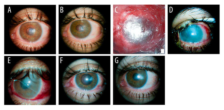
A 36-year-old male patient was referred to our center for management of a corneal ulcer of his left eye caused by a herpetic keratitis (Figure 1A) that perforated after regular administration of topical indomethacin (Indocollyre®, Chauvin Laboratories, France) for a week.
Figure 1.
Stages of the corneal defect from initial presentation till 1 year after surgery. (A) Corneal ulcer a week prior to the patient’s presentation to our clinic and prior to indomethacin instillation. (B) Perforated corneal ulcer upon presentation. (C) Intraoperative photo at the end of surgery using a lyophilized amniotic membrane. (D) A week after intervention. (E) Two weeks after intervention. (F) Three months after intervention. (G) Twelve months after intervention.
On ophthalmological examination, the patient’s best corrected visual acuity of the left eye was 0.05 (decimal notation). Slit-lamp biomicroscopy revealed a 2-mm-wide perforated corneal ulcer with positive seidel test, situated close to the pupil margins at 3 o’clock and surrounded by melted corneal tissue. Iris corectopia (Figure 1B) was also observed, as some iris fibers were drawn into the corneal defect. No iris incarceration or prolapse were noted.
The patients record before presentation suggested that he was treated with a combination of topical antibiotics (tobramycine and ciprofloxacine) and systemic anti-viral therapy (valaciclovir) for a herpetic keratitis, and his already-thin cornea (secondary to prior refractive surgery) perforated after he was administered non-steroidal anti-inflammatory (NSAID, indomethacin) drops to calm down the inflammation.
In fact, the patient had a LASIK surgery approximatively 8 years prior to his presentation and a kidney transplantation secondary to an anti-neutrophil cytoplasmic autoantibody (ANCA) vasculitis 5 years prior to his presentation. He was currently under various immunosuppressant medications, including mycophenolic acid (Cellcept®, Roche Laboratories, Switzerland). Anamnesis revealed a history a blepharitis and recurrent eye infections in the perforated eye.
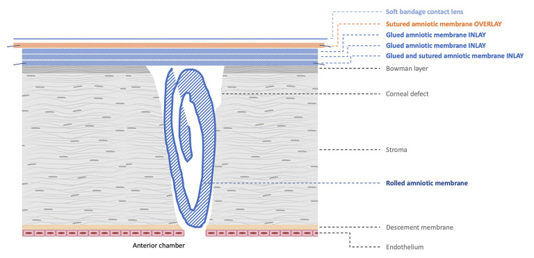
Upon examination, the diagnosis of corneal perforation was evident, and the patient was immediately hospitalized. Piperacillinofloxacin antibiotics were administered intravenously, and an emergency surgical intervention was performed under general anesthesia using what we called the “plug and patch” technique (Figure 2). After careful epithelial scraping of the ulcer margins, a spongious lyophilized amniotic membrane (Visio Amtrix®, TBF, France) was hydrated with balanced salt solution and rolled into a ball shape to fill the corneal defect. A drop of the human fibrinogen component of a biological glue (Tisseel®, Baxter Laboratories, USA) was then administered on top of the amniotic membrane and covered with a second 11-mm-wide amniotic membrane positioned epithelial-side up (inlay) and soaked in the thrombin component of the glue. When in contact, the interaction between the 2 components yielded a fibrin plug sealant that prevents future leakage. The inlay membrane was further fixated at the limbus and the surrounding conjunctiva using 4 interrupted vicryl 10-0 sutures. For further safety, 2 additional inlays grafts were glued over the first one and were covered by an overlay amniotic membrane patch that was sutured to the conjunctiva using a running vicryl 10-0 suture. A soft bandage contact lens was placed at the end of the procedure (Figure 1C). Postoperatively, the patient received 48 h of intravenous antibiotics (ofloxacine + piperacilline). He was then discharged on topical antibiotic and corticosteroid eyedrops (tobramycin + dexamethasone) for 4 weeks, along with a 10-day course of oral antibiotics (ofloxacin) and antiviral therapy (valaciclovir).
Figure 2.
Schematic representation of the plug and patch technique.
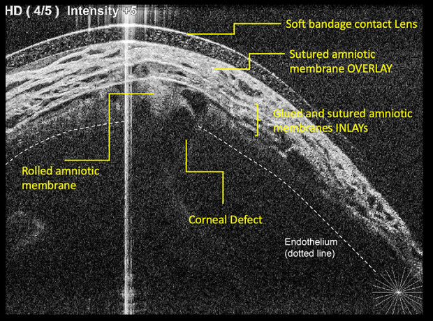
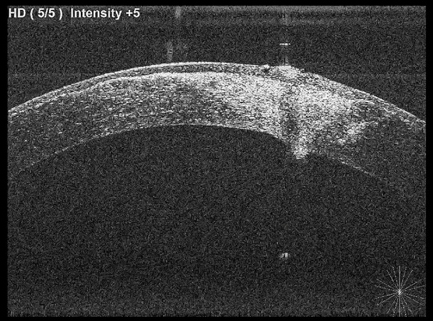
The patient was examined the day following surgery and a week after (Figure 1D) to make sure the overlay amniotic membrane was still in place. An anterior segment optical coherence tomography (OCT) was done to visualize the different layers and confirm the position of the plug (Figure 3). The membrane started dissolving 2 weeks after the surgery (Figure 1E). At month 3, examination revealed a slightly hazy cornea, a formed anterior chamber with no seidel, and a closed corneal defect (Figure 1F). White scarring was visible at the site of the lesion. Decimal best corrected visual acuity improved from 0.3 at month 3 to 0.4 at month 6 and remained stable for up to 12 months despite the scarred area. Upon coming for his latest routine check-up 1 year after initial presentation, an anterior segment photo (Figure 1G) and OCT (Figure 4) were performed, which showed a large scarred but healed cornea.
Figure 3.

Anterior segment optical coherence tomography the day following surgery visualizing the different layers and confirming the position of the plug.
Figure 4.

Anterior segment optical coherence tomography performed at 12 months showing a healed and scarred cornea.
Discussion
In this article, we present a case of herpetic keratitis with corneal perforation secondary to topical NSAID administration, surgically treated with multi-layered lyophilized amniotic membrane transplantation using a novel plug and patch technique. The technique described is actually a variation of existing techniques that are used in the management of corneal ulceration and perforation to restore corneal integrity without the need for keratoplasty. Amniotic membrane transplantation in such indications usually involves multilayered inlay grafts stacked as flat sheets to fill in the defect [17,18], or rolling, folding, or “stuffing” the membrane into the defect before covering it with further layers and fixating it with sutures and/or fibrin glue [11,19], or even rolling and directly securing the membrane into the defect with sutures [15], with or without the use of a contact lens.
The patient we describe here had a corneal perforation that was likely the result of several factors: prior refractive surgery, ocular surface disease, and herpetic keratitis, which most likely presented due to immune system down-regulation. Ocular herpetic keratitis can be a complication of the patient’s kidney transplantation [20], and the corneal melt secondary to infectious inflammation can lead to corneal perforation with severe ocular morbidity, including loss of vision if left untreated. While NSAIDs are important for the treatment of a wide range of ophthalmological conditions, including ocular inflammation, they have also been associated with corneal melt since 1999 [21–23]. The challenge in our case was therefore to find the right balance between controlling surface inflammation and reducing corneal melt.
Corneal melt consists of collagen breakdown initiated by a corneal epithelial defect. When the defect fails to re-epithelialize, it is followed by an enzyme-mediated stromal thinning, ultimately leading to corneal perforation [24]. The topical use of NSAIDs significantly decreases corneal sensation, delaying corneal wound re-epithelialization. NSAIDs also upregulate some matrix metalloproteinases (MMPs), hence disrupting the balanced interaction between MMPs, MMP inhibitors, prostaglandins, and cytokines and interrupting the healing process leading to keratolysis [25,26]. In addition, high doses of NSAIDs can paradoxically exacerbate the prostaglandin-mediated inflammatory effect [27].
In our case in particular, the adjunctive use of NSAIDs led to corneal perforation. While indomethacin 0.1% solution is a commercially available ophthalmic anti-inflammatory agent widely distributed in Europe, severe complications associated with its use have been described [23,28]. In order to fill the corneal defect and treat the concomitant surface inflammation, the choice was made to treat the patient surgically with an amniotic membrane, known for its healing, anti-inflammatory, anti-fibrotic, and anti-angiogenic properties [7]. Until recently, amniotic membranes were cryopreserved and needed special storage conditions that made them unavailable for corneal emergencies. Lyophilized amniotic membranes, on the other hand, can be stored at room temperature and have an extended shelf-life. Recent publications have shown that they are as effective as cryopreserved amniotic membranes when used as an overlay patch [29]. In our patient, their use led to preservation of the globe integrity, stopped further tissue loss, and was associated with a rapid visual recovery.
Conclusions
We report the successful use of the combination of a single round-shaped rolled amniotic membrane with a multilayered amniotic membrane transplantation for the treatment of corneal perforation, secondary to the use of topical indomethacin 0.1%. We would like to emphasize the dangers of using topical NSAIDs in cases of corneal surface disease and keratitis as well as the advantages of having lyophilized amniotic membrane at our disposal.
Acknowledgments
We would like to thank Marwan Sahyoun, M.D. for his insight and help in medical writing.
Footnotes
Publisher’s note: All claims expressed in this article are solely those of the authors and do not necessarily represent those of their affiliated organizations, or those of the publisher, the editors and the reviewers. Any product that may be evaluated in this article, or claim that may be made by its manufacturer, is not guaranteed or endorsed by the publisher
Department and Institution Where Work Was Done
Work was done at the Ophthalmology Department of Metz-Thionville Regional Hospital Center, Lorraine University, Mercy Hospital, Metz, France.
Declaration of Figures’ Authenticity
All figures submitted have been created by the authors who confirm that the images are original with no duplication and have not been previously published in whole or in part.
References:
- 1.Malhotra C, Jain AK. Human amniotic membrane transplantation: Different modalities of its use in ophthalmology. World J Transplant. 2014;4:111. doi: 10.5500/wjt.v4.i2.111. [DOI] [PMC free article] [PubMed] [Google Scholar]
- 2.Gupta A, Kedige SD, Jain K. Amnion and chorion membranes: Potential stem cell reservoir with wide applications in periodontics. Int J Biomater. 2015;2015:274082. doi: 10.1155/2015/274082. [DOI] [PMC free article] [PubMed] [Google Scholar]
- 3.Davis JS. Skin transplantation: With a review of 550 cases at the Johns Hopkins Hospital. Johns Hopkins Med J. 1910;15:307–96. [Google Scholar]
- 4.de Rotth A. Plastic repair of conjunctival defects with fetal membranes. Archives of Ophthalmology. 1940;23:522–25. [Google Scholar]
- 5.Meller D, Pauklin M, Thomasen H, et al. Amniotic membrane transplantation in the human eye. Dtsch Arztebl Int. 2011;108:243–48. doi: 10.3238/arztebl.2011.0243. [DOI] [PMC free article] [PubMed] [Google Scholar]
- 6.Kruse FE, Meller D. [Amniotic membrane transplantation for reconstruction of the ocular surface.] Ophthalmologe. 2001;98:801–10. doi: 10.1007/s003470170055. [in German] [DOI] [PubMed] [Google Scholar]
- 7.Jirsova K, Jones GLA. Amniotic membrane in ophthalmology: Properties, preparation, storage and indications for grafting – a review. Cell Tissue Bank. 2017;18:193–204. doi: 10.1007/s10561-017-9618-5. [DOI] [PubMed] [Google Scholar]
- 8.Yin J, Singh RB, Karmi RA, et al. Outcomes of cyanoacrylate tissue adhesive application in corneal thinning and perforation. Cornea. 2019;38:668–73. doi: 10.1097/ICO.0000000000001919. [DOI] [PMC free article] [PubMed] [Google Scholar]
- 9.Deshmukh R, Stevenson LJ, Vajpayee R. Management of corneal perforations: An update. Indian J Ophthalmol. 2020;68:7–14. doi: 10.4103/ijo.IJO_1151_19. [DOI] [PMC free article] [PubMed] [Google Scholar]
- 10.Solomon A, Meller D, Prabhasawat P, et al. Amniotic membrane grafts for nontraumatic corneal perforations, descemetoceles, and deep ulcers. Ophthalmology. 2002;109(4):694–703. doi: 10.1016/s0161-6420(01)01032-6. [DOI] [PubMed] [Google Scholar]
- 11.Hanada K, Shimazaki J, Shimmura S, Tsubota K. Multilayered amniotic membrane transplantation for severe ulceration of the cornea and sclera. Am J Ophthalmol. 2001;131:324–31. doi: 10.1016/s0002-9394(00)00825-4. [DOI] [PubMed] [Google Scholar]
- 12.Acar U. Amniotic membrane transplantation for spontaneous corneal perforation in a case of rheumatoid arthritis. Beyoglu Eye J. 2020;5:238–41. doi: 10.14744/bej.2020.40327. [DOI] [PMC free article] [PubMed] [Google Scholar]
- 13.Lavaris A, Elanwar MFM, Al-Zyiadi M, et al. Glueless and sutureless multilayer amniotic membrane transplantation in a patient with pending corneal perforation. Cureus. 2021;13:e16678. doi: 10.7759/cureus.16678. [DOI] [PMC free article] [PubMed] [Google Scholar]
- 14.Eslami M, Benito-Pascual B, Goolam S, et al. Case report: Use of amniotic membrane for tectonic repair of peripheral ulcerative keratitis with corneal perforation. Front Med (Lausanne) 2022;9:836873. doi: 10.3389/fmed.2022.836873. [DOI] [PMC free article] [PubMed] [Google Scholar]
- 15.Chan E, Shah AN, O’Brart DPS. “Swiss roll” amniotic membrane technique for the management of corneal perforations. Cornea. 2011;30:838–41. doi: 10.1097/ICO.0b013e31820ce80f. [DOI] [PubMed] [Google Scholar]
- 16.Hick S, Demers PE, Brunette I, et al. Amniotic membrane transplantation and fibrin glue in the management of corneal ulcers and perforations: a review of 33 cases. Cornea. 2005;24:369–77. doi: 10.1097/01.ico.0000151547.08113.d1. [DOI] [PubMed] [Google Scholar]
- 17.Prabhasawat P, Tesavibul N, Komolsuradej W. Single and multilayer amniotic membrane transplantation for persistent corneal epithelial defect with and without stromal thinning and perforation. Br J Ophthalmol. 2001;85:1455–63. doi: 10.1136/bjo.85.12.1455. [DOI] [PMC free article] [PubMed] [Google Scholar]
- 18.Chen HJ, Pires RT, Tseng SC. Amniotic membrane transplantation for severe neurotrophic corneal ulcers. Br J Ophthalmol. 2000;84:826–33. doi: 10.1136/bjo.84.8.826. [DOI] [PMC free article] [PubMed] [Google Scholar]
- 19.Kawakita T, Sumi T, Dogru M, et al. Amniotic membrane transplantation for wound dehiscence after deep lamellar keratoplasty: A case report. J Med Case Rep. 2007;1:28. doi: 10.1186/1752-1947-1-28. [DOI] [PMC free article] [PubMed] [Google Scholar]
- 20.Kremer I, Wagner A, Shmuel D, et al. Herpes simplex keratitis in renal transplant patients. Br J Ophthalmol. 1991;75:94–96. doi: 10.1136/bjo.75.2.94. [DOI] [PMC free article] [PubMed] [Google Scholar]
- 21.Congdon NG, Schein OD, von Kulajta P, et al. Corneal complications associated with topical ophthalmic use of nonsteroidal antiinflammatory drugs. J Cataract Refract Surg. 2001;27:622–31. doi: 10.1016/s0886-3350(01)00801-x. [DOI] [PubMed] [Google Scholar]
- 22.Flach AJ. Corneal melts associated with topically applied nonsteroidal anti-inflammatory drugs. Trans Am Ophthalmol Soc. 2001;99:205–10. discussion 210–12. [PMC free article] [PubMed] [Google Scholar]
- 23.Gueudry J, Lebel H, Muraine M. Severe corneal complications associated with topical indomethacin use. Br J Ophthalmol. 2010;94:133–34. doi: 10.1136/bjo.2008.155432. [DOI] [PubMed] [Google Scholar]
- 24.Rigas B, Huang W, Honkanen R. NSAID-induced corneal melt: Clinical importance, pathogenesis, and risk mitigation. Surv Ophthalmol. 2020;65:1–11. doi: 10.1016/j.survophthal.2019.07.001. [DOI] [PubMed] [Google Scholar]
- 25.Reviglio VE, Rana TS, Li QJ, et al. Effects of topical nonsteroidal antiinflammatory drugs on the expression of matrix metalloproteinases in the cornea. J Cataract Refract Surg. 2003;29:989–97. doi: 10.1016/s0886-3350(02)01737-6. [DOI] [PubMed] [Google Scholar]
- 26.Mian SI, Gupta A, Pineda R. Corneal ulceration and perforation with ketorolac tromethamine (Acular) use after PRK. Cornea. 2006;25:232–34. doi: 10.1097/01.ico.0000179931.05275.dd. [DOI] [PubMed] [Google Scholar]
- 27.Waterbury L, Kunysz EA, Beuerman R. Effects of steroidal and non-steroidal anti-inflammatory agents on corneal wound healing. J Ocul Pharmacol. 1987;3:43–54. doi: 10.1089/jop.1987.3.43. [DOI] [PubMed] [Google Scholar]
- 28.Guidera AC, Luchs JI, Udell IJ. Keratitis, ulceration, and perforation associated with topical nonsteroidal anti-inflammatory drugs. Ophthalmology. 2001;108:936–44. doi: 10.1016/s0161-6420(00)00538-8. [DOI] [PubMed] [Google Scholar]
- 29.Memmi B, Leveziel L, Knoeri J, et al. Freeze-dried versus cryopreserved amniotic membranes in corneal ulcers treated by overlay transplantation: A case-control study. Cornea. 2022;41:280–85. doi: 10.1097/ICO.0000000000002794. [DOI] [PubMed] [Google Scholar]


