Abstract
Menisci are crescent-shaped fibrocartilagenous structures that increase the tibiofemoral congruity, act as shock absorbers, and provide secondary anteroposterior stability. The root tears affect the biomechanical integrity of the whole meniscus, simulating a total meniscectomy, which can lead to early degeneration of the joint. Most of the root tears affect the posterior part rather than the anterior root. Very few reports have been described in the literature regarding anterior root tears and repairs. We present two such patients with anterior meniscal root tears, one of the lateral meniscus and one of the medial meniscus.
Keywords: Knee, Meniscus, Anterior root
Introduction
Menisci function as shock absorbers, provide anteroposterior stabilization and also have a proprioceptive role [1–3]. They are composed of collagen fibers, glycoproteins, and proteoglycans and transmit 50–70% of the load during weight bearing. The tears of the meniscus can be traumatic or degenerative, can have different shapes, and occur at different sites on the meniscus [4, 5]. The root tears affect the biomechanical integrity of the whole meniscus, due to loss of hoop stress, simulating a total meniscectomy, and can lead to early degeneration of the joint [6]. The root tears can be avulsions at the root or within 1 cm of the root [5, 6]. Most of the root tears affect the posterior part rather than the anterior root. The anterior root tears are the rarest of the meniscal tears and only a handful of such cases are described [7–9]. We present two patients who had traumatic anterior root tears.
Case 1
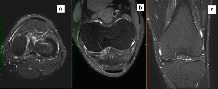
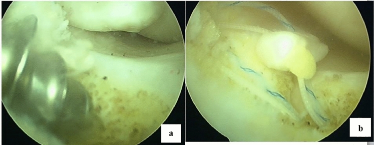
A 28-year-old gentleman sustained a twisting injury to the knee while playing football. He had complaints of giving away of the knee on brisk walking and climbing stairs. He had no episodes of locking. The clinical examination was suggestive of an anterior cruciate ligament (ACL) tear and medial meniscus tear. The anterior drawer test and the pivot shift were positive, there was no medial joint line tenderness, the mcmurray test was positive for medial meniscus at terminal extension as was the thesaly test at 20 degrees flexion. MRI showed an anterior cruciate ligament tear and a tear of the anterior root of the medial meniscus (Fig. 1). Arthroscopy showed a complete anterior cruciate ligament tear along with anterior root avulsion of the medial meniscus (Fig. 2). The semitendinosus graft was harvested and quadrupled for anterior cruciate ligament reconstruction. The anterior root of the medial meniscus was fixed with a double-loaded 5 mm metal anchor (TWINFIX Ti anchor; Smith & Nephew; Massachusetts; USA). The anchor was placed in the intercondylar region over the footprint of the anterior horn of the medial meniscus. The sutures of the anchor were used to fix the anterior root of the medial meniscus (Fig. 3). This was followed by trans-portal anterior cruciate ligament reconstruction with a continuous loop endobutton (Smith & Nephew; Massachusetts; USA) as a fixation device over the femoral side and an interference screw (Smith & Nephew; Massachusetts; USA) and post over the tibial side (Fig. 4). The patient was placed in a long knee brace for the first 2 weeks. The closed chain knee exercises and weight bearing were started after 2 weeks.
Fig. 1.
Axial T2W fat sat (a), oblique coronal PDW fat sat, and coronal STIR images showing avulsion of the detached anterior root of the medial meniscus from its attachment (arrows)
Fig. 2.
a Tear of the anterior horn of the medial meniscus. b Anterior horn of the medial meniscus could be reduced to its footprint
Fig. 3.
a 5 mm double-loaded anchors are placed in the footprint of the anterior horn of the medial meniscus. b Sutures passed in the anterior horn of the medial meniscus
Fig. 4.
Post-operative radiograph showing ACL reconstruction with endobutton, PEEK screw, and post. Anchor for medial meniscus anterior root fixation
Case 2
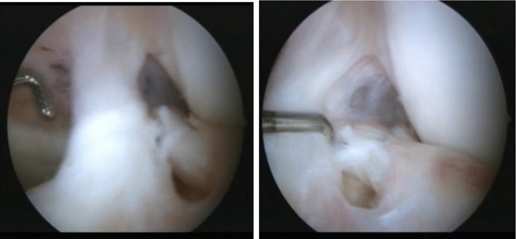
A 23-year-old female sustained a motor vehicle accident and presented to us 4 weeks later. She had a positive anterior drawer test, posterior drawer test, posterior tibial sag, and positive valgus stress test. There was joint line tenderness both on the medial and lateral side and the mcmurray test was not elicited. The MRI was suggestive of an anterior cruciate ligament tear, posterior cruciate ligament tear, medial collateral ligament tear, and vertical tear of the posterior horn of the medial meniscus and lateral meniscus anterior root avulsion (Fig. 5). Arthroscopy revealed an anterior cruciate ligament tear, posterior cruciate ligament tear, and longitudinal tear of the medial meniscus in its posterior horn. The lateral meniscus had an anterior root avulsion (Fig. 6). Quadriceps tendon graft was harvested for anterior cruciate ligament reconstruction and tibial inlay posterior cruciate ligament reconstruction. Semitendinosus graft was harvested for medial collateral ligament reconstruction. The anterior cruciate ligament reconstruction was done using one-half of the quadriceps tendon graft via the trans-portal conventional method with PEEK (Smith & Nephew; Massachusetts; USA) screw as a fixation on both the tibial and femoral side. The tibial side was additionally fixed with a staple (11 × 20 mm. Arthrex, Naples, Florida). The posterior cruciate ligament reconstruction was done using the tibial inlay technique. Staple (11 × 20 mm. Arthrex, Naples, Florida) was used for fixation of the patella bone plug on the tibial side, and a PEEK (Smith & Nephew; Massachusetts; USA) screw on the femoral side. The medial collateral ligament was reconstructed using semitendinosus and fixation on the femoral side was done using a PEEK screw (Smith & Nephew; Massachusetts; USA) and staple (11 × 20 mm. Arthrex, Naples, Florida) for the tibial side. The medial meniscus was repaired with #2 ethibond (Ethicon Inc., Johnson & Johnson Inc., New Brunswick, NJ, USA) using the inside-out technique. The anterior root of the lateral meniscus was fixed with two #2 ultra-braid sutures (Smith & Nephew; Massachusetts; USA) sutures in a cinch fashion (Fig. 7) and the sutures were taken out through the tibial tunnel created for anterior cruciate ligament reconstruction. As the footprint of the anterior root of the lateral meniscus and anterior cruciate ligament reconstruction are adjacent to each other, the tibial tunnel created for anterior cruciate ligament reconstruction was used for pulling out of repair sutures of the lateral meniscus anterior root. The sutures were tied over a staple which was used as a post for anterior cruciate ligament reconstruction (Fig. 8). Postoperatively, she was placed in a long knee brace with posterior support for 3 weeks. Range of movement exercises were started after three weeks with nighttime bracing and partial weight bearing which progressed to full weight bearing over one month.
Fig. 5.
Coronal STIR (A) MRI images showing root avulsion of the anterior horn of lateral meniscus (A) (arrow), Coronal STIR (B) a complete tear of the medial collateral ligament (B) (arrow), and Sagittal STIR (C) complete tear of anterior cruciate ligament (curved arrow) and posterior cruciate ligaments (arrowhead)
Fig. 6.
Arthroscopy showing lateral meniscus anterior root tear
Fig. 7.

Lateral meniscus anterior root fixed with two high tensile sutures and colored sutures seen for shuttling graft for ACL reconstruction
Fig. 8.
Postoperative radiograph
Discussion
This article presents two patients with tears of the anterior root of the meniscus, one of the lateral and the other of the medial meniscus. Root tears lead to meniscal extrusion and altered biomechanics which increases the contact pressures. With the repair of root tears, the meniscal integrity is restored and the contact pressures return to normal [5, 6]. They are also responsible for secondary anterior–posterior knee stability of the knee [4–6].
The root tears are broadly classified as avulsions or radial tears within a centimeter of the meniscus root [6]. The tears can be repaired by side-to-side technique if a remnant is still present at the root. The repairs that have been described for avulsions or tears at the root level without remnant include repair with anchors or trans-osseous suture pull-out repairs [9–12].
The medial meniscus anterior root is the anterior-most structure in the intercondylar region and anterior to tibial attachment of the anterior cruciate ligament. The anterior root of the medial meniscus has the largest surface area and is in line with the medial tibial spine at a distance of 7 mm to 12 mm from the ACL tibial footprint. The anterior root attachment of the medial meniscus has been classified into four types and the most common type being its attachment at the anterior intercondylar region near the tibial slope. Treatment of such tears involves fixation of the avulsed root to the insertion at the anterior intercondylar region near the slope with either suture anchors or transosseous repair [8–12].
Feucht et al. [9] and Matthias J et al. [10] used sutures and knotless anchors, whereas Wang et al. [11] used double-loaded titanium anchors for fixation of the anterior medial meniscus roots. The anchor is fixed at the downward slope of the tibial spine. The technique was similar to ours but he used sutures and knotless anchors. Ellman et al. [12] described the repair of iatrogenic medial meniscus root tear post-nailing of the tibial with transtibial pull-out technique along with a suture anchor as a supplementary fixation.
We have used double loaded metal anchor for the repair of the medial meniscus. Though suture anchor repair and transosseous repair have been described for anterior meniscal root repairs, each has its pros and cons. The trans-osseous repairs risks tunnel convergence when planned with ligament reconstructions, and suture abrasion in the tunnels. The suture anchor repair risks pulling out of the anchor in an osteoporotic tibia and suture dislodgement. In our patient who required concomitant anterior cruciate ligament reconstruction, the transtibial pull-out technique had the risk of tunnel convergence. Hence, we used a suture anchor for the repair.
The lateral meniscus anterior root attachment is anterior to the lateral tibial spine and lateral to ACL tibial footprint [1]. Travis et al. [13] used the transtibial pull-out technique for anterior root avulsion of the lateral meniscus. Sutures were used for securing the meniscal root which was pulled out through the tibial tunnel and secured on the tibia over a post. In our patients, the lateral meniscus root avulsion was a part of a concomitant multi-ligamentous knee injury including ACL injury. Since the footprint of the lateral meniscus anterior root and ACL tibial footprint are very close, the tibial tunnel of ACL reconstruction was used for trans-osseous repair of the lateral meniscus anterior root without the need for additional tunnels or anchors. When ACL reconstruction is not required, the anterior root tears of the lateral meniscus can be repaired using anchors or a suture pullout technique through the tibial tunnel at the anterior footprint.
Funding
This research did not receive any specific grant from funding agencies in the public, commercial or not-for-profit sectors.
Declarations
Conflict of Interest
The authors of this paper have no conflicts of interest to declare in the preparation and submission of this manuscript.
Ethical Approval
This article does not contain any studies with human or animal subjects performed by the any of the authors.
Informed Consent
Consent has been obtained from the patient regarding publishing his radiographs, MRI and intraoperative pictures excluding his identity details as a case report for learning purposes.
Footnotes
Publisher's Note
Springer Nature remains neutral with regard to jurisdictional claims in published maps and institutional affiliations.
Contributor Information
M. L. V. Sai Krishna, Email: krishna.mlv.sai@gmail.com
Nitin Chauhan, Email: nitinpgi286@gmail.com.
Ravi Kiran Vanapalli, Email: dr.ravikiranvanapalli@gmail.com.
Ravi Mittal, Email: ravimittal66@hotmail.com.
References
- 1.Bhatia S, LaPrade CM, Ellman MB, LaPrade RF. Meniscal root tears: Significance, diagnosis, and treatment. American Journal of Sports Medicine. 2014;42(12):3016–3030. doi: 10.1177/0363546514524162. [DOI] [PubMed] [Google Scholar]
- 2.Okazaki Y, Furumatsu T, Kodama Y, Matsumoto Y, Takahashi M, Ozaki T. Medial and lateral meniscus posterior root tears with an intact anterior cruciate ligament. Case Reports in Orthopedics. 2020;2020:8842167. doi: 10.1155/2020/8842167. [DOI] [PMC free article] [PubMed] [Google Scholar]
- 3.Feucht MJ, Izadpanah K, Lacheta L, Südkamp NP, Imhoff AB, Forkel P. Arthroscopic transtibial pullout repair for posterior meniscus root tears Arthroskopische transtibiale Auszugsnaht von posterioren Meniskuswurzelrissen. Operative Orthopädie und Traumatologie. 2019;31(3):248–260. doi: 10.1007/s00064-018-0574-4. [DOI] [PubMed] [Google Scholar]
- 4.Koo JH, Choi SH, Lee SA, Wang JH. Comparison of medial and lateral meniscus root tears. PLoS ONE. 2015;10(10):e0141021. doi: 10.1371/journal.pone.0141021. [DOI] [PMC free article] [PubMed] [Google Scholar]
- 5.Steineman BD, LaPrade RF, Santangelo KS, Warner BT, Goodrich LR, Haut Donahue TL. Early osteoarthritis after untreated anterior meniscal root tears: an in vivo animal study. Orthopaedic Journal of Sports Medicine. 2017;5(4):2325967117702452. doi: 10.1177/2325967117702452. [DOI] [PMC free article] [PubMed] [Google Scholar]
- 6.Palisch AR, Winters RR, Willis MH, Bray CD, Shybut TB. Posterior root meniscal tears: preoperative, intraoperative, and postoperative imaging for transtibial pullout repair. Radiographics. 2016;36(6):1792–1806. doi: 10.1148/rg.2016160026. [DOI] [PubMed] [Google Scholar]
- 7.Osti L, Del Buono A, Maffulli N. Anterior medial meniscal root tears: a novel arthroscopic all inside repair. Translational Medicine UniSa. 2014;12:41–46. [PMC free article] [PubMed] [Google Scholar]
- 8.Menge TJ, Chahla J, Dean CS, Mitchell JJ, Moatshe G, LaPrade RF. Anterior meniscal root repair using a transtibial double-tunnel pullout technique. Arthroscopy Techniques. 2016;5(3):e679–e684. doi: 10.1016/j.eats.2016.02.026. [DOI] [PMC free article] [PubMed] [Google Scholar]
- 9.Feucht MJ, Minzlaff P, Saier T, Lenich A, Imhoff AB, Hinterwimmer S. Avulsion of the anterior medial meniscus root: Case report and surgical technique. Knee Surgery, Sports Traumatology, Arthroscopy. 2015;23(1):146–151. doi: 10.1007/s00167-013-2462-7. [DOI] [PubMed] [Google Scholar]
- 10.MatthiasFeucht J, Minzlaff P, Saier T, Lenich A, Imhoff AB. Avulsion of the anterior medial meniscus root: case report and surgical technique. Knee Surgery, Sports Traumatology, Arthroscopy. 2015;23(1):146–151. doi: 10.1007/s00167-013-2462-7. [DOI] [PubMed] [Google Scholar]
- 11.Wang A, Lu H. Traumatic avulsion of the anterior medial meniscus root combined with PCL injury: A case report. BMC Musculoskeletal Disorders. 2020;21:642. doi: 10.1186/s12891-020-03671-x. [DOI] [PMC free article] [PubMed] [Google Scholar]
- 12.Ellman MB, James EW, LaPrade CM, et al. Anterior meniscus root avulsion following intramedullary nailing for a tibial shaft fracture. Knee Surgery, Sports Traumatology, Arthroscopy. 2015;23:1188–1191. doi: 10.1007/s00167-014-2941-5. [DOI] [PubMed] [Google Scholar]
- 13.Menge TJ, Chahla J, Mitchell JJ, Dean CS, LaPrade RF. Avulsion of the anterior lateral meniscal root secondary to tibial eminence fracture. The American Journal of Orthopedics. 2018 doi: 10.12788/ajo.2018.0024. [DOI] [PubMed] [Google Scholar]


