FIG 3.
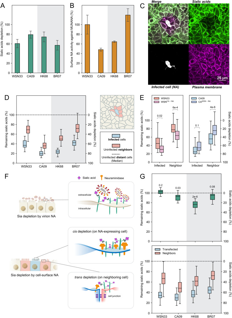
Cell-surface NA depletes sialic acid (Sia) in cis and in trans. (A) Cell-surface Sia depletion on cells infected by each viral strain at 16 h postinfection (h.p.i.). Quantifications are based on HA-positive cells. A value of 100% corresponds to complete depletion. (B) Surface NA activity against 2′-(4-methylumbelliferyl)-α-d-N-acetylneuraminic acid (MUNANA), normalized to data for WSN33. The experimental details are provided in the Materials and Methods section under “Quantification of NA activity with MUNANA.” (C) Image showing Sia, cell-surface NA, and plasma membrane (labeled with CellMask Orange) in the proximity of an isolated cell infected with HK68 at 16 h.p.i. (D) Quantification of remaining Sia on the surface of infected cells and uninfected neighbors at 16 h.p.i. The cells were selected based on NA expression by incubating with the anti-NA antibody 1G01 immediately before imaging. The data are combined from three biological replicates (at least eight sites of infection/replicate). (E) Quantification of remaining Sia on the surface of infected cells (determined by HA expression) and uninfected neighbors for viruses with exchanged NA segments at 16 h.p.i. The data are combined from three biological replicates (at least 14 sites of infection/replicate). The P values are determined by independent t tests. (F) Illustration of Sia depletion by virion-associated NA (top) and cell-associated NA (bottom). (G, top) Quantification of Sia depletion following incubation with virions. The results are normalized to Sia levels on untreated cell surfaces. The P values are determined from independent t tests relative to the untreated group. (Bottom) Quantification of Sia depletion by cell-surface NA (delivered through transfection, imaged at 48 h after transfection). The cells were selected based on NA expression. The data are combined from three biological replicates (at least 24 cells/replicate).

