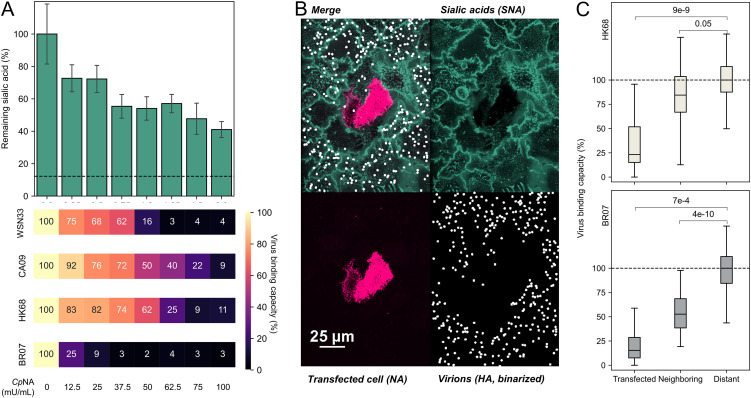
FIG 4.
Sialic acid depletion reduces viral attachment in a strain-dependent manner. (A, top) Cell-surface Sia abundance following treatment with CpNA. The concentrations are specified below. The dashed line indicates signal from CMP-sialic acid transporter SLC35A1 knockout cells treated with the highest CpNA concentration, representing the background of Sia labeling reaction. (Bottom) Relative binding capacities for each viral strain following CpNA treatment. The data are combined from three biological replicates. A replicate is defined by eight individual cell cultures treated by the indicated concentration of CpNA. (B) Representative image showing virus attachment to cell monolayers with sparse expression of HK68 NA (virus strain BR07). (C) Quantification of virus binding capacity on HK68 NA-expressing (Transfected) cells, their adjacent cells (Neighboring), and other cells (Distant). The data for HK68 and BR07 virions, which show different binding avidity, are shown in the top and bottom panels. The data are from three biological replicates (at least 21 NA-expressing cells/replicate) and are normalized to the distant cell data. The P values are determined by independent t tests. SNA, S. nigra lectin.

