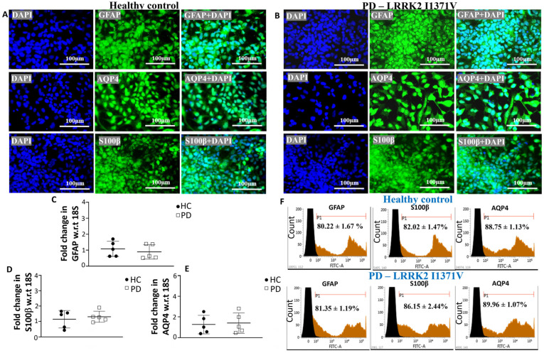
Figure 3.
Characterization of astrocytes derived from HC and PD LRRK2-I1371V GPCs: (A,B) ICC images of astrocytes from HC (A) and PD (B) iPSC lines staining positive for mature astrocyte markers GFAP, AQP4, and S100β. The nuclei were counterstained with DAPI. (C,E) qPCR analysis of mature astrocytes markers GFAP (C), S100β (D), and AQP4 (E) of mature astrocytes derived from HC and PD iPSCs; w.r.t, with respect to. (F) Flow cytometry histograms of astrocytes differentiated from the HC and PD iPSC lines immunolabeled with GFAP, S100β and AQP4. n = 5; mean ± SD.

