

Abstract
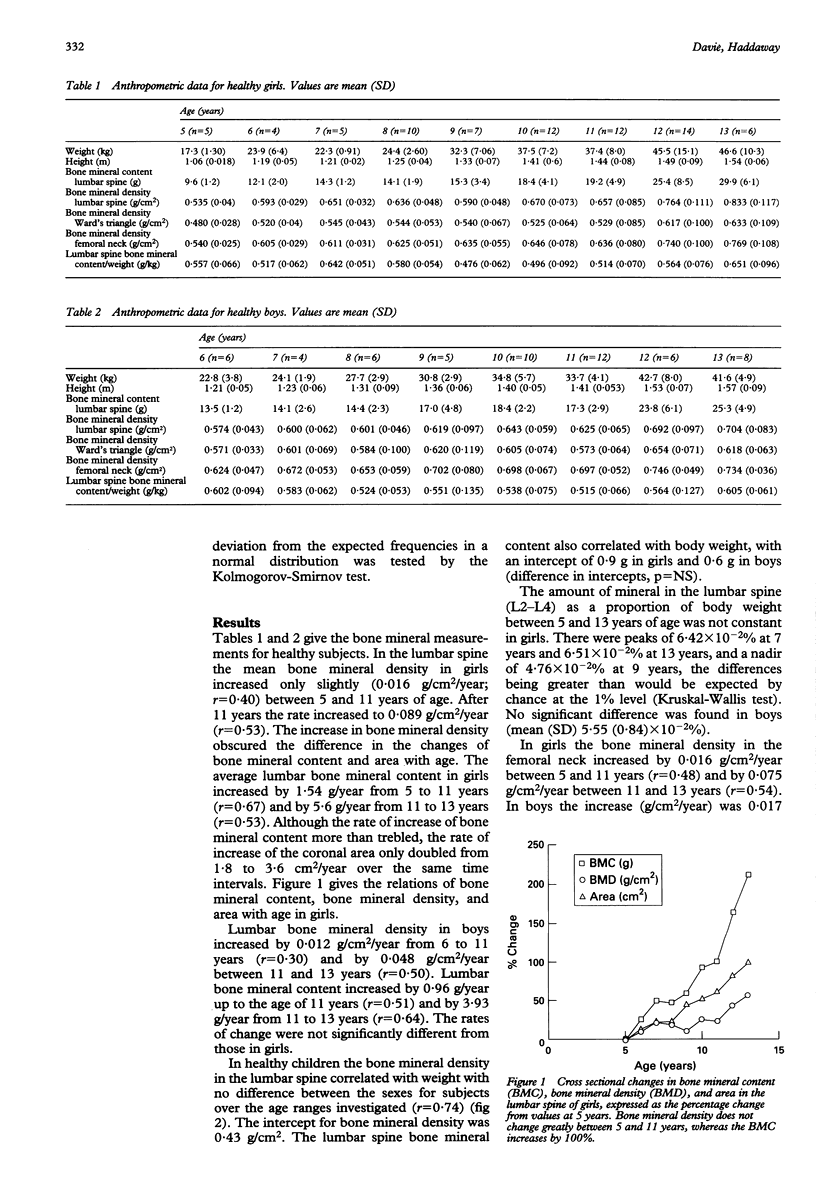
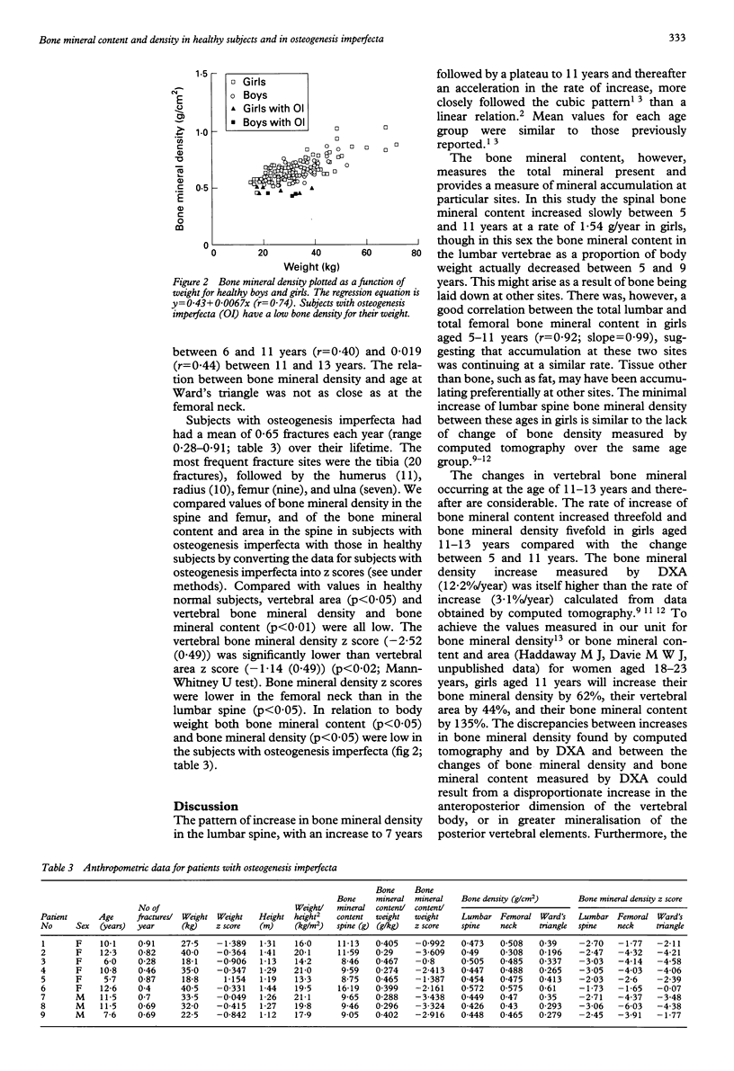
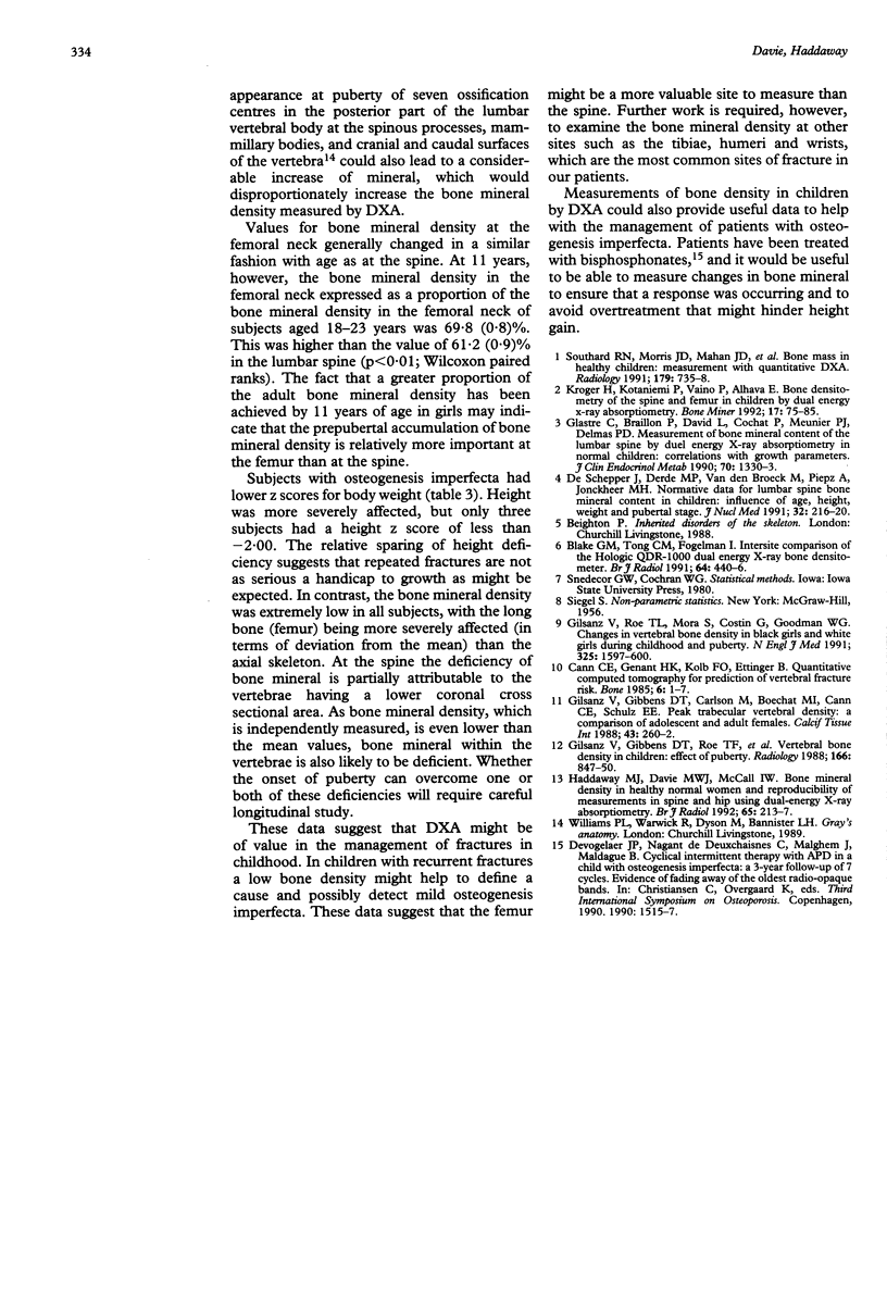
Lumbar spine bone mineral density in a cross sectional study of healthy subjects increased by 0.012 and 0.016 g/cm2/year in boys and girls respectively between 5 and 11 years of age. These rates increased five-fold in girls and threefold in boys between the ages of 11 and 13 years as a result of the bone mineral content increasing more rapidly than the coronal area at this age. By the age of 11 years the girls had 66% of the coronal area, 61% of the bone mineral density, and 41% of the bone mineral content of subjects aged 18-23 years. The ratio (lumbar spine bone mineral content/body weight) was constant in boys aged 6-13 years, but there were significant variations in girls. Femoral neck bone mineral density in both sexes changed little between 6 and 11 years and at 11 years was 69% of the adult values. Subjects with osteogenesis imperfecta had a low bone mineral density and bone mineral content for their age and weight. The z score of bone mineral density at the femoral neck was significantly lower than at the lumbar spine. In patients with recurrent fractures a low bone mineral density may help in identifying those with osteogenesis imperfecta and assist in their subsequent management.
Full text
PDF
Selected References
These references are in PubMed. This may not be the complete list of references from this article.
- Blake G. M., Tong C. M., Fogelman I. Intersite comparison of the Hologic QDR-1000 dual energy X-ray bone densitometer. Br J Radiol. 1991 May;64(761):440–446. doi: 10.1259/0007-1285-64-761-440. [DOI] [PubMed] [Google Scholar]
- Cann C. E., Genant H. K., Kolb F. O., Ettinger B. Quantitative computed tomography for prediction of vertebral fracture risk. Bone. 1985;6(1):1–7. doi: 10.1016/8756-3282(85)90399-0. [DOI] [PubMed] [Google Scholar]
- De Schepper J., Derde M. P., Van den Broeck M., Piepsz A., Jonckheer M. H. Normative data for lumbar spine bone mineral content in children: influence of age, height, weight, and pubertal stage. J Nucl Med. 1991 Feb;32(2):216–220. [PubMed] [Google Scholar]
- Gilsanz V., Gibbens D. T., Carlson M., Boechat M. I., Cann C. E., Schulz E. E. Peak trabecular vertebral density: a comparison of adolescent and adult females. Calcif Tissue Int. 1988 Oct;43(4):260–262. doi: 10.1007/BF02555144. [DOI] [PubMed] [Google Scholar]
- Gilsanz V., Gibbens D. T., Roe T. F., Carlson M., Senac M. O., Boechat M. I., Huang H. K., Schulz E. E., Libanati C. R., Cann C. C. Vertebral bone density in children: effect of puberty. Radiology. 1988 Mar;166(3):847–850. doi: 10.1148/radiology.166.3.3340782. [DOI] [PubMed] [Google Scholar]
- Gilsanz V., Roe T. F., Mora S., Costin G., Goodman W. G. Changes in vertebral bone density in black girls and white girls during childhood and puberty. N Engl J Med. 1991 Dec 5;325(23):1597–1600. doi: 10.1056/NEJM199112053252302. [DOI] [PubMed] [Google Scholar]
- Glastre C., Braillon P., David L., Cochat P., Meunier P. J., Delmas P. D. Measurement of bone mineral content of the lumbar spine by dual energy x-ray absorptiometry in normal children: correlations with growth parameters. J Clin Endocrinol Metab. 1990 May;70(5):1330–1333. doi: 10.1210/jcem-70-5-1330. [DOI] [PubMed] [Google Scholar]
- Haddaway M. J., Davie M. W., McCall I. W. Bone mineral density in healthy normal women and reproducibility of measurements in spine and hip using dual-energy X-ray absorptiometry. Br J Radiol. 1992 Mar;65(771):213–217. doi: 10.1259/0007-1285-65-771-213. [DOI] [PubMed] [Google Scholar]
- Kröger H., Kotaniemi A., Vainio P., Alhava E. Bone densitometry of the spine and femur in children by dual-energy x-ray absorptiometry. Bone Miner. 1992 Apr;17(1):75–85. doi: 10.1016/0169-6009(92)90712-m. [DOI] [PubMed] [Google Scholar]
- Southard R. N., Morris J. D., Mahan J. D., Hayes J. R., Torch M. A., Sommer A., Zipf W. B. Bone mass in healthy children: measurement with quantitative DXA. Radiology. 1991 Jun;179(3):735–738. doi: 10.1148/radiology.179.3.2027984. [DOI] [PubMed] [Google Scholar]