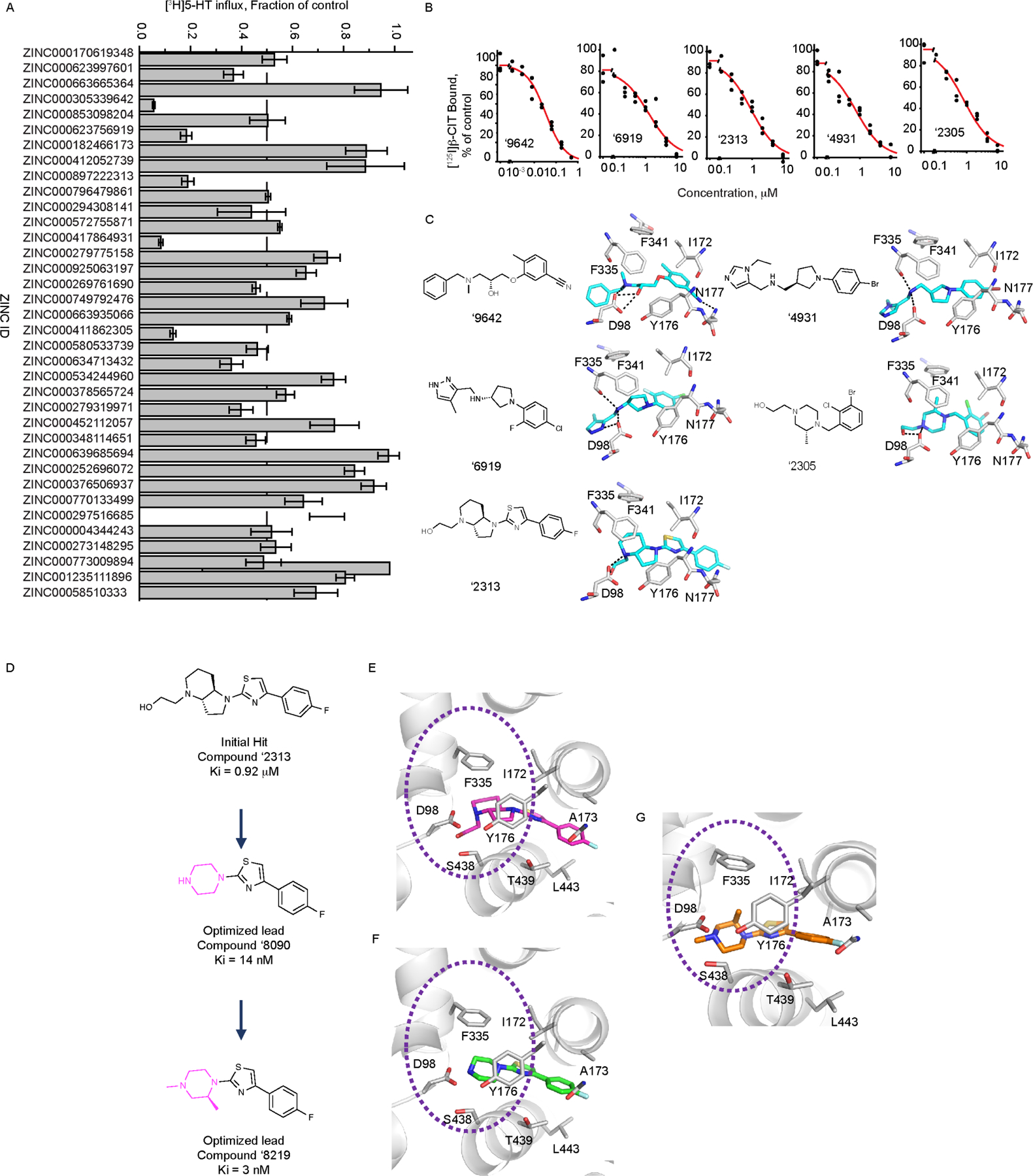
Figure 1. Docking-derived inhibitors of serotonin transporter.

(A) Inhibition of [3H]5-HT transport by docking-derived molecules, tested at 30 μM (Table S1). (B) Radio-ligand displacement of the top five docking hits (representative curves; summary data in Table S2). (C) 2D Chemical structures of the top five docking hits and their docked poses. Interactions are depicted as black dashed lines, ligand carbons in cyan and protein carbons in grey. Oxygens for both protein and ligand are red, nitrogen blue, and sulfur yellow. (D) Chemical structures of the parent compound, ‘2313 and its optimized analogs ‘8090 (Ki 14 nM) and ‘8219 (Ki 3 nM). The variable group in the analogs versus the parent are colored in magenta. Comparing the docked poses of (E) the parent ‘2313 (F) ‘8090 and (G) ‘8219. Dashed circles represent modeled improved stacking of F335 with ring substitutions in going from parent to the optimized analogs.
