Abstract
Microplastics have been considered a new type of pollutant in the marine environment and have attracted widespread attention worldwide in recent years. Plastic particles with particle size less than 5 mm are usually defined as microplastics. Because of their similar size to plankton, marine organisms easily ingest microplastics and can threaten higher organisms and even human health through the food chain. Most of the current studies have focused on the investigation of the abundance of microplastics in the environment. However, due to the limitations of analytical methods and instruments, the number of microplastics in the environment can easily lead to overestimation or underestimation. Microplastics in each environment have different detection techniques. To investigate the current status, hot spots, and research trends of microplastics detection techniques, this review analyzed the papers related to microplastics detection using bibliometric software CiteSpace and COOC. A total of 696 articles were analyzed, spanning 2012 to 2021. The contributions and cooperation of different countries and institutions in this field have been analyzed in detail. This topic has formed two main important networks of cooperation. International cooperation has been a common pattern in this topic. The various analytical methods of this topic were discussed through keyword and clustering analysis. Among them, fluorescent, FTIR and micro-Raman spectroscopy are commonly used optical techniques for the detection of microplastics. The identification of microplastics can also be achieved by the combination of other techniques such as mass spectrometry/thermal cracking gas chromatography. However, these techniques still have limitations and cannot be applied to all environmental samples. We provide a detailed analysis of the detection of microplastics in different environmental samples and list the challenges that need to be addressed in the future.
Keywords: microplastics, nanoplastics, marine pollutant, detection, analysis methods, bibliometrics
Introduction
Since the invention of plastic in the 1950s, plastic products have been widely used, and the environment is flooded with plastic waste.1,2 It is estimated that by 2060 there will be approximately 1.55–26.5 billion tons of plastic waste. 3 In 2004, the term “microplastics” was first introduced, referring to plastic particles smaller than 5 mm. 4 Compared to large plastics, tiny microplastics are widely distributed in the environment and are more easily ingested by organisms. Microplastics are prevalent in aquatic organisms such as fish and shellfish and are subject to food chain transport, 5 thus posing a potential threat to the health of organisms and ecosystems. Microplastics are broadly divided into two categories, primary microplastics, and secondary microplastics. The former refers to nanoplastics added to toiletries, biomedical products, waterproof coatings, and nanomedicines. 6 The latter refers to plastic particles formed from plastic waste under the external effects of light, mechanical action, chemical and biological degradation. 7 Table 1 shows the main categories of microplastics. Investigations have shown that micro/nanoplastics are widely present in water, sediment, soil, and atmosphere.8,9 In addition to areas of intense human activity, micro/nanoplastics have been found in uninhabited mountainous areas and even in surface water, sea ice, benthic organisms, and penguin gastrointestinal tracts in the North and South Poles. 10 The global plastic pollution situation is becoming increasingly critical, and it is estimated that microplastics in the subtropical convergence zone will increase by a factor of two or four by 2030 or 2060 compared to the present. 11
Table 1.
Source of microplastics.
| Microplastics category | Source | Ref. |
|---|---|---|
| Polyethylene (PE) | Facial cleanser, toothpaste, etc | 12,13 |
| Low density polyethylene (LDPE) | Plastic bags, bottles, fishing nets, straws, etc | 14,15 |
| High density polyethylene (HDPE) | Milk, juice cans, cosmetic packaging, etc | 16,17 |
| Polyvinyl chloride (PVC) | Plastic film, plastic cup, etc | 18,19 |
| Polyethylene terephthalate (PET) | Bottles etc. | 20 |
| Polypropylene (PP) | Rope, bottle caps, etc | 21,22 |
| Polystyrene (PS) | Food containers, plastic utensils, etc | 23 |
| Polyamide (PA) | Fishing nets etc | 24 |
| Foam polystyrene (EPS) | Buoys, bait boxes, disposable cups, etc | 25 |
Studying the ecological and environmental health impacts of micro- and nano-sized plastics requires effective detection and monitoring methods. The extraction, separation, and determination of plastic particles of all sizes, especially nanoscale particles, from complex environmental and biological samples is a hot topic of current research. Although there are many techniques and methods, there is a lack of uniform standards for the extraction, qualitative and quantitative analysis of microplastics from different sample matrices. 26 Therefore, there is an urgent need to develop efficient and pervasive extraction, identification, and quantification methods to obtain comparable data. 27 Bibliometric analysis is a literature and information mining method based on mathematical statistics. It can reflect research trends and hotspots through clustering relationships of keywords in the literature and has become an important tool for global analysis in various scientific fields28–37. As an emerging environmental pollutant, microplastics are receiving increasing attention worldwide. Therefore, it is necessary to update the bibliometrics of this topic. To date, bibliometric analyses on microplastics have focused on the development of the entire field. We believe that how microplastics are detected is a very important part of its assessment. Only an accurate measurement of microplastics can give an indication of its impact on the environment. Therefore, we have paid special attention to the development of detection techniques in microplastics in this bibliometric work.
Materials and methods
Two bibliometrics software have been used in this systematic literature review. The first is CiteSpace, developed by Dr Chaomei Chen, a professor at the Drexel University School of Information Science and Technology38–41. CiteSpace 6.1R2 was used to calculate and analyze all documents. COOC is another emerging bibliometrics software. 42 COOC12.6 was used to analysis of annual publications and keywords co-occurrence. We used the core collection on Web of Science as a database to assure the integrity and academic quality of the studied material. “microplastics detection”, “microplastics sensor” and “microplastics quantification” have been used as a “Topic.” The retrieval period was indefinite, and the date of retrieval was December 30, 2021. 696 articles were retrieved, spanning the years 2012 to 2021.
Developments in the research field
Literature development trends
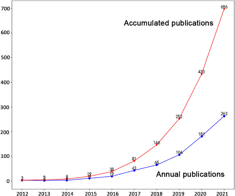
Figure 1 shows the annual and the cumulative number of publications on microplastic detection techniques between 2012 and 2021. As seen from the figure, the detection of microplastics did not become an immediate object of research with its conceptualization. Since the introduction of microplastics in 2004, no detection techniques for it were reported until 2012 (this does not mean that microplastics could not be detected in previous work). Harrison et al. 43 investigated the applicability of Fourier transform infrared spectroscopy in detecting microplastics. Fossi et al. 44 proposed phthalates as a tracer of microplastic ingestion when investigating whether baleen whales ingest microplastics during their filter-feeding activities. Imhof et al. 45 constructed a precipitate separator to improve the density separation method. This method allows the separation of different ecologically relevant size classes of plastic particles from sediment samples. At the same time, they identified and quantified microplastics using micro-Raman (μ-Raman) spectroscopy, verifying that the recovery rate of this separation technique is significantly higher than that of classical density separation devices and froth flotation commonly used in the industry.
Figure 1.
Annual and accumulated publications from 2012 to 2021 searched in the web of science about microplastic detection techniques.
The topic of microplastic detection has gradually gained attention since 2015, and the number of annual publications has started to exceed 10. After that, the topic started to enter a very rapid development, with more than 100 annual publications in 2019. By 2021, the annual number of articles on this topic has reached 263. Although we did not include data on new publications in 2022 in this bibliometric survey, the trend of continued increasing publications has not been diminished based on the available data (as of June 2022). From a bibliometric analysis, this topic is entering a phase of rapid development, attracting many scholars. There is no doubt that a large amount of interest in microplastic detection technology is inseparable from the fact that microplastics are a hot topic in the environmental field today. The update of old detection techniques and the establishment of new methods are generally based on the widespread interest in the analyte.
Journals, cited journals and research subjects
Figure 2 shows the top 10 journals that published the most papers regarding microplastic detection techniques. It can be seen that Marine Pollution Bulletin published the most significant number of papers, accounting for 12.64% of all papers on this topic. In second place was Science of the Total Environment, with 77 papers accounting for 11.06% of the total. More than half of the journals in Figure 2 are affiliated with environmental science. In addition, Analytical and Bioanalytical Chemistry and Analytical Methods are journals related to analytical chemistry, and Analytical Methods in particular focuses on new analytical assay techniques. This demonstrates that the detection of microplastics has now attracted the attention of not only environmental scientists but has also involved analytical chemists. Figure 2 also includes the Journal of Hazardous Materials, which published 23 papers on this topic. This journal mainly publishes papers related to materials harmful to humans and the environment. Microplastics, a series of tiny forms of polymeric materials, have received so much attention in recent years precisely because of the pollution and toxicity they produce in the environment.
Figure 2.
Top 10 journals that published articles on the microplastic detection techniques (the size of the box is proportional to the number of papers published in the journal).
In addition to the number of papers published by the journal on the topic, the frequency with which the journal is cited papers related to the theme is also an important indicator. Table 2 shows the top 15 cited journals on microplastic detection techniques. It can be seen that most of the journals in Figure 2 are also included in Table 2, except for the Journal of Hazardous Materials. The papers published in the Journal of Hazardous Material are most likely about the analysis of different environmental samples with different detection techniques. These works do not necessarily provide improvements and innovations in the methodology of detection. Therefore, they are not widely cited in papers on the topics we set. Journals in the analytical chemistry are further represented in Table 2 with the additional inclusion of TrAC Trends in Analytical Chemistry and Analytical Chemistry. On the other hand, comprehensive journals are also covered in Table 2, including Scientific Reports, Science, and PLOS ONE. These journals do not necessarily publish a large number of papers on the topic, but the articles that appear in them have an indirect impact on the topic. For example, the detection of other substances published in analytical chemistry-related journals indirectly inspired the detection of microplastics. The analysis results in Figure 2 and Table 2 show that microplastic detection techniques mainly attract scholars from two fields: environmental science and analytical chemistry. In addition to the journals related to these two fields, the coverage of this microplastic in comprehensive journals will significantly impact the investigation of this topic.
Table 2.
Top 15 cited journals on the microplastic detection techniques.
| No. | Citation | Cited Journal |
|---|---|---|
| 1 | 437 | Environmental Science & Technology |
| 2 | 425 | Marine Pollution Bulletin |
| 3 | 419 | Environmental Pollution |
| 4 | 374 | Science of The Total Environment |
| 5 | 292 | Scientific Reports |
| 6 | 267 | Water Research |
| 7 | 253 | Science |
| 8 | 232 | Chemosphere |
| 9 | 210 | Analytical Methods |
| 10 | 185 | TrAC Trends in Analytical Chemistry |
| 11 | 178 | Environmental Science and Pollution Research |
| 12 | 178 | Marine Environmental Research |
| 13 | 159 | Analytical and Bioanalytical Chemistry |
| 14 | 154 | PLOS ONE |
| 15 | 142 | Analytical Chemistry |
Although the most important journals on this topic and the fields to which they belong can be known from Figure 2 and Table 2, they do not present the most cutting-edge advances. Therefore, we analyzed journals that published this topic for the first time in 2020 and 2021 (Table 3). Environmental science and analytical chemistry-related journals remain the most dominant areas in the table. It is also worth noting that the new journals appearing in 2020 include a series of journals related to food science, including Food Bioscience, Food Chemistry, Food Control, and Food Packaging and Shelf Life. This reflects that detecting microplastics in food has become a more important direction of investigation in 2021. Gündoğdu et al. 46 investigated the presence of microplastics in mussels sold in five cities in Turkey. μ-Raman has been used for the quantitative detection of microplastics. Plastic-wrapped beverages may be a source of microplastics in the diet. Prata et al. 47 proposed an improved detection method for the detection of microplastics in white wines. μ-Raman has been used for the first time to identify microplastic particles in complex beverages. Huang et al. 48 examined the extent of PS and PVC contamination in chicken-based on attenuated total reflection mid-infrared spectroscopy (ATR-MIR) combined with chemometric techniques. Kedzierski et al. 49 investigated whether food trays made of extruded PS could generate microplastic contamination between the meat and the sealing film. Several microscopy and instrumentation-related journals appeared in 2021, including Micron, Microscopy Research and Technique, Microsystems & Nanoengineering. Schmidt et al. 50 proposed a detection method for nanoplastics smaller than 1 μm. Using a correlation between scanning electron microscopy (SEM) and μ-Raman, they identified nanoplastics in the 100 nm range in various environments. Qian et al. 51 tried to combine sparse particle localization and miniaturized mass sensing functions on a microelectromechanical system (MEMS) chip to realize the analysis of sparse particles. The use of 4-dimethylamino-4′-nitrostilbene (DANS) fluorescent dyes for detecting microplastics was proposed by Sancataldo et al. 52 DANS staining can provide access to different detection and analysis strategies based on fluorescence microscopy. Meanwhile, some pharmacology and toxicology journals also appeared in 2021, including Regulatory Toxicology and Pharmacology, Pharmaceutics.
Table 3.
List of journals published paper for the first time regards the microplastic detection techniques in the last two years.
| Year | Journals |
|---|---|
| 2020 | Advanced Intelligent Systems; Analyst; Applied Physics B-Lasers and Optics; Chemosensors; Clinical Hemorheology and Microcirculation; Earth System Science Data; Ecological Research; Food Bioscience; Food Chemistry; Food Control; Food Packaging and Shelf Life; Geochemical Perspectives Letters; Global Challenges; Heliyon; International Journal of Environmental Research; Journal of Analytical Atomic Spectrometry; Journal of Chemical Education; Journal of Coastal Research; Journal of Materials Chemistry B; Journal of Oceanology and Limnologyl; Ocean Science; Optical Review; RSC Advances; Sains Malaysiana; Spectroscopy; Surface and Interface Analysis |
| 2021 | ACS Photonics; ACS Sustainable Chemistry & Engineering; Aerosol Science and Technology; Agriculture-Basel; American Biology Teacher; Analytical Sciences; Biosensors-Basel; Carbohydrate Polymers; Chemical Communications; Chemico-Biological Interactions; Chimia; Current Analytical Chemistry; Environmental Chemistry Letters; Environmental Geotechnics; Exposure and Health; Global Change Biology; Indian Journal of Geo-Marine Sciences; Journal of Colloid and Interface Science; Journal of Environmental Quality; Journal of Marine Science and Engineering; Journal of Sensors; Journal of Soils and Sediments; Microchemical Journal; Micron; Microscopy Research and Technique; Microsystems & Nanoengineering; Molecular & Cellular Toxicology; Peerj; Pharmaceutics; Polymer Degradation and Stability; Regulatory Toxicology and Pharmacology; Small; Soil Biology & Biochemistry; Spectroscopy and Spectral Analysis; Sustainable Production and Consumption; Water Resources Research |
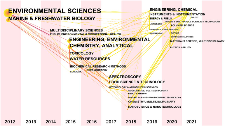
The category of the published paper can reflect the evolution of the topic. Figure 3 shows the evolution of the category of the microplastics detection techniques over time. This topic originated from the two categories most related to microplastics (Environmental Science, Marine & Freshwater Biology). Two years later, the topic was further covered in Public, Environmental & Occupational Health. Starting in 2015, this topic was formally explored methodologically, so Chemistry, Analytical and Biochemical Research Methods were included. In 2017, some niche analytical methods were included in the category, including Spectroscopy and Electrochemistry (not shown in the figure because the count is too small). In 2018, this theme was further associated with several analytics categories, including Remote Sensing and Imaging Science & Photographic Technology. From 2018 onwards, this topic started covering a wide range of categories. In 2021, this topic covered a range of engineering fields, including Engineering (Geological, Marine, Mechanical, Ocean) for the first time. In addition, Agronomy, Anatomy & Morphology, Biochemistry & Molecular Biology, Biology, Education, Scientific Disciplines, Metallurgy & Metallurgical Engineering, Pharmacology & Pharmacy, Physics, Condensed Matter, and Soil Science were also published for the first time in 2021 on this topic. It is worth noting that the papers surveyed here are on microplastics detection techniques rather than microplastics. Papers related to microplastics should be published earlier in journals related to environmental science and geology.
Figure 3.
Time-zone view of research categories for microplastic detection techniques (the year of the node in the graph is the time when a paper on this topic was first retrieved by a particular category in WOS. The size of a node is proportional to the number of connections between this paper and other nodes).
Geographic distribution
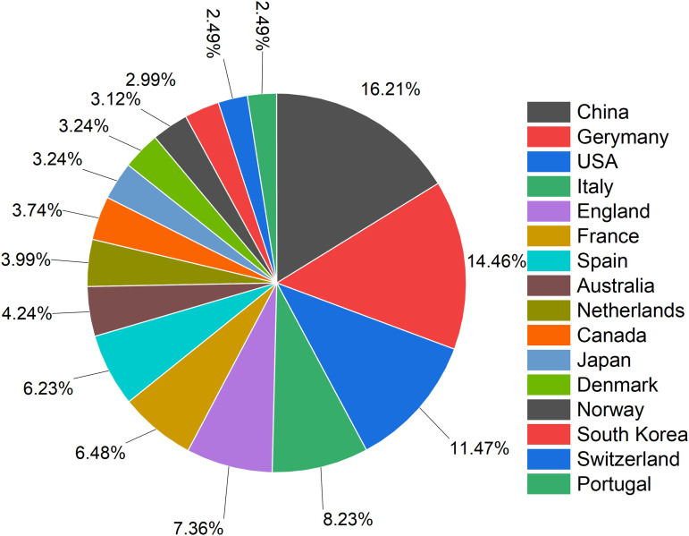
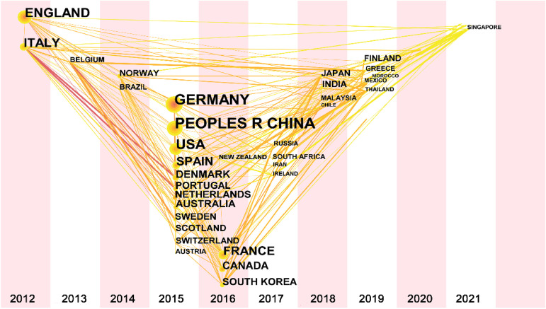
Figure 4 shows the top 16 countries with the most publications in microplastics detection techniques. Although Chinese scientists published the most articles, they only accounted for about 15% of all numbers. Both Germany and USA contributed more than 10% of the papers. This is due to the fact that this topic has attracted a great deal of academic attention and thus different countries have made considerable contributions to the topic. On the other hand, although a range of countries contributes to this topic, it is clear from the figure that Europe is the most actively involved region. This enthusiasm can also be observed in the time-zone view (Figure 5). The first countries to study this topic in 2012 were Italy and England. China and USA were added to the team in 2015. In Asia, Japan and South Korea are also interested in this topic. South Korea entered the survey on this topic in 2016, while Japan will not join in until 2018. Since the topic is still on the rise, many countries are getting involved every year. Singapore, Turkey, Iceland, U Arab Emirates, Ukraine, Romania, Peru, Croatia, Lithuania, and Cyprus published their first papers on this topic in 2021.
Figure 4.
Pie chart of papers related to microplastic detection techniques contributed by different countries.
Figure 5.
Time-zone view of geographic distribution for microplastic detection techniques.
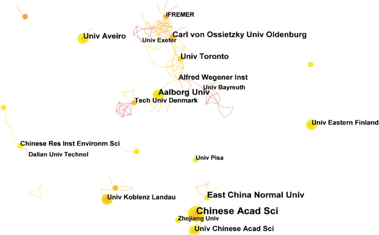
Figure 6 illustrates the cooperation network between the different institutions on this topic. As can be seen, this topic has formed two main important networks of cooperation. The first cooperative network was mainly led by the Institut Francais de Recherche pour l’exploitation de la mer(IFREMER), Carl von Ossietzky Universitat Oldenburg, University of Toronto, Aalborg University and Technical University of Denmark. This collaborative network covers many research institutions and universities in European countries and North America. Another collaborative network is led by the Chinese Academy of Sciences, the University of the Chinese Academy of Sciences, and East China Normal University. This network includes a range of research institutes and universities in China, Netherlands, and England.
Figure 6.
Institution cooperation network for microplastics detection techniques.
Keyword analysis and evolution of the field
A keyword analysis is often used in bibliometrics to understand the different directions under a topic. However, the fifteen most frequently occurring keywords in microplastic detection techniques do not include techniques and analytical methods. This is because a paper often contains five or more keywords. Microplastic detection techniques belong to one of the directions of microplastics, so this type of paper contains many keywords about environment and sample related. For this case, we purposely screened the keywords and listed only those related to detection technology (Table 4).
Table 4.
List of top eight keywords about the detection of microplastics.
| No. | Freq | Keywords |
|---|---|---|
| 1 | 30 | Nile red |
| 2 | 24 | Spectroscopy |
| 3 | 20 | Raman spectroscopy |
| 4 | 16 | FTIR |
| 5 | 13 | Microscopy |
| 6 | 8 | Density separation |
| 7 | 6 | Microspectroscopy |
| 8 | 5 | Mass spectrometry |
Visual classification is commonly used to identify microplastics in the environment, but the method has poor reliability and low accuracy. To obtain an accurate number of microplastics in the environment, fluorescent staining methods are used to aid in identifying microplastics. Usually, Nile red is used to fluorescently label microplastics. This method has the advantages of fast staining and a strong fluorescence signal. Therefore, it appears very frequently in the keywords in this topic. For example, Shim et al. 53 proposed a method for staining microplastics with Nile Red in 2016. They found that 5 mg/L of Nile Red in hexane can effectively stain plastics and identify them in green fluorescence. This technology can identify PE, PP, PS, polycarbonate, polyurethane (PU), and poly(ethylene-vinyl acetate). However, PVC, PA, and polyester cannot be identified. For a more efficient identification using fluorescent staining, the counting of microplastics in fluorescent images can be done by the corresponding developed software. 54 Other study has proposed staining microplastics with fluorescent dyes55–57. In these works, the Nile Red staining technique is often used as a control group to verify the feasibility and advancement of the new technique.
FTIR and μ-Raman are the two most commonly used spectroscopic techniques for microplastics. This technique has been widely used for the chemical identification of microplastics in water, sediment, and organism samples. In a study by Lusher et al., 58 FTIR spectroscopy was used to identify the presence of microplastics in fish successfully. The main components of these microplastics include PA, semi-synthetic cellulose materials, and rayon. However, this method is unable to identify irregular microplastics (reflectance FTIR measurements of irregularly shaped materials can have refractive errors 43 ). Attenuated total reflection (ATR)-FTIR can facilitate the identification of irregularly shaped microplastics but only applies to the analysis of plastic particles larger than 500 μm. 43 To address this problem, Löder et al. 59 applied focal plane array-based micro FT-IR imaging to identify microplastics in environmental samples to address this problem. This technique can detect plastic particles smaller than 20 μm and cover a larger filter surface area than conventional FTIR. However, analyzing the entire sample filter surface with high spatial resolution can be very time-consuming. Therefore, further optimization of FTIR spectroscopy is essential for detecting small particles of microplastics in complex environmental samples. Like FTIR spectroscopy, μ-Raman is a common technique for the analysis of microplastics. μ-Raman can detect plastic particles down to 1 µm and responds better to nonpolar plastic such as PP and PE. 60 The use of μ-Raman combined with microscopy allows the identification of microplastics of various sizes. 61 This technique does not destroy the sample while meeting the analytical needs of complex samples but cannot detect samples with fluorescence (e.g. residues from biological sample sources). In this case, purifying the sample before using μ-Raman measurements is recommended to prevent residuals in the fluorescent sample.
Mass spectrometry is often used with thermal cracking gas chromatography (Pyr-GC/MS). Pyr-GC/MS can identify the chemical composition of microplastics by analyzing their characteristic thermal degradation products. It accurately identifies the polymer type by comparing it to a pyrolysis reference map of a known pure polymer. 62 The advantage of this technique is that it does not require sample pretreatment and allows simultaneous determination of the type of plastic polymer and associated plastic additive. However, this technique only allows the analysis of one particle per cycle, which is not suitable for analyzing samples in complex environments. Mass spectrometry can also be used with thermogravimetric analysis and thermal desorption gas chromatography (TD-GC/MS). This technique is able to handle complex environmental samples. For example, Dümichen et al. 63 successfully identified PE microplastics from the soil, suspended solids, and mussels using TED-GC/MS.
Effectively separating microplastics from samples such as water, sediment, and organic matter is a crucial step for subsequent detection and analysis. Density separation is widely used to separate low-density plastic particles from dense sand, slurry, sediment, and other samples. Therefore, it becomes a high-frequency keyword in this topic. A variety of high-density solutions have been used to separate microplastics from environmental samples. The most commonly used solution is saturated NaCl. 64 It is inexpensive and non-toxic, but only for microplastics with densities below 1.20 g/cm3. However, some high-density microplastics, such as PVC and PET could not be completely separated. 65 To overcome this drawback, Nuelle et al. 66 developed a two-step method using air-induced NaCl solution overflow for pre-extraction followed by additional flotation using sodium iodide solution. The results showed that the recovery rates of all plastic pellets (PE, PP, PVC, PET, PS, PUR) ranged from 91% to 99%, except for the recovery rate of expanded PE, which was 68%. In addition, saturated sodium polytungstate solutions have been shown to isolate certain high-density microplastics. 67 Similar results can be achieved with other high-density solutions. For example, ZnCl2 solution has a density range of 1.50–1.70 g/cm3 and can extract almost all microplastic particles, but it is toxic and relatively expensive.45,68 NaI alone can also separate high-density microplastics, but it readily reacts with cellulose filter growth to darken them and thus affect visual recognition. 69
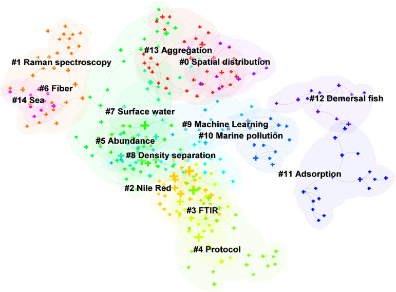
Cluster analysis of keywords can further understand the different directions of investigation in this topic. Figure 7 shows that 15 clusters were formed after clustering the keywords. Table 5 describes the clusters and their ID, size, silhouette, and respective keywords.
Figure 7.
Grouping of keywords for microplastics detection techniques.
Table 5.
Cluster analysis of keywords in topic of detection of microplastics deduced from citeSpace (g-index: k = 20; pruning method: pathfinder + pruning sliced networks + pruning)he merged networks).
| Cluster ID | Size | Silhouette | Keywords | Selected References |
|---|---|---|---|---|
| 0 | 36 | 0.849 | Environmental Sample, Degradation, Plastics, Spatial distribution, Nanoplastics, Ecosystem | 70–76 |
| 1 | 30 | 0.934 | Raman spectroscopy, Fresh water, Microscopy, Removal, Sample, Release, River, Activated sludge process | 70,77–79 |
| 2 | 29 | 0.907 | Pollution, Marine environment, Plastic debri, Ingestion, Sea, Nile Red, Organism | 44,80–87 |
| 3 | 27 | 0.920 | Sediment, Microplastics, Environment, Transport, Fate, FTIR | 43,88–93 |
| 4 | 26 | 0.932 | Identification, Extraction, Impact, Mytilus edulis L., Protocol, North sea, Toxicity, Polymer | 80,81,85,94–99 |
| 5 | 26 | 0.857 | Quantification, Particle, Accumulation, Abundance, Fish, | 68,83,86,88,100–104 |
| 6 | 24 | 0.902 | Fiber, Lost, Soil, Bivalve, Deposition, Sewage sludge, China | 69,85,105–108 |
| 7 | 22 | 0.911 | Water, Debri, Surface water, Persistent organic pollutant, Beach, Great lake | 87,99,104,109–112 |
| 8 | 20 | 0.919 | Litter, Zooplankton, Density separation, Aquatic environment, Monitoring microplastics | 82,113 |
| 9 | 19 | 0.957 | Mussel, Machine Learning, Field flow fractionation, Southeast coast, Plastic marine debri | 88,109,114,115 |
| 10 | 18 | 0.928 | Contamination, Polyethylene, Beach sediment, Marine pollution, Polystyrene microplastics, Pearl river | 63,94,116 |
| 11 | 17 | 0.956 | Spectroscopy, Sorption, Toxic chemical, Adsorption, Complex, Size | 98,117–119 |
| 12 | 13 | 0.927 | Plastic pollution, Demersal fish, Fresh water ecosystem, FT-IR, Gastrointestinal tract | 93,96,120,121 |
| 13 | 13 | 0.972 | Exposure, Nanoparticle, 20 μM, Aggregation | 122–124 |
| 14 | 10 | 0.996 | Baltic sea, Bohai sea, Microparticle | 58,80,125,126 |
Based on the bibliometric analysis, the detection of microplastics currently encompasses the following techniques:
The most basic method used for microplastic identification is the visual inspection method. This method uses the human eye or microscope to observe the microplastics and count them according to their size and dimensions. Microplastics with particle diameters of 1 mm to 5 mm can be directly identified and analyzed using visual inspection. Due to the differences in color, structure, and other characteristics of the microplastics, there will be some influence on the identification results of the visual inspection method.
If microplastic samples cannot be observed by visual inspection, they can be identified by microscopic techniques. Compared with a visual inspection, light microscopy is more convenient and efficient, but at the same time, the error is also more significant. Microplastics are analyzed and identified under ordinary light microscopy with an error rate of about 20%. In the case of colorless, transparent microplastics, the error rate would be over 70%. Scanning electron microscopy uses electron microscopy for analysis and identification, and the magnification of electron microscopy is greater than that of ordinary light microscopy. However, this method requires the sample to be solid, non-toxic, and non-radioactive. Also, this method has high requirements for the laboratory environment.
Microplastics can be cleaved to produce unique pyrolysis profiles, so their chemical composition can be analyzed using Pyr-GC/MS. However, different polymers may also produce the same or similar pyrolysis products, which can overlap in the pyrolysis profile and produce errors in the final results.
The best current method for determining microplastics is FTIR. It can obtain specific polymer information from the characteristic spectra of microplastics and can identify the type of microplastics in the environment. However, the sample must be dried when using this method to avoid interference from moisture and impurities in the sample.
Due to the diversity of microplastic types, different microplastics have their own unique Raman spectral profiles. Compared with FTIR, μ-Raman has a broader range of applications and better sensing ability. However, μ-Raman is susceptible to the interference of fluorescent substances.
Conclusion and perspectives
The detection of microplastic detection has gradually gained attention since 2015. So far, this topic has shown a growing trend year by year. The detection of microplastics has now attracted the attention of not only environmental scientists but has also involved analytical chemists. Environmental science and analytical chemistry-related journals remain the most dominant areas in this topic. It is also worth noting that the new journals appearing in 2020 include a series of journals related to food science. The results of the geographical analysis point to European scholars being the most active in this topic. This topic has formed two main important networks of cooperation.
Reliable identification of microplastic particles in various environmental matrices is still limited. First, some microplastics are present in trace level in the environment, thus requiring high sensitivity for detection techniques. At the same time, these microplastics present a mixed state, containing different types, which are difficult to distinguish quickly by detection techniques. Separation and concentration become effective methods to solve the above two challenges, but there is no well-established protocol.
Based on the bibliometric analysis, we believe that the following directions are the priorities that need to be overcome for the future development of microplastic detection technology.
Challenges remain in the concentration and detection of microplastics in water and air samples. The development of detection techniques requires new strategies for the analysis of these two types of environmental samples.
It is challenging to meet the requirements of sensitivity and resolution using a single analytical technology. Analytical techniques based on thermal cracking reactions coupled with GC-MS are most likely to be developed as a method that satisfies both detection sensitivity and specificity when supported by appropriate sample pre-treatment.
It is important to establish a standard protocol for the detection of microplastics. Detection techniques at this stage do not even use uniform concentration units for microplastics. Therefore, the establishment of a uniform set of criteria would allow for comparability between the results of different studies.
Acknowledgements
This work was financially supported by Zhejiang Key Laboratory of Ecological and Environmental Monitoring, Forewarning and Quality Control (EEMFQ-2021-2), National Science Foundation of China (No. 42173073) and National Natural Science Foundation of China (No. 22108265).
Footnotes
The author(s) declared no potential conflicts of interest with respect to the research, authorship, and/or publication of this article.
Funding: The author(s) disclosed receipt of the following financial support for the research, authorship, and/or publication of this article: This work was supported by the Zhejiang Key Laboratory of Ecological and Environmental Monitoring, Forewarning and Quality Control, National Natural Science Foundation of China, (grant number EEMFQ-2021-2, 22108265, 42173073)
ORCID iD: Li Fu https://orcid.org/0000-0002-5957-7790
References
- 1.Pathak S, Sneha C, Mathew BB. Bioplastics: its timeline based scenario & challenges. J Polym Biopolym Phys Chem 2014; 2: 84–90. [Google Scholar]
- 2.Alimba CG, Faggio C. Microplastics in the marine environment: current trends in environmental pollution and mechanisms of toxicological profile. Environ Toxicol Pharmacol 2019; 68: 61–74. [DOI] [PubMed] [Google Scholar]
- 3.Lebreton L, Andrady A. Future scenarios of global plastic waste generation and disposal. Palgrave Communications 2019; 5: 6. [Google Scholar]
- 4.Law KL, Thompson RC. Microplastics in the seas. Science 2014; 345: 144–145. [DOI] [PubMed] [Google Scholar]
- 5.Van Cauwenberghe L, Janssen CR. Microplastics in bivalves cultured for human consumption. Environ Pollut 2014; 193: 65–70. [DOI] [PubMed] [Google Scholar]
- 6.Cole M, Lindeque P, Halsband Cet al. et al. Microplastics as contaminants in the marine environment: a review. Mar Pollut Bull 2011; 62: 2588–2597. [DOI] [PubMed] [Google Scholar]
- 7.Wagner S, Reemtsma T. Things we know and don’t know about nanoplastic in the environment. Nat Nanotechnol 2019; 14: 300–301. [DOI] [PubMed] [Google Scholar]
- 8.Di M, Wang J. Microplastics in surface waters and sediments of the three gorges reservoir, China. Sci Total Environ 2018; 616: 1620–1627. [DOI] [PubMed] [Google Scholar]
- 9.Dris R, Gasperi J, Saad Met al. et al. Synthetic fibers in atmospheric fallout: a source of microplastics in the environment? Mar Pollut Bull 2016; 104: 290–293. [DOI] [PubMed] [Google Scholar]
- 10.Bessa F, Ratcliffe N, Otero V, et al. Microplastics in gentoo penguins from the antarctic region. Sci Rep 2019; 9: 1–7. [DOI] [PMC free article] [PubMed] [Google Scholar]
- 11.Isobe A, Iwasaki S, Uchida Ket al. et al. Abundance of non-conservative microplastics in the upper ocean from 1957 to 2066. Nat Commun 2019; 10: 417. [DOI] [PMC free article] [PubMed] [Google Scholar]
- 12.Julienne F, Delorme N, Lagarde F. From macroplastics to microplastics: Role of water in the fragmentation of polyethylene. Chemosphere 2019; 236: 124409. [DOI] [PubMed] [Google Scholar]
- 13.Araújo AP dC, de Melo NFS, de Oliveira Junior AG, et al. How much are microplastics harmful to the health of amphibians? A study with pristine polyethylene microplastics and Physalaemus cuvieri. J Hazard Mater 2020; 382: 121066. [DOI] [PubMed] [Google Scholar]
- 14.Harrison JP, Schratzberger M, Sapp Met al. et al. Rapid bacterial colonization of low-density polyethylene microplastics in coastal sediment microcosms. BMC Microbiol 2014; 14: 232. [DOI] [PMC free article] [PubMed] [Google Scholar]
- 15.Chen Y, Liu X, Leng Yet al. et al. Defense responses in earthworms (eisenia fetida) exposed to low-density polyethylene microplastics in soils. Ecotoxicol Environ Saf 2020; 187: 109788. [DOI] [PubMed] [Google Scholar]
- 16.Bringer A, Thomas H, Prunier G, et al. High density polyethylene (HDPE) microplastics impair development and swimming activity of pacific oyster D-larvae, crassostrea gigas, depending on particle size. Environ Pollut 2020; 260: 113978. [DOI] [PubMed] [Google Scholar]
- 17.Cheng Y, Song W, Tian H, et al. The effects of high-density polyethylene and polypropylene microplastics on the soil and earthworm metaphire guillelmi gut microbiota. Chemosphere 2021; 267: 129219. [DOI] [PubMed] [Google Scholar]
- 18.Wakkaf T, Allouche M, Harrath AH, et al. The individual and combined effects of cadmium, polyvinyl chloride (PVC) microplastics and their polyalkylamines modified forms on meiobenthic features in a microcosm. Environ Pollut 2020; 266: 115263. [DOI] [PubMed] [Google Scholar]
- 19.Ouyang Z, Zhang Z, Jing Y, et al. The photo-aging of polyvinyl chloride microplastics under different UV irradiations. Gondwana Res 2022; 108: 72–80. [Google Scholar]
- 20.Bai B, Liu Y, Zhang H, et al. Experimental investigation on gasification characteristics of polyethylene terephthalate (PET) microplastics in supercritical water. Fuel 2020; 262: 116630. [Google Scholar]
- 21.Hwang J, Choi D, Han Set al. et al. An assessment of the toxicity of polypropylene microplastics in human derived cells. Sci Total Environ 2019; 684: 657–669. [DOI] [PubMed] [Google Scholar]
- 22.Jiang Y, Yin X, Xi Xet al. et al. Effect of surfactants on the transport of polyethylene and polypropylene microplastics in porous media. Water Res 2021; 196: 117016. [DOI] [PubMed] [Google Scholar]
- 23.Mao Y, Ai H, Chen Y, et al. Phytoplankton response to polystyrene microplastics: perspective from an entire growth period. Chemosphere 2018; 208: 59–68. [DOI] [PubMed] [Google Scholar]
- 24.Rehse S, Kloas W, Zarfl C. Microplastics reduce short-term effects of environmental contaminants. Part I: effects of bisphenol A on freshwater zooplankton are lower in presence of polyamide particles. Int J Environ Res Public Health 2018; 15: 280. [DOI] [PMC free article] [PubMed] [Google Scholar]
- 25.Jang M, Shim WJ, Han GMet al. et al. Formation of microplastics by polychaetes (marphysa sanguinea) inhabiting expanded polystyrene marine debris. Mar Pollut Bull 2018; 131: 365–369. [DOI] [PubMed] [Google Scholar]
- 26.Ivleva NP, Wiesheu AC, Niessner R. Microplastic in aquatic ecosystems. Angew Chem, Int Ed. 2017; 56: 1720–1739. [DOI] [PubMed] [Google Scholar]
- 27.Vandermeersch G, Van Cauwenberghe L, Janssen CR, et al. A critical view on microplastic quantification in aquatic organisms. Environ Res 2015; 143: 46–55. [DOI] [PubMed] [Google Scholar]
- 28.Zheng Y, Karimi-Maleh H, Fu L. Advances in electrochemical techniques for the detection and analysis of genetically modified organisms: an analysis based on bibliometrics. Chemosensors 2022; 10: 194. [Google Scholar]
- 29.Zheng Y, Mao S, Zhu Jet al. et al. Current status of electrochemical detection of sunset yellow based on bibliometrics. Food Chem Toxicol 2022; 164: 113019. [DOI] [PubMed] [Google Scholar]
- 30.Shen Y, Mao S, Chen F, et al. Electrochemical detection of Sudan red series azo dyes: bibliometrics based analysis. Food Chem Toxicol 2022; 163: 112960. [DOI] [PubMed] [Google Scholar]
- 31.Zheng Y, Karimi-Maleh H, Fu L. Evaluation of antioxidants using electrochemical sensors: a bibliometric analysis. Sensors 2022; 22: 3238. [DOI] [PMC free article] [PubMed] [Google Scholar]
- 32.Fu L, Mao S, Chen F, et al. Graphene-based electrochemical sensors for antibiotic detection in water, food and soil: a scientometric analysis in CiteSpace (2011–2021). Chemosphere 2022; 297: 134127. [DOI] [PubMed] [Google Scholar]
- 33.Jin M, Liu J, Wu W, et al. Relationship between graphene and pedosphere: a scientometric analysis. Chemosphere 2022; 300: 134599. [DOI] [PubMed] [Google Scholar]
- 34.Zhang Y, Pu S, Lv Xet al. et al. Global trends and prospects in microplastics research: a bibliometric analysis. J Hazard Mater 2020; 400: 123110. [DOI] [PubMed] [Google Scholar]
- 35.Pauna VH, Buonocore E, Renzi Met al. et al. The issue of microplastics in marine ecosystems: a bibliometric network analysis. Mar Pollut Bull 2019; 149: 110612. [Google Scholar]
- 36.Ebrahimi P, Abbasi S, Pashaei Ret al. et al. Investigating impact of physicochemical properties of microplastics on human health: a short bibliometric analysis and review. Chemosphere 2022; 289: 133146. [DOI] [PubMed] [Google Scholar]
- 37.Qin F, Du J, Gao J, et al. Bibliometric profile of global microplastics research from 2004 to 2019. Int J Environ Res Public Health 2020; 17: 5639. [DOI] [PMC free article] [PubMed] [Google Scholar]
- 38.Börner K, Chen C, Boyack KW. Visualizing knowledge domains. Annu Rev Inf Sci Technol 2003; 37: 179–255. [Google Scholar]
- 39.Chen C. Citespace II: detecting and visualizing emerging trends and transient patterns in scientific literature. J Am Soc Inf Sci Technol 2006; 57: 359–377. [Google Scholar]
- 40.Chen C. Searching for intellectual turning points: progressive knowledge domain visualization. Proc Natl Acad Sci U S A. 2004;101: 5303–5310. [DOI] [PMC free article] [PubMed] [Google Scholar]
- 41.Chen C, Ibekwe-SanJuan F, Hou J. The structure and dynamics of cocitation clusters: a multiple-perspective cocitation analysis. J Am Soc Inf Sci Technol 2010; 61: 1386–1409. [Google Scholar]
- 42.Xueshu D W. COOC is a software for bibliometrics and knowledge mapping[CP/OL]. Published 2022. https://github.com/2088904822. (Accessed February 15, 2022).
- 43.Harrison JP, Ojeda JJ, Romero-González ME. The applicability of reflectance micro-Fourier-transform infrared spectroscopy for the detection of synthetic microplastics in marine sediments. Sci Total Environ 2012; 416: 455–463. [DOI] [PubMed] [Google Scholar]
- 44.Fossi MC, Panti C, Guerranti C, et al. Are baleen whales exposed to the threat of microplastics? A case study of the Mediterranean fin whale (balaenoptera physalus). Mar Pollut Bull 2012; 64: 2374–2379. [DOI] [PubMed] [Google Scholar]
- 45.Imhof HK, Schmid J, Niessner Ret al. et al. A novel, highly efficient method for the separation and quantification of plastic particles in sediments of aquatic environments. Limnology and Oceanography: Methods 2012; 10: 524–537. [Google Scholar]
- 46.Gündoğdu S, Çevik C, Ataş NT. Stuffed with microplastics: microplastic occurrence in traditional stuffed mussels sold in the turkish market. Food Biosci 2020; 37: 100715. [Google Scholar]
- 47.Prata JC, Paço A, Reis V, et al. Identification of microplastics in white wines capped with polyethylene stoppers using micro-Raman spectroscopy. Food Chem 2020; 331: 127323. [DOI] [PubMed] [Google Scholar]
- 48.Huang Y, Chapman J, Deng Yet al. et al. Rapid measurement of microplastic contamination in chicken meat by mid infrared spectroscopy and chemometrics: a feasibility study. Food Control 2020; 113: 107187. [Google Scholar]
- 49.Kedzierski M, Lechat B, Sire Oet al. et al. Microplastic contamination of packaged meat: occurrence and associated risks. Food Packaging and Shelf Life 2020; 24: 100489. [Google Scholar]
- 50.Schmidt R, Nachtnebel M, Dienstleder M, et al. Correlative SEM-Raman microscopy to reveal nanoplastics in complex environments. Micron 2021; 144: 103034. [DOI] [PubMed] [Google Scholar]
- 51.Qian J, Begum H, Lee JEY. Acoustofluidic localization of sparse particles on a piezoelectric resonant sensor for nanogram-scale mass measurements. Microsystems & Nanoengineering 2021; 7: 61. [DOI] [PMC free article] [PubMed] [Google Scholar]
- 52.Sancataldo G, Ferrara V, Bonomo FP, et al. Identification of microplastics using 4-dimethylamino-4′-nitrostilbene solvatochromic fluorescence. Microsc Res Tech 2021; 84: 2820–2831. [DOI] [PMC free article] [PubMed] [Google Scholar]
- 53.Shim WJ, Song YK, Hong SHet al. et al. Identification and quantification of microplastics using Nile red staining. Mar Pollut Bull 2016; 113: 469–476. [DOI] [PubMed] [Google Scholar]
- 54.Prata JC, Reis V, Matos JTVet al. et al. A new approach for routine quantification of microplastics using Nile red and automated software (MP-VAT). Sci Total Environ 2019; 690: 1277–1283. [DOI] [PubMed] [Google Scholar]
- 55.Karakolis EG, Nguyen B, You JBet al. et al. Fluorescent dyes for visualizing microplastic particles and fibers in laboratory-based studies. Environ Sci Technol Lett 2019; 6: 334–340. [Google Scholar]
- 56.Lv L, Qu J, Yu Z, et al. A simple method for detecting and quantifying microplastics utilizing fluorescent dyes-safranine T, fluorescein isophosphate, Nile red based on thermal expansion and contraction property. Environ Pollut 2019; 255: 113283. [DOI] [PubMed] [Google Scholar]
- 57.Tarafdar A, Choi SH, Kwon JH. Differential staining lowers the false positive detection in a novel volumetric measurement technique of microplastics. J Hazard Mater 2022; 432: 128755. [DOI] [PubMed] [Google Scholar]
- 58.Lusher AL, Mchugh M, Thompson RC. Occurrence of microplastics in the gastrointestinal tract of pelagic and demersal fish from the English channel. Mar Pollut Bull 2013; 67: 94–99. [DOI] [PubMed] [Google Scholar]
- 59.Löder MG, Gerdts G. Methodology used for the detection and identification of microplastics—a critical appraisal. Marine Anthropogenic Litter. Published online2015: 201–227. [Google Scholar]
- 60.Anger PM, von der Esch E, Baumann Tet al. et al. Raman Microspectroscopy as a tool for microplastic particle analysis. TrAC, Trends Anal Chem. 2018; 109: 214–226. [Google Scholar]
- 61.Araujo CF, Nolasco MM, Ribeiro AMet al. et al. Identification of microplastics using Raman spectroscopy: latest developments and future prospects. Water Res 2018; 142: 426–440. [DOI] [PubMed] [Google Scholar]
- 62.Fischer M, Scholz-Böttcher BM. Simultaneous trace identification and quantification of common types of microplastics in environmental samples by pyrolysis-gas chromatography–mass spectrometry. Environ Sci Technol 2017; 51: 5052–5060. [DOI] [PubMed] [Google Scholar]
- 63.Dümichen E, Barthel AK, Braun U, et al. Analysis of polyethylene microplastics in environmental samples, using a thermal decomposition method. Water Res 2015; 85: 451–457. [DOI] [PubMed] [Google Scholar]
- 64.Fries E, Dekiff JH, Willmeyer Jet al. et al. Identification of polymer types and additives in marine microplastic particles using pyrolysis-GC/MS and scanning electron microscopy. Environmental Science: Processes & Impacts 2013; 15: 1949–1956. [DOI] [PubMed] [Google Scholar]
- 65.Browne MA, Galloway TS, Thompson RC. Spatial patterns of plastic debris along estuarine shorelines. Environ Sci Technol 2010; 44: 3404–3409. [DOI] [PubMed] [Google Scholar]
- 66.Nuelle MT, Dekiff JH, Remy Det al. et al. A new analytical approach for monitoring microplastics in marine sediments. Environ Pollut 2014; 184: 161–169. [DOI] [PubMed] [Google Scholar]
- 67.Corcoran PL, Biesinger MC, Grifi M. Plastics and beaches: a degrading relationship. Mar Pollut Bull 2009; 58: 80–84. [DOI] [PubMed] [Google Scholar]
- 68.Liebezeit G, Dubaish F. Microplastics in beaches of the east frisian islands spiekeroog and kachelotplate. Bull Environ Contam Toxicol 2012; 89: 213–217. [DOI] [PubMed] [Google Scholar]
- 69.Crichton EM, Noël M, Gies EAet al. et al. A novel, density-independent and FTIR-compatible approach for the rapid extraction of microplastics from aquatic sediments. Anal Methods 2017; 9: 1419–1428. [Google Scholar]
- 70.Asamoah BO, Roussey M, Peiponen KE. On optical sensing of surface roughness of flat and curved microplastics in water. Chemosphere 2020; 254: 126789. [DOI] [PubMed] [Google Scholar]
- 71.Sun J, Zhu ZR, Li WH, et al. Revisiting microplastics in landfill leachate: unnoticed tiny microplastics and their fate in treatment works. Water Res 2021; 190: 116784. [DOI] [PubMed] [Google Scholar]
- 72.Toapanta T, Okoffo ED, Ede S, et al. Influence of surface oxidation on the quantification of polypropylene microplastics by pyrolysis gas chromatography mass spectrometry. Sci Total Environ 2021; 796: 148835. [DOI] [PubMed] [Google Scholar]
- 73.Vivekanand AC, Mohapatra S, Tyagi VK. Microplastics in aquatic environment: challenges and perspectives. Chemosphere 2021; 282: 131151. [DOI] [PubMed] [Google Scholar]
- 74.Prosenc F, Leban P, Šunta Uet al. et al. Extraction and identification of a wide range of microplastic polymers in soil and Compost Polymers (Basel) 2021; 13: 4069. [DOI] [PMC free article] [PubMed] [Google Scholar]
- 75.Du C, Wu J, Gong Jet al. et al. Tof-SIMS characterization of microplastics in soils. Surf Interface Anal 2020; 52: 293–300. [Google Scholar]
- 76.Mintenig S, Bäuerlein PS, Koelmans AAet al. et al. Closing the gap between small and smaller: towards a framework to analyse nano-and microplastics in aqueous environmental samples. Environmental Science: Nano 2018; 5: 1640–1649. [Google Scholar]
- 77.He P, Chen L, Shao Let al. et al. Municipal solid waste (MSW) landfill: a source of microplastics?-evidence of microplastics in landfill leachate. Water Res 2019; 159: 38–45. [DOI] [PubMed] [Google Scholar]
- 78.Rasmussen LA, Iordachescu L, Tumlin Set al. et al. A complete mass balance for plastics in a wastewater treatment plant-macroplastics contributes more than microplastics. Water Res 2021; 201: 117307. [DOI] [PubMed] [Google Scholar]
- 79.Castelvetro V, Corti A, Bianchi Set al. et al. Microplastics in fish meal: contamination level analyzed by polymer type, including polyester (PET), polyolefins, and polystyrene. Environ Pollut 2021; 273: 115792. [DOI] [PubMed] [Google Scholar]
- 80.Kirstein IV, Kirmizi S, Wichels A, et al. Dangerous hitchhikers? Evidence for potentially pathogenic Vibrio spp. On microplastic particles. Mar Environ Res 2016; 120: 1–8. [DOI] [PubMed] [Google Scholar]
- 81.Herrera A, Garrido-Amador P, Martínez I, et al. Novel methodology to isolate microplastics from vegetal-rich samples. Mar Pollut Bull 2018; 129: 61–69. [DOI] [PubMed] [Google Scholar]
- 82.Lima A, Costa M, Barletta M. Distribution patterns of microplastics within the plankton of a tropical estuary. Environ Res 2014; 132: 146–155. [DOI] [PubMed] [Google Scholar]
- 83.Käppler A, Windrich F, Löder MG, et al. Identification of microplastics by FTIR and Raman microscopy: a novel silicon filter substrate opens the important spectral range below 1300 cm − 1 for FTIR transmission measurements. Anal Bioanal Chem 2015; 407: 6791–6801. [DOI] [PubMed] [Google Scholar]
- 84.Claessens M, Van Cauwenberghe L, Vandegehuchte MBet al. et al. New techniques for the detection of microplastics in sediments and field collected organisms. Mar Pollut Bull 2013; 70: 227–233. [DOI] [PubMed] [Google Scholar]
- 85.Wiggin KJ, Holland EB. Validation and application of cost and time effective methods for the detection of 3–500 μm sized microplastics in the urban marine and estuarine environments surrounding long beach, California. Mar Pollut Bull 2019; 143: 152–162. [DOI] [PubMed] [Google Scholar]
- 86.Olivatto GP, Martins MCT, Montagner CCet al. et al. Microplastic contamination in surface waters in guanabara bay, rio de janeiro, Brazil. Mar Pollut Bull 2019; 139: 157–162. [DOI] [PubMed] [Google Scholar]
- 87.Jang M, Shim WJ, Han GMet al. et al. Widespread detection of a brominated flame retardant, hexabromocyclododecane, in expanded polystyrene marine debris and microplastics from South Korea and the Asia-pacific coastal region. Environ Pollut 2017; 231: 785–794. [DOI] [PubMed] [Google Scholar]
- 88.Li L, Li M, Deng H, et al. A straightforward method for measuring the range of apparent density of microplastics. Sci Total Environ 2018; 639: 367–373. [DOI] [PubMed] [Google Scholar]
- 89.Scopetani C, Chelazzi D, Martellini T, et al. Occurrence and characterization of microplastic and mesoplastic pollution in the migliarino san rossore, massaciuccoli nature park (Italy). Mar Pollut Bull 2021; 171: 112712. [DOI] [PubMed] [Google Scholar]
- 90.I LF, J PP. and, et al. M. A semi-automated Raman micro-spectroscopy method for morphological and chemical characterizations of microplastic litter. Mar Pollut Bull 2016; 113: 461–468. [DOI] [PubMed] [Google Scholar]
- 91.Hardesty BD, Holdsworth D, Revill ATet al. et al. A biochemical approach for identifying plastics exposure in live wildlife. Methods Ecol Evol 2015; 6: 92–98. [Google Scholar]
- 92.Chubarenko I, Stepanova N. Microplastics in sea coastal zone: lessons learned from the baltic amber. Environ Pollut 2017; 224: 243–254. [DOI] [PubMed] [Google Scholar]
- 93.Jaafar N, Musa SM, Azfaralariff Aet al. et al. Improving the efficiency of post-digestion method in extracting microplastics from gastrointestinal tract and gills of fish. Chemosphere 2020; 260: 127649. [DOI] [PubMed] [Google Scholar]
- 94.Santillo D, Miller K, Johnston P. Microplastics as contaminants in commercially important seafood species. Integr Environ Assess Manag 2017; 13: 516–521. [DOI] [PubMed] [Google Scholar]
- 95.Hara J, Frias J, Nash R. Quantification of microplastic ingestion by the decapod crustacean nephrops norvegicus from Irish waters. Mar Pollut Bull 2020; 152: 110905. [DOI] [PubMed] [Google Scholar]
- 96.Akhbarizadeh R, Dobaradaran S, Nabipour Iet al. et al. Abundance, composition, and potential intake of microplastics in canned fish. Mar Pollut Bull 2020; 160: 111633. [DOI] [PubMed] [Google Scholar]
- 97.Kedzierski M, Le Tilly V, Bourseau Pet al. et al. Microplastics elutriation system: part B: insight of the next generation. Mar Pollut Bull 2018; 133: 9–17. [DOI] [PubMed] [Google Scholar]
- 98.Lee J, Choi Y, Jeong Jet al. et al. Eye-glass polishing wastewater as significant microplastic source: microplastic identification and quantification. J Hazard Mater 2021; 403: 123991. [DOI] [PubMed] [Google Scholar]
- 99.Munno K, Helm PA, Jackson DAet al. et al. Impacts of temperature and selected chemical digestion methods on microplastic particles. Environ Toxicol Chem 2018; 37: 91–98. [DOI] [PubMed] [Google Scholar]
- 100.Alomar C, Estarellas F, Deudero S. Microplastics in the Mediterranean sea: deposition in coastal shallow sediments, spatial variation and preferential grain size. Mar Environ Res 2016; 115: 1–10. [DOI] [PubMed] [Google Scholar]
- 101.Vidal C, Pasquini C. A comprehensive and fast microplastics identification based on near-infrared hyperspectral imaging (HSI-NIR) and chemometrics. Environ Pollut 2021; 285: 117251. [DOI] [PubMed] [Google Scholar]
- 102.Mu J, Qu L, Jin F, et al. Abundance and distribution of microplastics in the surface sediments from the northern bering and chukchi seas. Environ Pollut 2019; 245: 122–130. [DOI] [PubMed] [Google Scholar]
- 103.Clunies-Ross P, Smith G, Gordon Ket al. et al. Synthetic shorelines in New Zealand? Quantification and characterisation of microplastic pollution on canterbury’s coastlines. Null 2016; 50: 317–325. [Google Scholar]
- 104.Rowley KH, Cucknell AC, Smith BDet al. et al. London’s river of plastic: high levels of microplastics in the thames water column. Sci Total Environ 2020; 740: 140018. [DOI] [PubMed] [Google Scholar]
- 105.Uddin S, Fowler SW, Saeed Tet al. et al. Standardized protocols for microplastics determinations in environmental samples from the gulf and marginal seas. Mar Pollut Bull 2020; 158: 111374. [DOI] [PubMed] [Google Scholar]
- 106.Huang Y, Liu Q, Jia Wet al. et al. Agricultural plastic mulching as a source of microplastics in the terrestrial environment. Environ Pollut 2020; 260: 114096. [DOI] [PubMed] [Google Scholar]
- 107.Rist S, Steensgaard IM, Guven O, et al. The fate of microplastics during uptake and depuration phases in a blue mussel exposure system. Environ Toxicol Chem 2019; 38: 99–105. [DOI] [PubMed] [Google Scholar]
- 108.Ma J, Chen F, Xu H, et al. Face masks as a source of nanoplastics and microplastics in the environment: quantification, characterization, and potential for bioaccumulation. Environ Pollut 2021; 288: 117748. [DOI] [PubMed] [Google Scholar]
- 109.Rodrigues MO, Abrantes N, Gonçalves FJMet al. et al. Spatial and temporal distribution of microplastics in water and sediments of a freshwater system (antuã river. Portugal). Science of The Total Environment 2018; 633: 1549–1559. [DOI] [PubMed] [Google Scholar]
- 110.Löder MGJ, Kuczera M, Mintenig Set al. et al. Focal plane array detector-based micro-Fourier-transform infrared imaging for the analysis of microplastics in environmental samples. Environ Chem 2015; 12: 563–581. [Google Scholar]
- 111.Naidoo T S, Thompson RC, Rajkaran A. Quantification and characterisation of microplastics ingested by selected juvenile fish species associated with mangroves in KwaZulu-natal, South Africa. Environ Pollut 2020; 257: 113635. [DOI] [PubMed] [Google Scholar]
- 112.Phuong NN, Poirier L, Lagarde Fet al. et al. Microplastic abundance and characteristics in French atlantic coastal sediments using a new extraction method. Environ Pollut 2018; 243: 228–237. [DOI] [PubMed] [Google Scholar]
- 113.Lusher AL, Munno K, Hermabessiere Let al. et al. Isolation and extraction of microplastics from environmental samples: an evaluation of practical approaches and recommendations for further harmonization. Appl Spectrosc 2020; 74: 1049–1065. [DOI] [PubMed] [Google Scholar]
- 114.Ahamed A, Ge L, Zhao Ket al. et al. Environmental footprint of voltammetric sensors based on screen-printed electrodes: an assessment towards “green” sensor manufacturing. Chemosphere 2021; 278: 130462. [DOI] [PubMed] [Google Scholar]
- 115.Behera DP, Kolandhasamy P, Sigamani Set al. et al. A preliminary investigation of marine litter pollution along mandvi beach, kachchh, gujarat. Mar Pollut Bull 2021; 165: 112100. [DOI] [PubMed] [Google Scholar]
- 116.Coppock RL, Cole M, Lindeque PKet al. et al. A small-scale, portable method for extracting microplastics from marine sediments. Environ Pollut 2017; 230: 829–837. [DOI] [PubMed] [Google Scholar]
- 117.Lavoy M, Crossman J. A novel method for organic matter removal from samples containing microplastics. Environ Pollut 2021; 286: 117357. [DOI] [PubMed] [Google Scholar]
- 118.Shan J, Zhang Y, Wang Jet al. et al. Microextraction based on microplastic followed by SERS for on-site detection of hydrophobic organic contaminants, an indicator of seawater pollution. J Hazard Mater 2020; 400: 123202. [DOI] [PubMed] [Google Scholar]
- 119.Elert AM, Becker R, Duemichen E, et al. Comparison of different methods for MP detection: what can we learn from them, and why asking the right question before measurements matters? Environ Pollut 2017; 231: 1256–1264. [DOI] [PubMed] [Google Scholar]
- 120.Picó Y, Alvarez-Ruiz R, Alfarhan AHet al. et al. Pharmaceuticals, pesticides, personal care products and microplastics contamination assessment of al-hassa irrigation network (Saudi Arabia) and its shallow lakes. Sci Total Environ 2020; 701: 135021. [DOI] [PubMed] [Google Scholar]
- 121.Zhu L, Wang H, Chen Bet al. et al. Microplastic ingestion in deep-sea fish from the South China Sea. Sci Total Environ 2019; 677: 493–501. [DOI] [PubMed] [Google Scholar]
- 122.Paul A, Wander L, Becker Ret al. et al. High-throughput NIR spectroscopic (NIRS) detection of microplastics in soil. Environmental Science and Pollution Research 2019; 26: 7364–7374. [DOI] [PubMed] [Google Scholar]
- 123.Vicentini DS, Nogueira DJ, Melegari SP, et al. Toxicological evaluation and quantification of ingested metal-core nanoplastic by daphnia magna through fluorescence and inductively coupled plasma-mass spectrometric methods. Environ Toxicol Chem 2019; 38: 2101–2110. [DOI] [PubMed] [Google Scholar]
- 124.Gillibert R, Balakrishnan G, Deshoules Q, et al. Raman Tweezers for small microplastics and nanoplastics identification in seawater. Environ Sci Technol 2019; 53: 9003–9013. [DOI] [PubMed] [Google Scholar]
- 125.Enders K, Käppler A, Biniasch O, et al. Tracing microplastics in aquatic environments based on sediment analogies. Sci Rep 2019; 9: 15207. [DOI] [PMC free article] [PubMed] [Google Scholar]
- 126.Hengstmann E, Weil E, Wallbott PCet al. et al. Microplastics in lakeshore and lakebed sediments–external influences and temporal and spatial variabilities of concentrations. Environ Res 2021; 197: 111141. [DOI] [PubMed] [Google Scholar]


