Abstract
Background
Early detection of BCR::ABL1-like ALL could impact treatment management and improve the overall survival/outcome. BCR::ABL1-like ALL cases are characterised by diverse genetic alterations activating cytokine receptors and kinase signalling. Its detection is still an unmet need in low–middle-income countries due to the unavailability of a patented TLDA assay.
Methods
This study’s rationale is to identify BCR::ABL1-like ALLs using the PHi-RACE classifier, followed by the characterisation of underlying adverse genetic alterations in recurrent gene abnormalities negative (RGAneg) B-ALLs (n = 108).
Results
We identified 34.25% (37/108) BCR::ABL1-like ALLs using PHi-RACE classifier, characterised by TSLPR/CRLF2 expression (11.58%), IKZF1 (Δ4–7) deletion (18.9%) and chimeric gene fusions (34.61%). In overexpressed TSLPR/CRLF2 BCR::ABL1-like ALLs, we identified 33.33% (1/3) CRLF2::IGH and 33.33% (1/3) EPOR::IGH rearrangements with concomitant JAK2 mutation R683S (50%). We identified 18.91% CD13 (P = 0.02) and 27.02% CD33 (P = 0.05) aberrant myeloid markers positivity, which was significantly higher in BCR::ABL1-like ALLs compared to non-BCR::ABL1-like ALLs. MRD positivity was considerably higher (40% in BCR::ABL1-like vs. 19.29% in non-BCR::ABL1-like ALLs).
Conclusions
With this practical approach, we reported a high incidence of BCR::ABL1-like ALLs, and a lower frequency of CRLF2 alteration & associated CGFs. Recognising this entity, early at diagnosis is crucial to optimise personalised treatment strategies.
Subject terms: Genetics research, PCR-based techniques
Background
“B-lymphoblastic leukaemia/lymphoma, BCR::ABL1-like acute lymphoblastic leukaemia (ALL)” has been considered an entity in the revised World Health Organization (WHO) (2022) classification of Haematolymphoid neoplasms [1, 2]. The unique gene expression profile (GEP) of this entity resembles BCR::ABL1-positive ALL cases without the presence of t(9:22)(q34;q11.2) translocation [3–24]. BCR::ABL1-like ALL cases encompass a diverse range of genomic alterations, including the deletions, and sequence mutations in lymphoid developmental transcription factors, most commonly IKZF1 (IKAROS Family Zinc Finger 1), rearrangement of CRLF2 (cytokine receptor-like factor 2) and kinase fusions including ABL (ABL Proto-Oncogene) & JAK (Janus Kinase) alterations [3, 5–12, 15, 19–21, 24, 25]. In BCR::ABL1-like ALL cases, two broad genetic categories have been classified. In the first category, approximately 50% of patients have overexpression of CRLF2, and half of those with CRLF2 overexpression have accompanying JAK-STAT mutations (JAK2R683G), which results in the activation of JAK-STAT signalling amenable to JAK inhibition. In the second category, cases without CRLF2 overexpression having kinase fusions involving JAK2 (Janus-kinase2), ABL1 (ABL Proto-Oncogene 1), ABL2 (ABL Proto-Oncogene 2), CSF1R (colony-stimulating factor-1), PDGFRB (Platelet-derived growth factor receptor beta), and EPOR (erythropoietin receptor), and many are amenable to ABL-type inhibitors or JAK inhibitors (tyrosine kinase inhibitors [TKIs]) [3–12, 14–16, 18, 21, 22, 24, 26–28]. The incidence of BCR::ABL1-like ALL cases comprises 10-15% of childhood cases, and its incidence increases with age to a high estimate of 33.1% in adults. These cases are associated with poorer clinical outcomes [3–33]. Initially, two independent research groups (2009) from high-income countries described BCR::ABL1-like ALL cases by means of >100 genes and different approaches. One group from the Children’s Oncology Group (COG) used predictive analysis of microarray (PAM) [3], and another group from the Dutch Children’s Oncology Group (DCOG) used hierarchical clustering (HC) [4] for its detection. The authors reported the frequent deletions & genomic alterations in B-cell developmental genes, including IKZF1, CRLF2 & PAX5, and these cases are associated with poorer clinical outcomes [3, 4].
In low–middle income countries, its detection is exceptionally challenging and complex as there is no easily well-defined approach for diagnosing BCR::ABL1-like ALLs among recurrent gene abnormalities negative (RGAneg) B-ALL cases. GEP is the gold standard for its detection. Authors used 15-8 patented TaqMan low-density (TLDA) array based upon GEP or multistep approach (RNA-sequencing, fluorescence in situ hybridisation (FISH), Multiplex RT-PCR, and flow cytometry) to detect this entity at baseline [5, 7, 8, 11, 12, 15, 16, 22, 23, 27, 28, 30, 33–36]. The multistep approach is costly and not practically feasible in resource-constraint laboratories, and TLDA is only present in certain specific CLIA-certified laboratories.
An extensive literature search has revealed the scarcity of literature from the low–middle-income countries on these cases, with only an occasional short report [37]. The study’s rationale is to propose a well-defined practical approach to identify BCR::ABL1-like ALL cases using our recently published PGIMER in-house rapid & cost-effective classifier (PHi-RACE), followed by its molecular characterisation in Indian patients [23]. The detection and characterisation of this clinical entity are crucial to start the tailored regimes among B-ALLs to improve the overall outcomes of these cases.
Materials and methods
The study was conducted in a North Indian Tertiary Referral Health-Care Centre, Postgraduate Institute of Medical Education and Research (PGIMER), Chandigarh, India. This study was approved (2019/000611, Dated 03.19.2019) by the Institutional Ethics Committee. Signed informed written consent was taken from all the adult participants, and all the procedures in the study were conducted in compliance with the Helsinki Declaration of 1975. In the present study, we examined adult RGAneg B-ALL cases (n = 108) negative for four RGA, namely t(9:22)(BCR::ABL1), t(1:19)(TCF3::PBX1), t(12:21) (ETV6::RUNX1) & t(4:11)(KMT2A::AFF1) diagnosed during 03.01.2019–03.01.2021. The haematological as well as clinico-biological characteristics of all the RGAneg B-ALL cases (n = 108) were noted at diagnosis. The baseline evaluation of newly diagnosed B-ALLs in the Department of Hematology is described in Supplementary Methods.
Flow cytometric immunophenotyping (FCM-IP)
In all acute leukaemia cases, 2–3 ml of aspirated bone marrow (BM) or 3–5 ml of peripheral blood (PB) were collected. The morphological assessment was performed in all acute leukaemia cases as a routine diagnostic workup. Further, samples were subjected to flow cytometric-immunophenotyping (FCM-IP) for diagnosing B-ALL cases according to the standardised ten colour/12-parameter protocol followed in the Department of Hematology [23, 38, 39]. The 10-colour/12-parameter panel used for diagnosing B-ALL is shown in Supplementary Table S1.
Minimal residual disease (MRD) analysis
For minimal residual disease (MRD), the adult B-ALL patients were treated with the modified Berlin–Frankfurt–Munich (BFM) protocol followed in the North Indian Tertiary Care Centre (PGIMER) [40]. BM samples were collected after induction therapy (Day 28) and processed for MRD using the standardised lyse-stain-wash method [41]. We acquired at least 1.6 million cells for MRD analysis using 10-colour/12-parameter flow cytometric immunophenotyping on Navios Ex (Beckman Coulter, Inc, USA). At the end of induction therapy, a threshold of ≥0.01% for MRD Positivity was considered as per the Institutional treatment protocol [42, 43].
Multiplex RT-PCR for four recurrent genetic abnormalities (RGAs)
Total RNA was extracted from newly diagnosed B-ALL cases using a Qiagen Mini-amp Blood RNA kit (Qiagen, Hilden, Germany), in accordance with the manufacturer’s instructions. Next, cDNA was synthesised using a cDNA synthesis kit (Thermo Scientific, Waltham, Massachusetts, United States). Finally, the cDNA (1 µg) was subjected to standardised multiplex RT-PCR assay using the primers specific for four RGA, namely t(9:22)(BCR::ABL1), t(1:19)(TCF3::PBX1), t(12:21)(ETV6::RUNX1) & t(4:11)(KMT2A::AFF1) according to the protocol followed in the Department of Hematology [44, 45]. The primers for four RGA are shown in Supplementary Table S2. The positive controls for four RGA were gifted by Christian Medical College, Vellore, India.
BCR::ABL1-Like ALL cases identification
For the identification of BCR::ABL1-like ALL cases, we have measured the expression of eight genes used in the PHi-RACE classifier. The primers of eight genes & HKG used for detecting the cases of BCR::ABL1-like ALL among B-ALLs have been shown in Supplementary Table S3. Primer3 Input (version 0.4.0) was used to design the primers, and the primers were conventionally standardised. The primers’ specificity was confirmed through melt curve analysis (Supplementary Figs. s1 and s2). The log2 normalised expression of eight genes was tested using a PGIMER in-house rapid & cost-effective (PHi-RACE) classifier for the identification of BCR::ABL1-like ALL instances at baseline [23]. In the PHi-RACE classifier, the tested RGAneg B-ALL cases with ≥0.28 cut-off were considered cases of BCR::ABL1-like ALL (Supplementary Fig. s3). The detailed methodology of the PHi-RACE classifier as described in Supplementary Methods & the predictions of BCR::ABL1-like ALL cases from RGAneg B-ALL cases are shown in Supplementary Appendix S1 (Supplementary Data).
Chimeric gene fusions (CGFs) evaluation in BCR::ABL1-like ALLs
CGFs were evaluated in identified BCR::ABL1-like ALL cases (n = 26) using dual-colour break-apart probes for ABL1, ABL2, PDGFRB, CSF1R, CRLF2, JAK2, EPOR, IGH. (CytoTest Inc, USA) and a deletion probe for P2RY8 (Cytocell, Oxford gene technology, Cambridge) according to the standardised conventional FISH protocol followed in the Department of Hematology [46]. A minimum of 200 interphase cells were counted, and a cut-off of at least 20 cells was used to consider positivity for all the probes.
TSLPR/CRLF2 positivity using flow cytometry and qPCR
The fluorescence minus one (FMO) approach was used to determine the expression of TSLPR/CRLF2 in identified BCR::ABL1-like ALL cases (n = 26). 1 × 10⁶ BM cells were lysed and processed using a lyse-stain-wash method and stained with a panel of pre-titrated antibodies, as shown in Supplementary Table S4. For the expression of CRLF2, CD45 dim positive (Blasts) cells were gated, followed by the gating of CD19 +/CD10+ cells. The PE channel negative region was documented for blasts, followed by the staining of cells with PE-conjugated anti-TSLPR antibodies (eBio1A6, biosciences, Thermo Fisher Scientific, Waltham, USA). TSLPR/CRLF2 was considered positive if 10% or more blast cells were outside the negative region. After surface staining, the cells were immediately acquired on BD FACS Canto II (Navios Ex (Beckman Coulter, Inc, USA) [36, 47]. The percentage of cells lying outside the negative region was documented. The positive TSLPR/CRLF2 cases in identified BCR::ABL1-like ALL cases were validated using qPCR.
JAK2 exon sequencing
In total, 50% of CRLF2-rearranged Ph-like ALL cases harboured concomitant JAK-STAT activating mutation, most commonly R683G mutation [5, 8, 19]. We performed the sequencing of exon 16 (JAK2) was performed in TSLPR/CRLF2 overexpressed BCR::ABL1-like ALL cases (n = 2) by Sanger Sequencing using exon 16 (JAK2) primers as shown in Supplementary Table S5.
IKZF1 copy number variation (CNV) using digital droplet PCR (DD-PCR)
In IKZF1, the most frequent deletion affects exons [4–7] in B-ALL cases [3, 48]. In this study, DNA was isolated from the samples of non-BCR::ABL1-like (n = 71) as well as BCR::ABL1-like ALL (n = 37) using Qiagen commercial QIAamp DNA Kits (Qiagen, Hilden, Germany). The quantitation of copy number variation of IKZF1 (Δ4–7) deletion was performed using DD-PCR. The primers and probes for IKZF1 (FAM) and RNase P probe (VIC) were taken from Hasiguchi et al. [49], and the sequence of primers and probes for IKZF1 (Δ4–7) deletion is shown in Supplementary Table S6. The samples were run in duplicates, and we used a DG8 cartridge for droplet generation followed by PCR reaction in QX200 Droplet Digital PCR System (Bio-Rad, Hercules, California, USA) according to manufacturers’ instructions. We only selected cases having ≥10,000 droplets for result analysis, as shown in Supplementary Fig. s4. The results were analysed using Quanta Soft (1.7.4) software (Bio-Rad, Hercules, California, USA).
Statistical analysis
Results have been presented as median & range in this study. The distribution of data was checked by the Shapiro–Wilk test. Various clinical & haematological features between groups were compared using Mann–Whitney U test (non-normally distributed) & Student’s t test (normally distributed). All the statistical analyses were performed using GraphPad Prism (v9.2). Statistical significances were evaluated at P values below <0.05 and all statistical tests were two-tailed.
Results
Patients’ characteristics
In this study, we studied 108 adult RGAneg B-ALL cases diagnosed during 03.01.2019–03.01.2021. In total, 31.48% (34/108) were females, and 68.51% (74/108) were males, with an overall male-to-female ratio of 1:0.45. The median age of RGAneg B-ALL cases was 20 years (range: 12–62). On complete blood count (CBC), the median (range) of haemoglobin (HB) (g/L), total leucocyte count (TLC) (×106/L), platelet count (×106/L) and bone marrow blasts (%) were 6.85 (24–134), 14.5 (0.6–317.5), 23.5 (4–324) and 86.5 (25–98) respectively. The clinico-biological features of RGAneg B-ALL cases (including BCR::ABL1-like and non-BCR::ABL1-like ALLs) are shown in Table 1.
Table 1.
Clinico-biological features of RGAneg B-ALL cases, including BCR::ABL1-like and non-BCR::ABL1-like ALL cases.
| Parameters | RGAneg B-ALL cases (n = 108) | non-BCR::ABL1-like ALLs (n = 71) | BCR::ABL1-like ALLs (n = 37) | Sig. |
|---|---|---|---|---|
| Age | 108 | 65.74% (71/108) | 34.25% (37/108) | ns |
| Median | 20 | 21 | 18 | |
| Range (min–max) | 12–62 | 12–61 | 12–62 | |
| Sex | ||||
| Male | 68.51% (74/108) | 69.01% (49/71) | 67.56% (25/37) | ns |
| Median (age) | 19 | 20 | 18 | |
| Range | 12–62 | 13–50 | 12–62 | |
| Females | 31.48% (34/108) | 30.98% (22/71) | 32.43% (12/37) | ns |
| Median (age) | 28 | 34.50 | 21.5 | |
| Range | 13–61 | 13–61 | 14–47 | |
| H.B. g/dl | ns | |||
| Median | 6.85 | 7.2 | 6.5 | |
| Range (min–max) | 2.4–13.4 | 3.3–9.3 | 2.4–13.4 | |
| PLT × 109/l | ns | |||
| Median | 23.5 | 23 | 24 | |
| Range (min–max) | 4–324 | 4–324 | 5–287 | |
| TLC × 109/l | 0.018* | |||
| Median | 14.5 | 12.4 | 16.60 | |
| Range (min–max) | 0.6–317.5 | 0.7–156 | 0.6–317.5 | |
| Blasts n (%) | 86.5 (25–98) | 85 (27–98) | 88 (25–97) | ns |
| M.R.D. positivity/induction failure | 80.55% (87/108) | 19.29% (11/57) | 40% (12/30) | 0.83 |
| IKZF1 (Δ 4–7 exon deletion) | 8.33% (9/108) | 2.81% (2/71) | 18.91% (7/37) | ns |
| BCR::ABL1-like ALL cases | ||||
| Negative | Positive | |||
| CGFs in BCR-ABL1-like ALL cases | 70.27% (26/37) | 65.38% (17/26) | 34.61% (9/26) | |
| TSLPR/CRLF2 Positivity | 70.27% (n = 26/37) | 88.46% (23/26) | 11.53% (3/26) | |
HB haemoglobin, TLC total leucocyte count.
*Statistical significant.
Normally distributed Data: Student’s t test, non-normally distributed data: Mann–Whitney test.
BCR::ABL1-like ALLs identification
For the identification of BCR::ABL1-like ALL cases, we tested RGAneg B-ALL cases (n = 108) using PHi-RACE classifier (described in Supplementary Methods). To classify BCR::ABL1-like ALL cases, we have calculated the Delta Ct values and converted the expression values in 2LogFC of eight genes. Further, we have tested eight genes normalised 2LogFC values in our recently published and in-house PHi-RACE classifier to detect BCR::ABL1-like ALL cases. We have identified 34.25% (37/108) cases of BCR::ABL1-like ALL out of the 108 tested RGAneg B-ALL cases. TLC was significantly higher in case of BCR::ABL1-like ALL (P = 0.018) in comparison to non-BCR::ABL1-like ALL. The clinico-biological features of identified 34.25% (37/108) BCR::ABL1-like ALL cases & 65.75% (71/108) non-BCR::ABL1-like ALLs are shown in Table 1.
Molecular characterisation of BCR::ABL1-like ALL cases
In BCR::ABL1-like ALL cases identified using the PHi-RACE classifier, CGFs could be tested by FISH in 70.27% (26/37) cases using ABL1, ABL2, PDGFRB, CSF1R, CRLF2, JAK2, EPOR, IGH. (Dual-colour break-apart probes, CytoTest Inc, USA) and P2RY8 (Deletion probe, Cytocell, Oxford gene technology, Cambridge). A BCR::ABL1-like ALL associated CGFs was identified in 34.61% (9/26) cases; and includes three cases (33.3%) of PDGFRB rearrangement (-r), two cases (22%) of ABL1-r, one case each (11%) of CRLF2::IGH, P2RY8::CRLF2, JAK2-r, and EPOR-IGH, cases as shown in Fig. 1.
Fig. 1.

FISH analysis of a BCR::ABL1-like ALL case showing deletion of a distal (red) portion of the P2RY8 gene, resulting in CRLF2-P2RY8 translocation (Cytocell dual-colour deletion probe, Oxford gene technology, Cambridge).
FCM TSLPR/CRLF2 expression and qPCR analysis
The expression of TSLPR/CRLF2 was tested in 70.27% (n = 26/37) BCR::ABL1-like ALL cases, respectively. A positivity for TSLPR/CRLF2 in ≥10% leukaemic cells was detected in 11.58% (3/26) cases, as shown in Fig. 2 and validated using RQ-PCR. Out of TSLPR/CRLF2 positive BCR::ABL1-like ALL cases, we identified CRLF2::IGH and EPOR::IGH translocations on one patient each.
Fig. 2. Flowcytometric analysis.
The gating strategy for identifying TSLPR/CRLF2 expression on blasts in BCR::ABL1-like ALL cases. Tube 1: without CRLF2 expression. Tube 2: with CRLF2 expression.
JAK2 exon sequencing analysis
Out of three TSLPR/CRLF2 positive BCR::ABL1-like ALL cases, we performed Sanger sequencing of JAK2 (exon 16) in two patients, among which a missense variant R683S was identified in one case (50%) (Supplementary Fig. s5).
IKZF1 (Δ4–7) deletion by DD-PCR
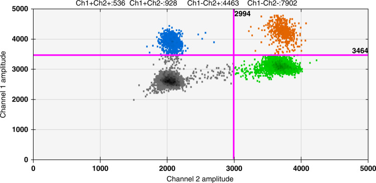
We examined the IKZF1 deletion (Δ4–7) in identified BCR::ABL1-like (n = 37) and non-BCR::ABL1-like ALL cases (n = 71). We identified IKZF1 (Δ4–7) CNV in 8.33% (9/108) with a range of 6.14–69.8% in RGAneg B-ALL cases (n = 108), as depicted in Table 2 and Fig. 3. We identified IKZF1 (Δ4–7) deletions in 18.91% (7/37) of BCR::ABL1-like and 2.81% (2/71) of non-BCR::ABL1-like ALLs. 22.22% (2/9) of BCR::ABL1-like ALL having CGFs had IKZF1 (Δ4–7) deletions. Although, the IKZF1 (Δ4–7) deletions were more frequent in BCR::ABL1-like ALL in comparison to non-BCR::ABL1-like ALL, but the difference in the frequency was not statistically significant (P = 0.44).
Table 2.
Ratio of IKZF1 (Δ4–7) deletion detected in 9/108 (8.33%) (range 6.14–69.8%) RGAneg B-ALL cases.
| IKZF1 copies/ul | RNase P copies/ul | Ratio*100 | Copy number variation (CNV) | Ratio |
|---|---|---|---|---|
| 1 | 777 | 0.168 | 0.336 | 16.8 |
| 1.1 | 815 | 0.165 | 0.329 | 16.5 |
| 2 | 880 | 0.66 | 1.32 | 66 |
| 2.1 | 945 | 0.70 | 1.396 | 69.8 |
| 3 | 144 | 0.49 | 0.99 | 49.4 |
| 3.1 | 141 | 0.54 | 1.08 | 53.9 |
| 4 | 284 | 0.41 | 0.82 | 41 |
| 4.1 | 311 | 0.43 | 0.86 | 42.8 |
| 5 | 1098 | 0.60 | 1.197 | 59.8 |
| 5.1 | 1093 | 0.63 | 1.263 | 63.2 |
| 6 | 2284 | 0.14 | 0.276 | 13.8 |
| 6.1 | 1469 | 0.16 | 0.313 | 15.6 |
| 7 | 1740 | 0.06 | 1.123 | 6.14 |
| 7.1 | 1085 | 0.06 | 0.124 | 6.2 |
| 8 | 826 | 0.26 | 0.523 | 26.1 |
| 8.1 | 932 | 0.25 | 0.49 | 24.5 |
| 9 | 650 | 0.56 | 1.13 | 56.4 |
| 9.1 | 322 | 0.60 | 1.19 | 59.5 |
Fig. 3. Scatter 2D plot acquired by Quanta Soft (1.7.4) software using CNV protocol.
The Blue signal is represented by mutated IKZF1 alleles (∆4–7) deletion in the FAM channel, and the green signal is represented by RNase P alleles (reference gene) in the VIC channel. The ratio was calculated by IKZF1 (4–7 deletion) copies/ul/ RNase P copies/µl*100 in RGAneg B-ALL cases.
Expression of myeloid-associated markers in BCR::ABL1-like ALL cases
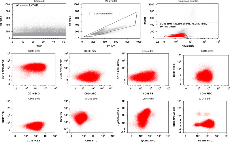
The expression of myeloid-associated markers, including 18.91% (7/37) CD13, 27.02% (10/37) CD33, and 5.40% (2/37) CD117, was significantly higher in BCR::ABL1-like ALL as compared to 12.67% (9/71) CD13, 25.35% (18/71) CD33, and 2.81% (2/71) CD117 in non-BCR::ABL1-like ALL (Supplementary Table S7). CD13 + BCR::ABL1-like ALL cases expressed higher HB value compared to CD13 + non-BCR::ABL1-like ALL and were statistically significant (P = 0.02). CD33 + BCR::ABL1-like ALL cases expressing higher TLC in comparison to CD33 + non-BCR::ABL1-like ALL were statistically significant (P = 0.05). A representative case of BCR::ABL1-like ALL expressing the myeloid-associated markers e.g., CD13 & CD33, as shown in Fig. 4.
Fig. 4. Flowcytometric analysis.
A representative case of BCR::ABL1-like ALL expressing myeloid-associated markers e.g., CD13 and CD33.
Outcomes of identified BCR::ABL1-like ALL patients
The adult B-ALL cases were treated using a modified BFM protocol. A high sensitivity MRD (sensitivity 0.01%) was evaluated using a ten colour/12 parameters flow cytometry at the end of induction therapy (after 4 weeks of therapy). The MRD data was available in 80.55% (87/108) of all RGAneg B-ALL cases. 19.29% (11/57) of cases showed MRD positivity in non-BCR::ABL1-like ALLs and 40% (12/30) in BCR::ABL1-like ALLs. The MRD positivity was higher in BCR::ABL1-like ALL cases (40%) as compared to non-BCR::ABL1-like ALL cases (19.29%) but did not show statistical significance (P = 0.78).
Discussion
From the high-income countries, the authors from COG & DCOG (2009) used a genome-wide GEP (> 100 genes) approach for the detection of this clinically significant entity [3, 4]. Further, the authors developed the 15-10-9–8 gene LDA and reported the incidence of 15–33.1% Ph-like ALL cases [8, 12, 15, 16, 30]. From the low–middle income countries, the identification of BCR::ABL1-like ALLs is still a pending matter, and it is associated with difficulties due to the diverse spectrum of genetic alterations and the unavailability of advanced technologies in most centres. The published literature is scarce on the well-defined methodology for its detection. There is an urgent need to adopt simpler and cost-effective technologies to identify these groups of RGAneg B-ALL cases effectively and to facilitate a personalised treatment regimen to improve the outcomes of these patients. With this rationale, this study proposed a step-wise well-defined methodology including a PHi-RACE classifier (GEP profiling) for the identification of BCR::ABL1-like ALL signature [23], followed by the characterisation of TSLPR/CRLF2 positivity and the identification of CGFs in BCR::ABL1-like ALL cases in this study cohort.
After an intensive literature search, we found three published studies in the context of BCR::ABL1-like ALLs from low–middle-income countries. Totadri et al. proposed a PACE approach to classify cases of BCR::ABL1-like ALL based on CRLF2 expression, Multiplex RT-PCR, MLPA, FISH, and Sanger Sequencing [27]. Their proposed strategy detected CRLF2 overexpression with associated mutations in a limited panel of B-developmental genes. However, the PACE algorithm cannot detect all mutations, and this algorithm still requires validation. So, implementing the PACE algorithm in resource-constraint laboratories is still a question. Patkar et al. proposed NARASIMHA assay, and detected 18 cases of BCR::ABL1-like ALL with CGFs based upon targeted RNA-Seq. RNA-seq is a powerful approach to classify BCR::ABL1-like ALL CGFs at baseline. However, the incidence of these cases comprises 10–33.1% and performing RNA-seq in all RGAneg B-ALL cases significantly increases the cost, and the data analysis requires a lot of bioinformatic skills. It is challenging to implement RNA-seq in all the RGAneg B-ALL cases in resource-constrained laboratories to classify BCR::ABL1-like ALL cases due to its high cost and complicated analysis [35]. Virk et al. implemented a strategy based on elucidating TSLPR/CRLF2 positivity & FISH to detect CGFs in BCR::ABL1-negative ALL cases. The authors detected 14.6% CRLF2 positivity in B-ALL, but this strategy does not elucidate other genetic alterations in BCR::ABL1-negative ALL cases, including IKZF1 alterations [36]. The authors, as mentioned above, reported the expression of CRLF2, CNV & CGFs in BCR::ABL1-negative ALL cases using a different approach with associated drawbacks. None of the authors use GEP to classify BCR::ABL1-like ALL cases and does not further characterise these cases. So, the true incidence of BCR::ABL1-like ALL cases remains unknown in low–middle income countries.
In one of our recent studies, we have published the PHi-RACE classifier based on GEP of eight selected overexpressed genes identified using transcriptome profiling (Affymetrix microarray U133 Plus 2.0 Array). We reported the incidence of 26.67% (143/536) BCR::ABL1-like ALL cases at diagnosis. PHi-RACE classifier (GEP) follows the gold standard to detect these cases, and we have calculated the cost of 42.00 USD/sample at diagnosis [23]. The proposed cost is more reasonable for patients, and this approach does not involve complicated calculations. This approach is straightforward, economical, and has less turnaround time (4~hour) compared to the above-mentioned strategies from low–middle-income countries. In this study, we have validated the PHi-RACE classifier and detected 34.25% (37/108) BCR::ABL1-like ALL cases in adult RGAneg B-ALL cases. Further, we have characterised the identified BCR::ABL1-like ALL cases and elucidated the prognostic gene alterations associated with poorer clinical outcomes, including 11.58% CRLF2 positivity, 18.91% IKZF1 alterations and 34.61% CGFs in BCR::ABL1-like ALLs for the time in low–middle income countries. None of the authors from low–middle income countries implemented GEP and characterised prognostic gene alterations (IKZF1) in BCR::ABL1-like ALL cases.
Several authors have suggested that IKZF1 plays a key role in leukemogenesis. IKZF1 (Δ4–7) deletions lead to the generation of a dominant negative IK6 isoform [50]. Various authors reported IKZF1 deletions, including IKZF1 (Δ4–7) deletion from 9.4% to 83.7% in B-ALLs, as shown in Table 3 [3, 22, 29, 49, 51–60]. All the mentioned studies used SNP array, MLPA & FISH to detect IKZF1 deletions in B-ALLs. Hashiguchi et al. established a simple, efficient, fast quantitative approach for detecting IKZF1 (∆4–7) deletion in ALL cases using DD-PCR [49]. In this study, we detected 8.33% (9/108) IKZF1 (Δ4–7) deletion in RGAneg B-ALL cases using DD-PCR and 18.91% (7/37) IKZF1 (Δ4–7) deletions in identified BCR::ABL1-like ALL cases. Furthermore, we found the positivity of TSLPR/CRLF2 in 11.58% (3/26) BCR::ABL1-like ALL cases, as the criterion is taken ≥10% leukaemic cells [36, 47]. Various authors reported the expression positivity of TSLPR/CRLF2 from 7.5 to 35.7% using different qPCR and FCM cut-offs as shown in Table 4 [1, 22, 25, 32, 36, 47, 54, 58, 60–64]. The variance in the reported frequency of TSLPR/CRLF2 positivity is due to the author’s use of different qPCR & FCM cut-offs. In one of CRLF2 overexpressed BCR::ABL1-like ALLs, we observed the IGH::EPOR translocation, a similar finding was reported by Tasian et al. The authors reported one case of IGH:EPOR and one case of IL7R indel with CRLF2 overexpression [65].
Table 3.
Reported incidence comparison of IKZF1 deletions, including IKZF1 (Δ4–7) deletion in B-ALLs, RGAneg B-ALL cases & BCR::ABL1-like ALL cases.
| Authors [Ref.] | Method | IKZF1& IKZF1 (Δ4–7) deletion. | Cases |
|---|---|---|---|
| Mullighan CG et al. [29] | 250k Sty and Nsp SNP array | 83.7% (14/23) IKZF1 deletion. | BCR::ABL1-positive ALLs |
| Mullighan CG et al. [3] | 250k Sty and Nsp SNP array | 28.50% (63/221) IKZF1 deletion | High-risk B-ALLs |
| Iacobucci I et al. [51] | 250k Sty and Nsp SNP array | 42% IKZF1(Δ4–7) | BCR::ABL1-positive ALLs |
| Kuiper RP et al. [53] | MLPA | 13.7% (18/31) IKZF1 deletion | Paediatric B-ALLs |
| Iacobucci I et al. [52] | 250k Sty and Nsp SNP array | 49% IKZF1(Δ4–7) | BCR::ABL1-positive ALLs |
| Yamashita Y et al. [54] | MLPA | 12% (22/177) IKZF1 deletion | BCR::ABL1-negative ALLs |
| Martinelli G et al. [55] | 250k Sty and Nsp SNP array | 63% (52/83) IKZF1 deletion | BCR::ABL1-positive ALLs |
| Caye A et al. [56] |
MLPA PCR |
4.49% (23/512) IKZF1(Δ4–7) 6.64% (34/512) IKZF1(Δ4–7) |
BCR::ABL1-negative ALLs |
| MI JQ et al. [57] | 250k Sty and Nsp SNP array |
14.7%) (56/607) IKZF1(Δ4–7) 31.3% (64/262)) IKZF1(Δ4–7) |
Paediatric B-ALLs Adult B-ALLs |
| Palmi C et al. [58] | MLPA |
13.2% (54/410) deletion. 2.19% (9/410) IKZF1 (Δ4–7). |
BCP-ALL cases |
| Liu X et al. [59] | MLPA |
20% (19/96) IKZF1 deletion 21.05% (4/19) IKZF1 (Δ4–7) deletion. |
B-ALL cases |
| Asai D et al. [60] | MLPA | 9.4% (19/202) IKZF1 deletion | B-ALL cases |
| Hashiguchi J et al. [49] | DD-PCR | 12% (22/177) IKZF1 deletion | BCP-ALL cases |
| Chiaretti S et al. [22] | MLPA | 63.96% (14/22) IKZF1 deletion | BCR::ABL1-like ALL cases |
| Present study | DD-PCR | 8.33% (9/108) IKZF1 (Δ4–7) deletion 18.91% (7/37) IKZF1 (Δ4–7) deletions |
RGAneg B-ALL cases BCR::ABL1-like ALL cases |
Table 4.
Comparison of reported incidence of TSLPR/CRLF2 positivity in B-ALLs.
| Authors [Ref.] | Method | Cutt-off | % TSLPR/CRLF2 positivity |
|---|---|---|---|
| Harvey et al. [25] | Affymetrix probe | ≥10× (median value) | 14% (29/207) |
| Asai et al. [60] | qPCR | ≥10× (median value) | 15.0% (16/107) |
| Yamashita et al. [54] | qPCR | ≥10× (median value) | 10.63% (15/141) |
| Cario et al. [63] | qPCR | ≥10× (median value | 8.82% (49/555) |
| Yoda et al. [61] | qPCR | Mean value | 12.5% (15/120) |
| Palmi et al. [58] | qPCR | >20× (median value) | 4.7% (22/464) |
| Chen et al. [62] | qPCR | ΔCt ≤ 8 expression | 17.5% (186/1061) |
| Chiaretti et al. [32] | qPCR | ΔCt ≤ 8 expression | 34.3% (35/102) |
| Chiaretti et al. [22] | qPCR | ΔCt ≤ 8 expression | 35.7% (10/28) |
| Bugarin et al. [47] | FCM | >10% | 9.6% (37/384) |
| Virk et al. [36] | FCM | >10% | 14.6% (70/478) |
| Pastorczak et al. [64] | FCM | >5% | 7.5% (29/386) |
| Present study | FCM | >10% | 11.58% (3/26) |
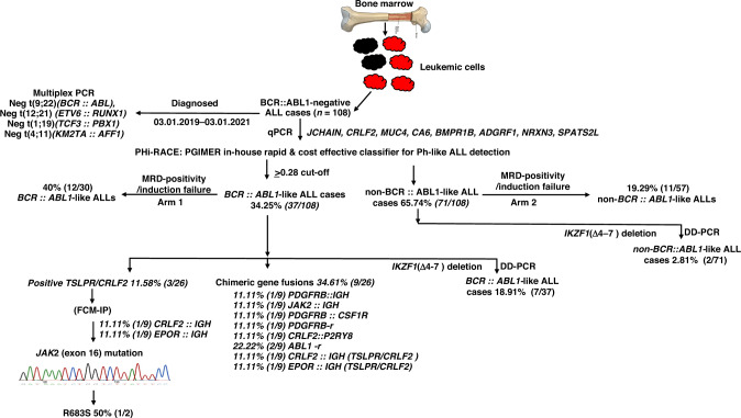
To conclude, in this study, we found a higher incidence (34.25%) of BCR::ABL1-like ALL cases, and these findings are in accordance with the findings of Jain et al. and Chiaretti et al. [10, 22]. Both authors reported a higher incidence of 33.1% in B-ALLs and 31.8% in BCR::ABL1-negative ALLs. We reported 11.58% TSLPR/CRLF2 positivity in the identified BCR::ABL1-like ALL cases. Our findings of TSLPR/CRLF2 positivity are in accordance with Yamashita et al. [54], Asai et al. [60], Harvey et al. [25] and Virk et al. [36] B-ALL cases. The authors reported the TSLPR/CRLF2 positivity of 10.63–15.0% in B-ALLs. These authors reported a lower frequency of CRLF2 positivity in Japanese & Indian population (Asian Ancestry). This indicates that Asian ancestry expresses a lower frequency of CRLF2 alterations in BCR::ABL1-like and B-ALL cases. We also reported a low frequency of 8.33% (9/108) and 18.91% (7/37) IKZF1 (Δ4–7) deletions in RGAneg B-ALL cases and identified BCR::ABL1-like ALLs. The reason could be that the studies mentioned above-analysed patients of different races and ethnicity; Hispanic/Latino and Japanese. In India, no study has so far evaluated the IKZF1 (Δ4–7) deletions using DD-PCR in BCR::ABL1-like ALLs. This is the first report to identify and characterise the genomic milieu of BCR::ABL1-like ALL patients of Indian ethnicity and added a well-defined methodology (PHi-RACE) to detect these cases in literature from low–middle income countries. Regarding the study’s shortcomings, we are evaluating the PHi-RACE classifier in a large RGAneg B-ALL cases of the Indian patient cohort for a better picture of BCR::ABL1-like ALL cases and the treatment outcomes/overall survival of these cases from our Institute. The schematic presentation of the diagnostic summary to detect and characterise the cases of BCR::ABL1-like ALL has been shown in Fig. 5.
Fig. 5.
Diagnostic summary of detection of BCR::ABL1-like ALL cases, based on our experience in Indian patients.
Supplementary information
Acknowledgements
I sincerely thank the faculty of Haematology, technical staff, and clerical staff, project staff (Mrs Sonia) for providing valuable support for this study. I would like to extend my sincere thanks to Dr. Praveen (PGIMER), Dr Pramod Singh (PGIMER), Dr Mathew Li Arwood (Ann Roberts H. Lurie Children’s Hospital), and Dr Loretta S Li (Ann Roberts H. Lurie Children’s Hospital) for their valuable suggestions in this manuscript.
Author contributions
DGG and NV designed the experiments and analysed the generated data; DGG, SS, JB and PB performed the experiments. DGG and NV wrote and prepared the manuscript; PM, AK and SV provided the adult B-ALL samples. SAA read, reviewed and helped us properly restructure the manuscript. All other authors read, provided intellectual comments, and approved the submitted manuscript.
Funding
This research study was supported by the Intramural Grant provided by PGIMER, Chandigarh, India (No.71/2-Edu-16/937/18/03/2019). Dr. Dikshat Gopal Gupta received financial assistance from the Indian Council of Medical Education and Research (ICMR), New Delhi, India.
Data availability
All research data generated during this study are included in this research article.
Competing interests
The authors declare no competing interests.
Ethics approval and consent to participate
PGIMER constituted Institutional Ethics Committee had approved this research study (vide no. INT/IEC/2019/000611 dated March 19, 2019). In all, 2–3 ml of aspirated bone marrow (BM) or 3–5 ml of peripheral blood (PB) samples were collected, after obtaining the informed consent.
Consent for publication
Not applicable.
Footnotes
Publisher’s note Springer Nature remains neutral with regard to jurisdictional claims in published maps and institutional affiliations.
Supplementary information
The online version contains supplementary material available at 10.1038/s41416-023-02294-y.
References
- 1.Arber DA, Orazi A, Hasserjian R, Thiele J, Borowitz MJ, Le Beau MM, et al. The 2016 revision to the World Health Organization classification of myeloid neoplasms and acute leukemia. Blood. 2016;127:2391–405. doi: 10.1182/blood-2016-03-643544. [DOI] [PubMed] [Google Scholar]
- 2.Alaggio R, Amador C, Anagnostopoulos I, Attygalle AD, Araujo IBO, Berti E, et al. The 5th edition of the World Health Organization Classification of haematolymphoid tumours: lymphoid neoplasms. Leukemia. 2022;36:1720–48. doi: 10.1038/s41375-022-01620-2. [DOI] [PMC free article] [PubMed] [Google Scholar]
- 3.Mullighan CG, Su X, Zhang J, Radtke I, Phillips LA, Miller CB, et al. Deletion of IKZF1 and prognosis in acute lymphoblastic leukemia. N Engl J Med. 2009;360:470–80. doi: 10.1056/NEJMoa0808253. [DOI] [PMC free article] [PubMed] [Google Scholar]
- 4.Den Boer ML, van Slegtenhorst M, De Menezes RX, Cheok MH, Buijs-Gladdines JG, Peters ST, et al. A subtype of childhood acute lymphoblastic leukaemia with poor treatment outcome: a genome-wide classification study. Lancet Oncol. 2009;10:125–34. doi: 10.1016/S1470-2045(08)70339-5. [DOI] [PMC free article] [PubMed] [Google Scholar]
- 5.Roberts KG, Li Y, Payne-Turner D, Harvey RC, Yang YL, Pei D, et al. Targetable kinase-activating lesions in Ph-like acute lymphoblastic leukemia. N Engl J Med. 2014;371:1005–15. doi: 10.1056/NEJMoa1403088. [DOI] [PMC free article] [PubMed] [Google Scholar]
- 6.Mullighan CG. The genomic landscape of acute lymphoblastic leukemia in children and young adults. Hematol Am Soc Hematol Educ Program. 2014;2014:174–80. doi: 10.1182/asheducation-2014.1.174. [DOI] [PubMed] [Google Scholar]
- 7.Roberts KG, Gu Z, Payne-Turner D, McCastlain K, Harvey RC, Chen IM, et al. High frequency and poor outcome of Philadelphia chromosome-like acute lymphoblastic leukemia in adults. J Clin Oncol. 2017;35:394–401. doi: 10.1200/JCO.2016.69.0073. [DOI] [PMC free article] [PubMed] [Google Scholar]
- 8.Reshmi SC, Harvey RC, Roberts KG, Stonerock E, Smith A, Jenkins H, et al. Targetable kinase gene fusions in high-risk B-ALL: a study from the Children’s Oncology Group. Blood. 2017;129:3352–61. doi: 10.1182/blood-2016-12-758979. [DOI] [PMC free article] [PubMed] [Google Scholar]
- 9.Ofran Y, Izraeli S. BCR-ABL (Ph)-like acute leukemia-pathogenesis, diagnosis and therapeutic options. Blood Rev. 2017;31:11–6. doi: 10.1016/j.blre.2016.09.001. [DOI] [PubMed] [Google Scholar]
- 10.Jain N, Roberts KG, Jabbour E, Patel K, Eterovic AK, Chen K, et al. Ph-like acute lymphoblastic leukemia: a high-risk subtype in adults. Blood. 2017;129:572–81. doi: 10.1182/blood-2016-07-726588. [DOI] [PMC free article] [PubMed] [Google Scholar]
- 11.Herold T, Schneider S, Metzeler KH, Neumann M, Hartmann L, Roberts KG, et al. Adults with Philadelphia chromosome-like acute lymphoblastic leukemia frequently have IGH-CRLF2 and JAK2 mutations, persistence of minimal residual disease and poor prognosis. Haematologica. 2017;102:130–8. doi: 10.3324/haematol.2015.136366. [DOI] [PMC free article] [PubMed] [Google Scholar]
- 12.Heatley SL, Sadras T, Kok CH, Nievergall E, Quek K, Dang P, et al. High prevalence of relapse in children with Philadelphia-like acute lymphoblastic leukemia despite risk-adapted treatment. Haematologica. 2017;102:e490–e3. doi: 10.3324/haematol.2016.162925. [DOI] [PMC free article] [PubMed] [Google Scholar]
- 13.Wells J, Jain N, Konopleva M. Philadelphia chromosome-like acute lymphoblastic leukemia: progress in a new cancer subtype. Clin Adv Hematol Oncol. 2017;15:554–61. [PubMed] [Google Scholar]
- 14.Tasian SK, Loh ML, Hunger SP. Philadelphia chromosome-like acute lymphoblastic leukemia. Blood. 2017;130:2064–72. doi: 10.1182/blood-2017-06-743252. [DOI] [PMC free article] [PubMed] [Google Scholar]
- 15.Roberts KG, Reshmi SC, Harvey RC, Chen IM, Patel K, Stonerock E, et al. Genomic and outcome analyses of Ph-like ALL in NCI standard-risk patients: a report from the Children’s Oncology Group. Blood. 2018;132:815–24. doi: 10.1182/blood-2018-04-841676. [DOI] [PMC free article] [PubMed] [Google Scholar]
- 16.Chiaretti S, Messina M, Grammatico S, Piciocchi A, Fedullo AL, Di Giacomo F, et al. Rapid identification of BCR/ABL1-like acute lymphoblastic leukaemia patients using a predictive statistical model based on quantitative real time-polymerase chain reaction: clinical, prognostic and therapeutic implications. Br J Haematol. 2018;181:642–52. doi: 10.1111/bjh.15251. [DOI] [PMC free article] [PubMed] [Google Scholar]
- 17.Siegele BJ, Nardi V. Laboratory testing in BCR-ABL1-like (Philadelphia-like) B-lymphoblastic leukemia/lymphoma. Am J Hematol. 2018;93:971–7. doi: 10.1002/ajh.25126. [DOI] [PubMed] [Google Scholar]
- 18.Chiaretti S, Messina M, Foa R. BCR/ABL1-like acute lymphoblastic leukemia: how to diagnose and treat? Cancer. 2019;125:194–204. doi: 10.1002/cncr.31848. [DOI] [PubMed] [Google Scholar]
- 19.Shiraz P, Payne KJ, Muffly L. The current genomic and molecular landscape of Philadelphia-like acute lymphoblastic leukemia. Int J Mol Sci. 2020;21:2193. doi: 10.3390/ijms21062193. [DOI] [PMC free article] [PubMed] [Google Scholar]
- 20.Jain S, Abraham A. BCR-ABL1-like B-acute lymphoblastic leukemia/lymphoma: a comprehensive review. Arch Pathol Lab Med. 2020;144:150–5. doi: 10.5858/arpa.2019-0194-RA. [DOI] [PubMed] [Google Scholar]
- 21.Ribera JM. Philadelphia chromosome-like acute lymphoblastic leukemia. Still a pending matter. Haematologica. 2021;106:1514–6. doi: 10.3324/haematol.2020.270645. [DOI] [PMC free article] [PubMed] [Google Scholar]
- 22.Chiaretti S, Messina M, Della Starza I, Piciocchi A, Cafforio L, Cavalli M, et al. Philadelphia-like acute lymphoblastic leukemia is associated with minimal residual disease persistence and poor outcome. First report of the minimal residual disease-oriented GIMEMA LAL1913. Haematologica. 2021;106:1559–68. doi: 10.3324/haematol.2020.247973. [DOI] [PMC free article] [PubMed] [Google Scholar]
- 23.Gupta DG, Varma N, Kumar A, Naseem S, Sachdeva MUS, Binota J, et al. PHi-RACE: PGIMER in-house rapid & cost effective classifier for the detection of BCR-ABL1-like acute lymphoblastic leukaemia in Indian patients. Leuk Lymphoma. 2022;63:633–43. doi: 10.1080/10428194.2021.1999439. [DOI] [PubMed] [Google Scholar]
- 24.Plotka A, Lewandowski K. BCR/ABL1-like acute lymphoblastic leukemia: from diagnostic approaches to molecularly targeted therapy. Acta Haematol. 2022;145:122–31. doi: 10.1159/000519782. [DOI] [PubMed] [Google Scholar]
- 25.Harvey RC, Mullighan CG, Chen IM, Wharton W, Mikhail FM, Carroll AJ, et al. Rearrangement of CRLF2 is associated with mutation of JAK kinases, alteration of IKZF1, Hispanic/Latino ethnicity, and a poor outcome in pediatric B-progenitor acute lymphoblastic leukemia. Blood. 2010;115:5312–21. doi: 10.1182/blood-2009-09-245944. [DOI] [PMC free article] [PubMed] [Google Scholar]
- 26.Chiaretti S, Gianfelici V, O’Brien SM, Mullighan CG. Advances in the genetics and therapy of acute lymphoblastic leukemia. Am Soc Clin Oncol Educ Book. 2016;35:e314–22. doi: 10.1200/EDBK_156628. [DOI] [PubMed] [Google Scholar]
- 27.Totadri S, Singh M, Trehan A, Varma N, Bhatia P. Keeping PACE with Ph positive to Ph-like detection in B-lineage acute lymphoblastic leukemia: a practical and cost effective (PACE) approach in a resource constrained setting. Indian J Hematol Blood Transfus. 2018;34:595–601. doi: 10.1007/s12288-018-0997-y. [DOI] [PMC free article] [PubMed] [Google Scholar]
- 28.Sanchez R, Ribera J, Morgades M, Ayala R, Onecha E, Ruiz-Heredia Y, et al. A novel targeted RNA-Seq panel identifies a subset of adult patients with acute lymphoblastic leukemia with BCR-ABL1-like characteristics. Blood Cancer J. 2020;10:43. doi: 10.1038/s41408-020-0308-3. [DOI] [PMC free article] [PubMed] [Google Scholar]
- 29.Mullighan CG, Miller CB, Radtke I, Phillips LA, Dalton J, Ma J, et al. BCR-ABL1 lymphoblastic leukaemia is characterized by the deletion of Ikaros. Nature. 2008;453:110–4. doi: 10.1038/nature06866. [DOI] [PubMed] [Google Scholar]
- 30.Harvey RC, Kang H, Roberts KG, Chen I-ML, Atlas SR, Bedrick EJ, et al. Development and validation of a highly sensitive and specific gene expression classifier to prospectively screen and identify B-precursor acute lymphoblastic leukemia (ALL) patients with a Philadelphia chromosome-like (“Ph-like” or “BCR-ABL1-Like”) signature for therapeutic targeting and clinical intervention. Blood. 2013;122:826. doi: 10.1182/blood.V122.21.826.826. [DOI] [Google Scholar]
- 31.Boer JM, Koenders JE, van der Holt B, Exalto C, Sanders MA, Cornelissen JJ, et al. Expression profiling of adult acute lymphoblastic leukemia identifies a BCR-ABL1-like subgroup characterized by high non-response and relapse rates. Haematologica. 2015;100:e261–4. doi: 10.3324/haematol.2014.117424. [DOI] [PMC free article] [PubMed] [Google Scholar]
- 32.Chiaretti S, Brugnoletti F, Messina M, Paoloni F, Fedullo AL, Piciocchi A, et al. CRLF2 overexpression identifies an unfavourable subgroup of adult B-cell precursor acute lymphoblastic leukemia lacking recurrent genetic abnormalities. Leuk Res. 2016;41:36–42. doi: 10.1016/j.leukres.2015.11.018. [DOI] [PubMed] [Google Scholar]
- 33.Tsaur G, Muhacheva T, Kovalev S, Druy A, Sibiryakov P, Olshanskaya Y, et al. Application of real-time PCR for the detection of BCR-ABL1-like group in pediatric acute lymphoblastic leukemia patients. Blood. 2018;132:1376. doi: 10.1182/blood-2018-99-115035. [DOI] [Google Scholar]
- 34.Fasan Annette KW, Nadarajah N, Weber S, Schindela S, Schlenther N, Schnittger S, et al. Three steps to the diagnosis of adult Ph-like ALL. Blood. 2015;126:2610. doi: 10.1182/blood.V126.23.2610.2610. [DOI] [Google Scholar]
- 35.Patkar N, Bhanshe P, Rajpal S, Joshi S, Chaudhary S, Chatterjee G, et al. NARASIMHA: novel assay based on targeted rna sequencing to identify chimeric gene fusions in hematological malignancies. Blood Cancer J. 2020;10:50. doi: 10.1038/s41408-020-0313-6. [DOI] [PMC free article] [PubMed] [Google Scholar]
- 36.Virk H, Rana S, Sharma P, Bose PL, Yadav DD, Sachdeva MUS, et al. Hematological characteristics, cytogenetic features, and post-induction measurable residual disease in thymic stromal lymphopoietin receptor (TSLPR) overexpressed B-cell acute lymphoblastic leukemia in an Indian cohort. Ann Hematol. 2021;100:2031–41. doi: 10.1007/s00277-021-04574-0. [DOI] [PubMed] [Google Scholar]
- 37.Sharma P, Rana S, Virk H, Sachdeva MUS, Sharma P, Varma N, et al. The frequency, hematological characteristics, and end-of induction residual disease in B-acute lymphoblastic leukemia with BCR-ABL1-like chimeric gene fusions in a high-risk cohort from India. Leuk Lymphoma. 2021;63:2474–8. [DOI] [PubMed]
- 38.Gupta DG, Varma N, Naseem S, Sachdeva MUS, Bose P, Binota J, et al. Characterization of immunophenotypic aberrancies with respect to common fusion transcripts in B-cell precursor acute lymphoblastic leukemia: a report of 986 Indian patients. Turk J Haematol. 2022;39:1–12. doi: 10.4274/tjh.galenos.2021.2021.0326. [DOI] [PMC free article] [PubMed] [Google Scholar]
- 39.Sharma M, Sachdeva MU, Varma N, Varma S, Marwaha RK. Characterization of immunophenotypic aberrancies in adult and childhood acute lymphoblastic leukemia: a study from Northern India. J Cancer Res Ther. 2016;12:620–6. doi: 10.4103/0973-1482.147716. [DOI] [PubMed] [Google Scholar]
- 40.Malhotra P, Varma S, Varma N, Kumari S, Das R, Jain S, et al. Outcome of adult acute lymphoblastic leukemia with BFM protocol in a resource-constrained setting. Leuk Lymphoma. 2007;48:1173–8. doi: 10.1080/10428190701343255. [DOI] [PubMed] [Google Scholar]
- 41.Bommannan K, Singh Sachdeva MU, Varma N, Bose P, Bansal D. Role of mid-induction peripheral blood minimal residual disease detection in pediatric B-lineage acute lymphoblastic leukemia. Indian Pediatr. 2016;53:1065–8. [PubMed] [Google Scholar]
- 42.Tembhare PR, Subramanian PgPG, Ghogale S, Chatterjee G, Patkar NV, Gupta A, et al. A high-sensitivity 10-color flow cytometric minimal residual disease assay in B-lymphoblastic leukemia/lymphoma can easily achieve the sensitivity of 2-in-10(6) and is superior to standard minimal residual disease assay: a study of 622 patients. Cytom B Clin Cytom. 2020;98:57–67. doi: 10.1002/cyto.b.21831. [DOI] [PubMed] [Google Scholar]
- 43.Stow P, Key L, Chen X, Pan Q, Neale GA, Coustan-Smith E, et al. Clinical significance of low levels of minimal residual disease at the end of remission induction therapy in childhood acute lymphoblastic leukemia. Blood. 2010;115:4657–63. doi: 10.1182/blood-2009-11-253435. [DOI] [PMC free article] [PubMed] [Google Scholar]
- 44.Pakakasama S, Kajanachumpol S, Kanjanapongkul S, Sirachainan N, Meekaewkunchorn A, Ningsanond V, et al. Simple multiplex RT-PCR for identifying common fusion transcripts in childhood acute leukemia. Int J Lab Hematol. 2008;30:286–91. doi: 10.1111/j.1751-553X.2007.00954.x. [DOI] [PubMed] [Google Scholar]
- 45.Bhatia P, Binota J, Varma N, Bansal D, Trehan A, Marwaha RK, et al. Incidence of common chimeric fusion transcripts in B-cell acute lymphoblastic leukemia: an Indian perspective. Acta Haematol. 2012;128:17–9. doi: 10.1159/000338260. [DOI] [PubMed] [Google Scholar]
- 46.Sharma P, Rana S, Sreedharanunni S, Gautam A, Sachdeva MUS, Naseem S, et al. An evaluation of a fluorescence in situ hybridization strategy using air-dried blood and bone-marrow smears in the risk stratification of pediatric B-lineage acute lymphoblastic leukemia in resource-limited settings. J Pediatr Hematol Oncol. 2021;43:e481–e5. doi: 10.1097/MPH.0000000000001892. [DOI] [PubMed] [Google Scholar]
- 47.Bugarin C, Sarno J, Palmi C, Savino AM, te Kronnie G, Dworzak M, et al. Fine tuning of surface CRLF2 expression and its associated signaling profile in childhood B-cell precursor acute lymphoblastic leukemia. Haematologica. 2015;100:e229–32. doi: 10.3324/haematol.2014.114447. [DOI] [PMC free article] [PubMed] [Google Scholar]
- 48.Boer JM, van der Veer A, Rizopoulos D, Fiocco M, Sonneveld E, de Groot-Kruseman HA, et al. Prognostic value of rare IKZF1 deletion in childhood B-cell precursor acute lymphoblastic leukemia: an international collaborative study. Leukemia. 2016;30:32–8. doi: 10.1038/leu.2015.199. [DOI] [PubMed] [Google Scholar]
- 49.Hashiguchi J, Onozawa M, Okada K, Amano T, Hatanaka KC, Nishihara H, et al. Quantitative detection of IKZF1 deletion by digital PCR in patients with acute lymphoblastic leukemia. Int J Lab Hematol. 2019;41:e38–e40. doi: 10.1111/ijlh.12945. [DOI] [PubMed] [Google Scholar]
- 50.Kastner P, Dupuis A, Gaub MP, Herbrecht R, Lutz P, Chan S. Function of Ikaros as a tumor suppressor in B cell acute lymphoblastic leukemia. Am J Blood Res. 2013;3:1–13. [PMC free article] [PubMed] [Google Scholar]
- 51.Iacobucci I, Storlazzi CT, Cilloni D, Lonetti A, Ottaviani E, Soverini S, et al. Identification and molecular characterization of recurrent genomic deletions on 7p12 in the IKZF1 gene in a large cohort of BCR-ABL1-positive acute lymphoblastic leukemia patients: on behalf of Gruppo Italiano Malattie Ematologiche dell’Adulto Acute Leukemia Working Party (GIMEMA AL WP) Blood. 2009;114:2159–67. doi: 10.1182/blood-2008-08-173963. [DOI] [PubMed] [Google Scholar]
- 52.Iacobucci I, Iraci N, Messina M, Lonetti A, Chiaretti S, Valli E, et al. IKAROS deletions dictate a unique gene expression signature in patients with adult B-cell acute lymphoblastic leukemia. PLoS ONE. 2012;7:e40934. doi: 10.1371/journal.pone.0040934. [DOI] [PMC free article] [PubMed] [Google Scholar]
- 53.Kuiper RP, Waanders E, van der Velden VH, van Reijmersdal SV, Venkatachalam R, Scheijen B, et al. IKZF1 deletions predict relapse in uniformly treated pediatric precursor B-ALL. Leukemia. 2010;24:1258–64. doi: 10.1038/leu.2010.87. [DOI] [PubMed] [Google Scholar]
- 54.Yamashita Y, Shimada A, Yamada T, Yamaji K, Hori T, Tsurusawa M, et al. IKZF1 and CRLF2 gene alterations correlate with poor prognosis in Japanese BCR-ABL1-negative high-risk B-cell precursor acute lymphoblastic leukemia. Pediatr Blood Cancer. 2013;60:1587–92. doi: 10.1002/pbc.24571. [DOI] [PubMed] [Google Scholar]
- 55.Martinelli G, Iacobucci I, Storlazzi CT, Vignetti M, Paoloni F, Cilloni D, et al. IKZF1 (Ikaros) deletions in BCR-ABL1-positive acute lymphoblastic leukemia are associated with short disease-free survival and high rate of cumulative incidence of relapse: a GIMEMA AL WP report. J Clin Oncol. 2009;27:5202–7. doi: 10.1200/JCO.2008.21.6408. [DOI] [PubMed] [Google Scholar]
- 56.Caye A, Beldjord K, Mass-Malo K, Drunat S, Soulier J, Gandemer V, et al. Breakpoint-specific multiplex polymerase chain reaction allows the detection of IKZF1 intragenic deletions and minimal residual disease monitoring in B-cell precursor acute lymphoblastic leukemia. Haematologica. 2013;98:597–601. doi: 10.3324/haematol.2012.073965. [DOI] [PMC free article] [PubMed] [Google Scholar]
- 57.Mi JQ, Wang X, Yao Y, Lu HJ, Jiang XX, Zhou JF, et al. Newly diagnosed acute lymphoblastic leukemia in China (II): prognosis related to genetic abnormalities in a series of 1091 cases. Leukemia. 2012;26:1507–16. doi: 10.1038/leu.2012.23. [DOI] [PubMed] [Google Scholar]
- 58.Palmi C, Vendramini E, Silvestri D, Longinotti G, Frison D, Cario G, et al. Poor prognosis for P2RY8-CRLF2 fusion but not for CRLF2 over-expression in children with intermediate risk B-cell precursor acute lymphoblastic leukemia. Leukemia. 2012;26:2245–53. doi: 10.1038/leu.2012.101. [DOI] [PubMed] [Google Scholar]
- 59.Liu X, Zhang L, Zou Y, Chang L, Wei W, Ruan M, et al. [Significance of ikaros family zinc finger 1 deletion in pediatric B-acute lymphoblastic leukemia without reproducible cytogenetic abnormalities] Zhonghua Er Ke Za Zhi. 2016;54:126–30. doi: 10.3760/cma.j.issn.0578-1310.2016.02.011. [DOI] [PubMed] [Google Scholar]
- 60.Asai D, Imamura T, Suenobu S, Saito A, Hasegawa D, Deguchi T, et al. IKZF1 deletion is associated with a poor outcome in pediatric B-cell precursor acute lymphoblastic leukemia in Japan. Cancer Med. 2013;2:412–9. doi: 10.1002/cam4.87. [DOI] [PMC free article] [PubMed] [Google Scholar]
- 61.Yoda A, Yoda Y, Chiaretti S, Bar-Natan M, Mani K, Rodig SJ, et al. Functional screening identifies CRLF2 in precursor B-cell acute lymphoblastic leukemia. Proc Natl Acad Sci USA. 2010;107:252–7. doi: 10.1073/pnas.0911726107. [DOI] [PMC free article] [PubMed] [Google Scholar]
- 62.Chen IM, Harvey RC, Mullighan CG, Gastier-Foster J, Wharton W, Kang H, et al. Outcome modeling with CRLF2, IKZF1, JAK, and minimal residual disease in pediatric acute lymphoblastic leukemia: a Children’s Oncology Group study. Blood. 2012;119:3512–22. doi: 10.1182/blood-2011-11-394221. [DOI] [PMC free article] [PubMed] [Google Scholar]
- 63.Cario G, Zimmermann M, Romey R, Gesk S, Vater I, Harbott J, et al. Presence of the P2RY8-CRLF2 rearrangement is associated with a poor prognosis in non-high-risk precursor B-cell acute lymphoblastic leukemia in children treated according to the ALL-BFM 2000 protocol. Blood. 2010;115:5393–7. doi: 10.1182/blood-2009-11-256131. [DOI] [PubMed] [Google Scholar]
- 64.Pastorczak A, Sedek L, Braun M, Madzio J, Sonsala A, Twardoch M, et al. Surface expression of cytokine receptor-like factor 2 increases risk of relapse in pediatric acute lymphoblastic leukemia patients harboring IKZF1 deletions. Oncotarget. 2018;9:25971–82. doi: 10.18632/oncotarget.25411. [DOI] [PMC free article] [PubMed] [Google Scholar]
- 65.Tasian SK, Dai Y, Devidas M, Roberts KG, Harvey RC, Chen I-ML, et al. Outcomes of patients with CRLF2-overexpressing acute lymphoblastic leukemia without down syndrome: a report from the Children’s Oncology Group. Blood. 2020;136:45–6. doi: 10.1182/blood-2020-134327. [DOI] [Google Scholar]
Associated Data
This section collects any data citations, data availability statements, or supplementary materials included in this article.
Supplementary Materials
Data Availability Statement
All research data generated during this study are included in this research article.


