



Abstract
Purpose:
To investigate the feasibility of combining simultaneous multi-slice (SMS) and region-optimized virtual coils (ROVir) for single breath-hold CINE imaging.
Method:
ROVir is a recent virtual coil approach that allows reduced-FOV imaging by localizing the signal from a region-of-interest (ROI) and/or suppressing the signal from unwanted spatial regions. In this work, ROVir is used for reduced-FOV SMS bSSFP CINE imaging, which enables whole heart CINE with a single breath-hold acquisition.
Results:
Reduced-FOV CINE with either SMS-only or ROVir-only resulted in significant aliasing, with severely reduced image quality when compared to the full FOV reference CINE, while the visual appearance of aliasing was substantially reduced with the proposed SMS+ROVir. The end diastolic volume, end systolic volume, and ejection fraction obtained using the proposed approach were similar to the clinical reference (correlations of 0.92, 0.94, and 0.88, respectively with in each case, and biases of 0.1ml, 1.6ml, and −0.6%, respectively). No statistically-significant differences for these parameters were found with a Wilcoxon rank test (, and 0.40, respectively).
Conclusion:
We demonstrated that reduced-FOV CINE imaging with SMS+ROVir enables single breath-hold whole-heart imaging without compromising visual image quality or quantitative cardiac function parameters.
Keywords: CINE MRI, Simultaneous-Multi-Slice (SMS), Virtual coils, Reduced Field-of-View Imaging, Accelerated Acquisition
INTRODUCTION
Conventional clinical cardiac CINE MRI protocols require a large number of breath-holds (typically > 20) for capturing the entire heart, which results in long scan durations. Patient fatigue during these multiple breath-holds results in decreased breath-hold quality and as a result degradation of the reconstructed images. In this context, recent advances such as simultaneous multi-slice (SMS) acquisition (1,2), parallel imaging (3,4) and compressed sensing (5) have been employed to drastically reduce CINE imaging scan time and the number of required breath-holds. One complementary approach that could potentially reduce the time required for each CINE acquisition even further would be to restrict the FOV to only include the heart, which could result in an additional 4 to 5-fold acceleration (6). However, due to the position of the heart within the body, current FOVs for CINE acquisitions generally include the entire torso, since the use of a smaller FOV would otherwise cause portions of the torso to alias onto the heart.
Recently, we developed a novel approach to localize signal from a region-of-interest (ROI) and/or suppress signal from unwanted spatial regions. This approach, which we call region-optimized virtual (ROVir) coils (7), linearly mixes the signals acquired from a multi-channel receiver to obtain a new set of “virtual coils.” Since ROVir linearly mixes the multi-channel data after it has been acquired, this is achieved without requiring any modification of imaging hardware or pulse sequences.
Similar to other virtual coil approaches (8–10), ROVir enables dimensionality reduction (coil compression) and improved computational efficiency by condensing the original receiver channels into a smaller set of channels that contain almost the same information. However, different from other approaches, ROVir does this while intentionally preserving the signal from a specific user-specified ROI and suppressing the signal energy from unwanted spatial regions. When ROVir channels are obtained as described in Ref. (7), the resulting virtual channels have optimal signal-to-interference ratio (SIR).The optimality of ROVir endows it with substantial SIR advantages compared to other virtual coil approaches and related techniques like coil selection (11). Though to the best of our knowledge, coil selection for Cartesian SMS applications have yet to be fully investigated. In previous retrospective analyses, ROVir has been shown to achieve substantial suppression of unwanted spatial regions and enable reduced FOV imaging in applications like brain, vocal tract and cardiac imaging (7).
In this work, we perform prospective experiments to investigate the feasibility of using ROVir to suppress unwanted signal from the torso in cardiac CINE imaging, which allows the use of a substantially smaller FOV than in conventional cardiac CINE. This represents the first prospective application of ROVir in a practical application. This ROVir-based reduced FOV approach is combined with SMS acquisition (which provides acceleration along the slice-dimension that complements the in-plane acceleration offered by ROVir and enables a multiplicative increase in acceleration) to achieve single breath-hold cardiac CINE imaging. Our results, based on the evaluation of prospectively acquired data from nine healthy subjects, suggest that single breath-hold whole-heart CINE imaging is feasible using SMS+ROVir, with no statistically-significant difference compared to conventional full FOV data obtained with multiple breath-holds. A preliminary account of portions of this work was previously presented at a recent conference (12).
METHODS
In Vivo Experiments
The proposed SMS+ROVir method was tested on nine healthy subjects recruited with the appropriate approval of institutional review board at the Massachusetts General Hospital. All volunteers were scanned on a clinical 3T MRI system (MAGNETOM Prisma, Siemens Healthcare, Erlangen, Germany) using a multi-channel coil array (30 channels for all subjects except for one that had 24 channels).
Whole ventricular CINE were acquired using a bSSFP CINE sequence comparing the clinical reference and the proposed SMS+ROVir accelerated approach. For clinical reference, we acquired a stack of short axis slices (12 slices) across the whole ventricle with a large FOV (typically 280–360mm × 360mm based on most body habitus) and a single slice being acquired per single breath-hold. Specifically, each single slice was acquired within 6.7s, which was compatible with the requirement for clinical applications that breath-holds should be less than or equal to 10s. For the proposed SMS+ROVir approach, we acquired data in a single breath-hold matching the ≤ 10s constraint on breath-hold duration and number of slices (12 slices) of the clinical reference using a research SMS bSSFP CINE acquisition with SMS factor 2, and with reduced FOV along the phase encoding direction (90mm x 360mm FOV around the heart, or 25%–32% of the clinical reference FOV). The acquisition parameters for both the clinical reference and the proposed SMS+ROVir accelerated approach were , 1.6 mm×1.6 mm×8 mm spatial resolution, 49.6 ms temporal resolution, and a 25% slice gap. For flip angle, the conventional reference was acquired at the and SMS+ROVir was acquired at due to SAR limitations.
Our SMS bSSFP CINE research sequence used RF phase cycling based CAIPIRINHA encoding (1) in conjunction with gradient-controlled local Larmor adjustment (GC-LOLA (2)) to restore the frequency response with respect with the single band bSSFP and and stabilize banding artifacts across SMS acquired slices. This allows for predictable band placement across all slices and removes banding artifacts that might otherwise disrupt image quality (i.e. banding in blood pool). For this study, we used identical SMS parameters (same RF pulse shape, RF pulse length, and SMS acceleration factor of 2) as in previous work (2). One exception is that we used a SMS slice gap of 48mm instead of 37mm. Additionally, a single cardiac phase from the full FOV measurement was used so that signal and interference regions could be drawn on unaliased images, and this data was also used as calibration for the ROVir linear mixing weights.
ROVir and SMS Reconstruction
ROVir and SMS reconstruction were applied sequentially. While in principle the techniques could be applied in either order, we chose to apply ROVir first and SMS second which reduces the computational complexity of SMS reconstruction because of the coil-compression offered by ROVir. Use of ROVir requires the specification of the spatial region of interest (the signal region) and interference regions, which can be chosen to have arbitrary shape and size in the image domain (7). These regions were drawn manually on the calibration scan for each subject, with the signal region designed to cover the heart and the interference regions designed to cover parts of the torso that would alias when using a reduced FOV. A representative example is shown in Fig. 1(a), with signal regions drawn in green and interference regions drawn in red.
Figure 1:
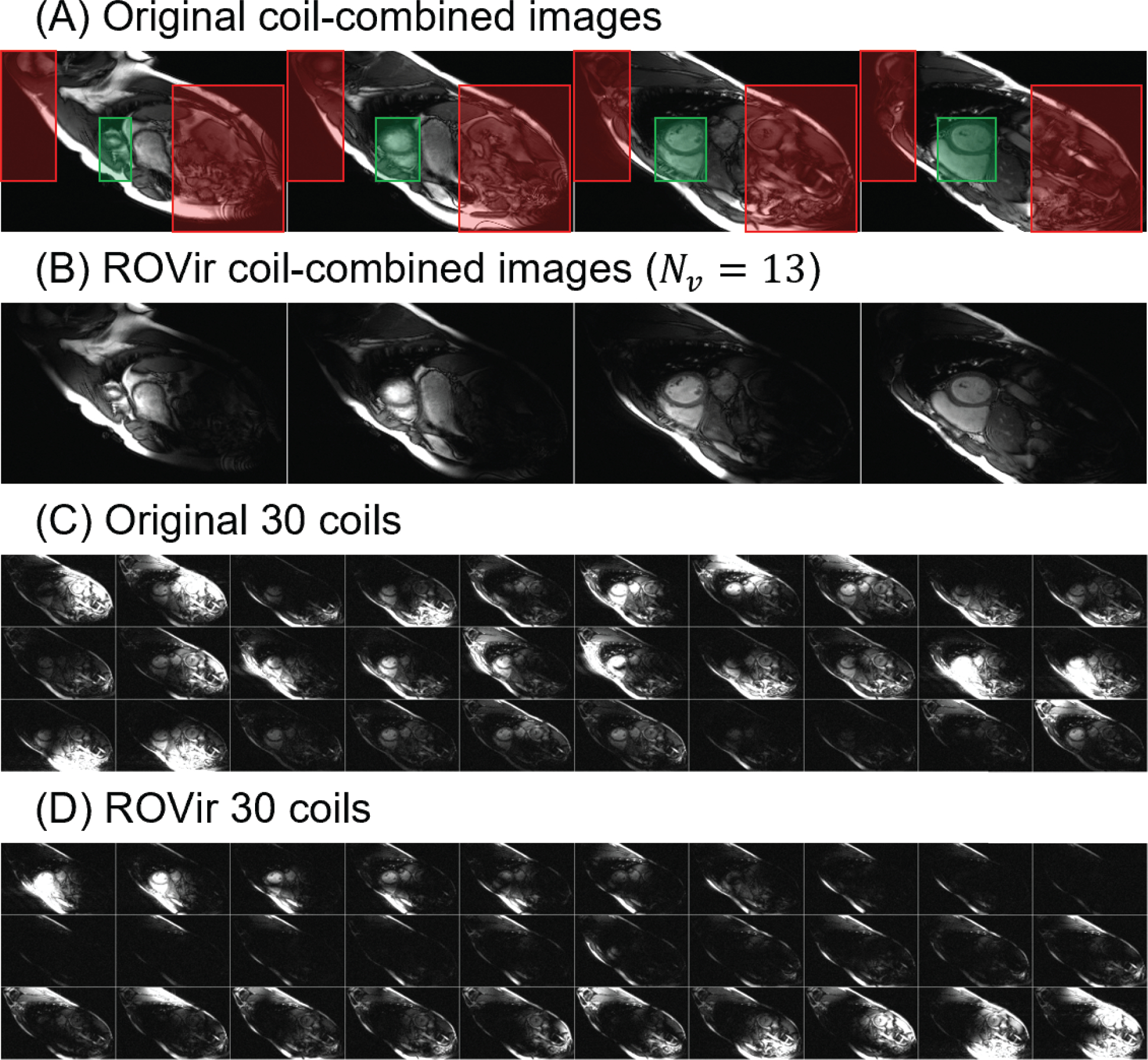
Illustration of applying ROVir to representative full FOV images. (a) Four slices of a representative set of images obtained from full FOV data using the original set of coils. The manually-drawn desired signal region and interference regions used for ROVir are overlaid in green and red, respectively. (b) The corresponding full FOV images obtained by applying ROVir with to the same data from (a). (c) Images showing the individual characteristics of the original 30 coils from the third slice. (d) The 30 individual virtual coils obtained after applying ROVir. It should be noted that the intensity of these images has been scaled to maximize the visibility of the heart. With this choice, the very large intensity of the fat signal is not depicted accurately, since our visualization saturates at the maximum value of the color scale.
Subsequently, optimal ROVir linear mixing weights were computed from the noise-whitened multi-channel calibration data using a generalized eigendecomposition approach with Gram-Schmidt orthonormalization, as described in more detail in the original ROVir paper (7). Note that because the k-space data from the reduced FOV SMS scan contains information from the mixture of multiple slices and because the ROVir weights are designed to be applied directly to that k-space data, it was necessary to optimize the signal-to-interference ratio for all of the simultaneous slices together.
Coil compression and interference suppression was achieved by arranging the optimal ROVir coils in order of descending signal-to-interference ratio, and then discarding all but the top- virtual channels. The number of retained virtual channels was determined automatically. While there are many possible automatic decision rules (7), in this work we selected to prioritize interference suppression. Specifically, we chose such that the retained interference was < 2% compared to the interference observed in the original full set of coils, which was always possible.
Once ROVir was applied, the resulting ROVir SMS k-space data was reconstructed with Split-Slice GRAPPA (13). For comparison, we also reconstructed the original reduced-FOV SMS k-space data without ROVir.
Image Analysis and Statistics
For both the full FOV and SMS+ROVir CINE images, the left-ventricles were manually segmented using Medviso Segment (http://segment.heiberg.se) to compute the ejection fraction (EF), end systolic and diastolic volumes (ESV and EDV) for each subject. Statistical comparisons were performed with Pearson correlation analysis, Bland-Altman plots, and Wilcoxon rank tests with a significance level of 0.05.
RESULTS
Figure 1 shows a representative example of the effects of applying ROVir to single-slice full-FOV images, which will provide insight into the use of ROVir with reduced FOV data. Figure 1(a) shows images generated by applying root-sum-of-squares coil combination to the original set of 30 coils (with signal and interference regions respectively marked with green and red overlays), while Fig. 1(b) shows images generated by applying root-sum-of-squares coil combination to the top ROVir coils. It is visually evident that ROVir has achieved substantial suppression of the interference regions while preserving information from the heart. Figure 1(c) shows images corresponding to the original set of 30 coils, while Fig. 1(d) shows the images corresponding to the 30 virtual coils generated by ROVir, where the ROVir coils are placed in order of descending signal-to-interference ratio. As can be seen in Fig. 1(d), ROVir has nicely separated the heart signal from the interference signal. Specifically, the first several virtual coils contain a substantial amount of signal energy from the heart, while the remaining virtual coils contain a substantial amount of energy from the interference regions. Substantial interference removal is achieved when all but the top- ROVir coils are discarded.
Figure 2 shows quantitative plots of signal and interference characteristics corresponding to the same set of representative data from subject shown in Fig. 1. In this case, the automatic choice of to achieve retained interference < 2% resulted in . As can be seen in Fig. 2(b), this choice retains 71.6 % of the desired signal and 1.8 % of the interference.
Figure 2:
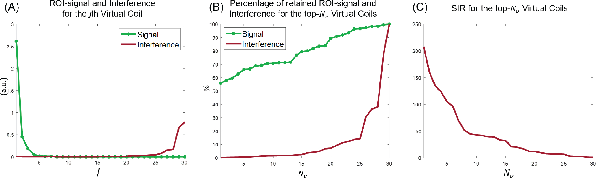
Plots of the quantitative signal and interference characteristics of ROVir corresponding to the data from Fig. 1. (a) Plots of the desired signal energy and the interference signal energy for each of the individual ROVir coils. (b) Plots of the percentage of retained desired signal energy and retained interference energy as a function of the total number of coils that is used, where percentages are computed relative to the original set of coils. (c) Plot of the signal-to-interference ratio (SIR) as a function of .
Figure 3 shows a representative result (from the same subject as in Fig. 1) of reconstructing the reduced FOV SMS data using direct Fourier transform, SMS only, ROVir only, and combined SMS+ROVir reconstruction. Specifically, Fig. 3(a) shows direct Fourier transform reconstruction of the reduced FOV SMS data without applying SMS reconstruction or ROVir; Fig. 3(b) shows the results of applying SMS reconstruction to the original coils without applying ROVir; Fig. 3(c) shows the results of applying ROVir without applying SMS reconstruction; and Fig. 3(d) shows the results obtained with both ROVir and SMS reconstruction. As can be seen, the combination of SMS and ROVir together provide good quality CINE images, while using SMS reconstruction by itself fails to resolve the aliasing artifacts that result from the torso because of the reduced FOV, and the results of ROVir alone fail to separate the simultaneously-excited slices.
Figure 3:

Illustration of applying different reconstruction methods to a set of representative reduced FOV SMS data. (a) Simple Fourier transform reconstruction of the original coils, without SMS reconstruction or ROVir; (b) SMS reconstruction of the original coils without ROVir; (c) Simple Fourier transform reconstruction of the ROVir coils, without SMS reconstruction; (d) the proposed approach combining SMS reconstruction with ROVir.
For single breath-hold whole LV CINE acquisition, Fig. 4 shows a representative case at end diastole compared directly with the clinical reference. The proposed method enables the whole heart imaging without substantial aliasing. Supporting Information Video S1 shows the whole sequence of time-resolved SMS+ROVir CINE compared to the conventional CINE for this case.
Figure 4:

Representative comparison of clinical reference CINE with the proposed single breath hold SMS+ROVir for whole LV 12-slice images. Only the heart is displayed from reduced FOV.
Across all subjects, the SMS+ROVir reduced FOV images resulted in similar quantitative cardiac function parameters as those obtained with the full FOV acquisitions. Figure 5 shows correlation plots and Bland-Altman analyses. There were no significant differences found between left ventricular EDV, ESDV, and EF obtained with proposed SMS+ROVir technique (142.4 ± 25.9ml, 57.6 ± 13.1ml, and 59.6 ± 3.0%) compared to the conventional CINE acquisition (142.3±26.0ml, 56.0±12.9ml, and 60.2±3.2%). Furthermore, the Pearson correlation coefficient between SMS+ROVir and conventional CINE was significant for EDV, ESV, and EF (0.92, 0.94, and 0.88).
Figure 5:

Cardiac function quantification comparison between the proposed SMS+ROVir and conventional reference with Correlation plots (a) and Bland-Altman plots (b) for EDV, ESV, SV, and EF. All parameters resulted in significant agreement between the two acquisitions, with a correlation of 0.92, 0.94, 0.88, and 0.88, respectively, and a bias of 0.1ml, 1.6ml, −1.5ml, and −1.2%, respectively.
DISCUSSION
In this work, we demonstrated that combining SMS and ROVir can enable highly accelerated CINE imaging (8-fold reduced scan time) for single breath-hold whole ventricular acquisition with minimal bias in characterizing cardiac function. The proposed technique uses optimized virtual coils to emphasize a desired region of interest around the heart while suppressing unwanted signal of the outer torso region that would otherwise result in aliasing due to the reduced FOV. Applying SMS or ROVir reconstructions only lead to 2-fold and 4-fold acceleration, respectively, which was not sufficient in fully resolving aliasing. However, we demonstrated a unique combination of the two, which resulted in acceptable image quality for 8-fold accelerated data with minimal SNR penalty. We prospectively validated the proposed technique’s feasibility in yielding cardiac functional parameters against a fully sampled conventional CINE scan in a cohort of normal volunteers demonstrating a non-significant bias in calculating ventricular volumes and ejection fraction.
One key inherent difference between the clinical reference and the proposed SMS+ROVir technique was the 2-fold reduction in flip angle for the SMS-based acquisition due to SAR limitations. This reduction in flip angle reduces the T2 contrast of the bSSFP signal and as a result reduces the blood to myocardium contrast ratio. Though this effect is qualitatively visible and may affect discerning small structures like trabeculae and papillary muscles, it did not result in any significant differences in the quantification of cardiac function. Future work will be to explore the potential to reduce such SAR limitations with more optimized RF waveforms.
While the data acquisition in this study is based on Cartesian k-space sampling, non-Cartesian k-space sampling could also benefit from ROVir in further pushing the limits of acceleration (7), and the combination of ROVir, SMS, and non-Cartesian acquisition may be a promising direction for future exploration. This paper also only considered the acceleration of bSSFP cardiac CINE experiments, although the same principles would also be expected to generalize in a straightforward way to other cardiac imaging sequences and applications. Furthermore, a commercial 30-channel flex body array coil was used for acquisition that was not optimized for cardiac ROVir applications. The array geometry is important for the performance of ROVir, so there is room to improve the performance of ROVir using customized array coil designs. Unfortunately, our current study design is limited in only being able to investigate a standard coil configuration. Repeating the acquired study protocols with various different cardiac coils would provide valuable insights into the potential impact of coil configurations on ROVir reconstruction. We hypothesize that cardiac ROVir may greatly benefit from a combination of some coils that specifically detect regions of interest centered on the heart with other coils tailored to isolate components of the unwanted interference signal. Conversely since ROVir requires multiple coil array elements, a minimum number of elements can also be explored in conjunction with optimal coil geometries. This kind of coil optimization would be an interesting topic for future study.
Similar to other acceleration techniques like parallel imaging (3,4), the use of SMS+ROVir is expected to be associated with some degree of SNR penalty. In particular, the reduction in time spent acquiring data should be expected to reduce the SNR by at least a factor of , where is the acceleration factor (3); the use of ROVir results in an easily calculated reduction in the retained signal energy (7) (which directly corresponds to a further reduction in SNR, similar to a parallel imaging g-factor); the use of SMS reconstruction leads to further g-factor related SNR reductions (13); and changes in MR pulse sequence parameters (e.g., flip angle, TR, and TE) will also modify the SNR. Similar to the case for other acceleration techniques, the speed improvements achieved by SMS+ROVir are often worth a small SNR penalty, and advanced reconstruction and denoising techniques can potentially be applied to mitigate noise in scenarios that are truly SNR-limited. However, it should also be observed that when using ROVir, users have direct control over the trade-off between retained signal energy and retained interference energy through the choice of (7). In this work, we have chosen to aggressively suppress interference while still retaining adequate SNR. In more SNR-limited scenarios, the choice of could have been performed differently to preserve more of the desired signal, at the expense of slightly more retained interference.
Our implementation of ROVir made use of manually-selected signal and interference regions, with the signal region designed to encompass the heart and the interference regions designed to target the portions of the chest wall and arms that may be likely to alias onto the heart along the phase encode direction. We expect the performance of ROVir to be relatively insensitive to small variations in how these regions are drawn. In particular, calculation of the ROVir weights involves performing a generalized eigendecomposition of large region-specific inter-coil correlation matrices formed by averaging over many different voxels contained in the signal and interference regions. We do not expect these correlation matrices or the resulting ROVir weights to change much if a small number of voxels are added to or deleted from the signal and interference regions. Furthermore, it should also be noted that if the signal and interference regions are ever drawn poorly, they are easy to change retrospectively (post-acquisition) without needing to rescan the subject. A preliminary analysis supporting this expectation is shown in the Supporting Information Figure S1 qualitatively demonstrating the impact of a small translational shift in ROI selection. Further analysis needs to be performed in a dedicated study and ideally with expert clinicians identifying the ROI.
In some subjects, residual aliasing is still seen outside the target cardiac ROI. This is entirely expected from ROVir, and could be easily mitigated by appropriate masking to the signal ROI as described in the previous ROVir paper (7). We have not done such masking in this article for the sake of full transparency. However, this residual aliasing may have implications for studies that also care about other organs beyond the heart, and mitigation strategies (e.g., using multiple ROVir-based reconstructions, where each reconstruction targets a different organ with a different ROI) may be valuable in such cases.
There were several limitations in the study, including a small sample size in normal volunteers, and the lack of testing in patients. A critical next step would be to validate if the proposed combination of SMS and ROVir maintains its performance in patients with especially large torsos, since the ability to substantially reduce the FOV would be especially beneficial in such cases. Furthermore, testing of other nuanced clinical indications such as regional wall motion abnormality and impact of the proposed technique on CINE feature tracking should also be carried out. Taken together, follow up clinical studies are needed to fully realize the potential of combining ROVir and SMS for accelerated CINE imaging and validate its clinical utility.
Finally, it should be noted that our comparisons focused on very simple image reconstruction approaches, and our results did not leverage the benefits of advanced constrained image reconstruction methods that are achievable using approaches like sparsity-based reconstruction (14–16), low-rank reconstruction (17–22), structured low-rank reconstruction (23–27), or machine-learning based reconstruction (28–30). SMS and ROVir are expected to be complementary to such approaches, and the combination of the proposed approach with more advanced reconstruction methods (including previous methods that are also capable of single breath-hold CINE imaging (31–33)) is another potentially-promising future direction.
CONCLUSION
We demonstrated the feasibility of combining SMS and ROVir with a reduced FOV acquisition for highly accelerated CINE imaging (8-fold reduced scan time), thus enabling a 10-second single breath-hold 12-slice whole ventricular acquisition. Single breath-hold SMS+ROVir whole-heart CINE yielded cardiac function parameters with no significant bias when compared to SMS CINE.
Supplementary Material
Supporting Information Figure S1. Representative example of a translational shift of the ROI selection on ROVir reconstruction. Minimal qualitative differences can be found up to 1.6cm shift.
Supporting Information Video S1. The video of 12-slice cardiac CINE comparing both the proposed single breath hold SMS+ROVir CINE and the clinial reference.

ACKNOWLEDGMENTS
This work was supported in part by National Institute of Health R01 HL151704, R01 HL159010, and R01 HL135242.
References
- 1.Stäb D, Ritter CO, Breuer FA, Weng AM, Hahn D, Köstler H. CAIPIRINHA accelerated SSFP imaging. Magn Reson Med 2011;65:157–164. [DOI] [PubMed] [Google Scholar]
- 2.Stäb D, Speier P. Gradient-controlled local larmor adjustment (GC-LOLA) for simultaneous multislice bSSFP imaging with improved banding behavior. Magn Reson Med 2019;81:129–139. [DOI] [PubMed] [Google Scholar]
- 3.Pruessmann KP, Weiger M, Boesiger P. Sensitivity encoded cardiac MRI. J Cardiovasc Magn Reson 2001;3:1–9. [DOI] [PubMed] [Google Scholar]
- 4.Griswold MA, Jakob PM, Heidemann RM, Nittka M, Jellus V, Wang J, Kiefer B, Haase A. Generalized autocalibrating partially parallel acquisitions (GRAPPA). Magn Reson Med 2002;47:1202–1210. [DOI] [PubMed] [Google Scholar]
- 5.Kido T, Kido T, Nakamura M, Watanabe K, Schmidt M, Forman C, Mochizuki T. Compressed sensing real-time cine cardiovascular magnetic resonance: accurate assessment of left ventricular function in a single-breath-hold. J Cardiovasc Magn Reson 2016;18:50. [DOI] [PMC free article] [PubMed] [Google Scholar]
- 6.Madore B, Fredrickson JO, Alley MT, Pelc NJ. A reduced field-of-view method to increase temporal resolution or reduce scan time in cine MRI. Magn Reson Med 2000; 43:549–558. [DOI] [PubMed] [Google Scholar]
- 7.Kim D, Stephen CF, Nayak KS, Leahy RM, Haldar JP. Region-optimized virtual (ROVir) coils: Localization and/or suppression of spatial regions using sensor-domain beamforming. Magn Reson Med 2021;86:197–212. [DOI] [PMC free article] [PubMed] [Google Scholar]
- 8.Buehrer M, Pruessmann KP, Boesiger P, Kozerke S. Array compression for MRI with large coil arrays. Magn Reson Med 2007;57:1131–1139. [DOI] [PubMed] [Google Scholar]
- 9.Huang F, Vijayakumar S, Li Y, Hertel S, Duensing GR. A software channel compression technique for faster reconstruction with many channels. Magn Reson Imag 2008;26:133–141. [DOI] [PubMed] [Google Scholar]
- 10.Zhang T, Pauly JM, Vasanawala SS, Lustig M. Coil compression for accelerated imaging with Cartesian sampling. Magn Reson Med 2013;69:571–582. [DOI] [PMC free article] [PubMed] [Google Scholar]
- 11.Xue Y, Yu J, Kang HS, Englander S, Rosen MA, Song HK. Automatic coil selection for streak artifact reduction in radial MRI. Magn Reson Med 2012;67:470–476. [DOI] [PubMed] [Google Scholar]
- 12.Kim D, Lobos RA, Coll-Font J, van den Boomen M, Conklin J, Pang J, Staeb D, Speier P, Bi X, Ghoshhajra B, Haldar JP, Nguyen CT. Feasibility of single breath-hold CINE with combined simultaneous multi-slice (SMS) and region-optimized virtual (ROVir) coils. In: Proc. Int. Soc. Magn. Reson. Med. 2021; p. 0025. [Google Scholar]
- 13.Cauley SF, Polimeni JR, Bhat H, Wald LL, Setsompop K. Interslice leakage artifact reduction technique for simultaneous multislice acquisitions. Magn Reson Med 2014; 72:93–102. [DOI] [PMC free article] [PubMed] [Google Scholar]
- 14.Lustig M, Santos JM, Donoho DL, Pauly JM. k-t SPARSE: High frame rate dynamic MRI exploiting spatio-temporal sparsity. In: Proc. Int. Soc. Magn. Reson. Med. 2006; p. 2420. [Google Scholar]
- 15.Jung H, Sung K, Nayak KS, Kim EY, Ye JC. k-t FOCUSS: A general compressed sensing framework for high resolution dynamic MRI. Magn Reson Med 2009;61:103–116. [DOI] [PubMed] [Google Scholar]
- 16.Feng L, Axel L, Chandarana H, Block KT, Sodickson DK, Otazo R. XD-GRASP: Golden-angle radial MRI with reconstruction of extra motion-state dimensions using compressed sensing. Magn Reson Med 2016;75:775–788. [DOI] [PMC free article] [PubMed] [Google Scholar]
- 17.Liang ZP. Spatiotemporal imaging with partially separable functions. In: Proc. IEEE Int. Symp. Biomed. Imag. 2007; pp. 988–991. [Google Scholar]
- 18.Haldar JP, Liang ZP. Spatiotemporal imaging with partially separable functions: A matrix recovery approach. In: Proc. IEEE Int. Symp. Biomed. Imag. 2010; pp. 716–719. [Google Scholar]
- 19.Lingala SG, Hu Y, DiBella E, Jacob M. Accelerated dynamic MRI exploiting sparsity and low-rank structure: k-t SLR. IEEE Trans Med Imag 2011;30:1042–1054. [DOI] [PMC free article] [PubMed] [Google Scholar]
- 20.Trzasko J, Manduca A. Local versus global low-rank promotion in dynamic MRI series reconstruction. In: Proc. Int. Soc. Magn. Reson. Med. 2011; p. 4371. [Google Scholar]
- 21.Zhao B, Haldar JP, Christodoulou AG, Liang ZP. Image reconstruction from highly undersampled (k,t)-space data with joint partial separability and sparsity constraints. IEEE Trans Med Imag 2012;31:1809–1820. [DOI] [PMC free article] [PubMed] [Google Scholar]
- 22.Otazo R, Candès E, Sodickson DK. Low-rank plus sparse matrix decomposition for accelerated dynamic MRI with separation of background and dynamic components. Magn Reson Med 2015;73:1125–1136. [DOI] [PMC free article] [PubMed] [Google Scholar]
- 23.Haldar JP, Setsompop K. Linear predictability in magnetic resonance imaging reconstruction: Leveraging shift-invariant Fourier structure for faster and better imaging. IEEE Signal Process Mag 2020;37:69–82. [DOI] [PMC free article] [PubMed] [Google Scholar]
- 24.Shin PJ, Larson PEZ, Ohliger MA, Elad M, Pauly JM, Vigneron DB, Lustig M. Calibrationless parallel imaging reconstruction based on structured low-rank matrix completion. Magn Reson Med 2014;72:959–970. [DOI] [PMC free article] [PubMed] [Google Scholar]
- 25.Haldar JP. Low-rank modeling of local k-space neighborhoods (LORAKS) for constrained MRI. IEEE Trans Med Imag 2014;33:668–681. [DOI] [PMC free article] [PubMed] [Google Scholar]
- 26.Jin KH, Lee D, Ye JC. A general framework for compressed sensing and parallel MRI using annihilating filter based low-rank Hankel matrix. IEEE Trans Comput Imaging 2016;2:480–495. [Google Scholar]
- 27.Ongie G, Jacob M. Off-the-grid recovery of piecewise constant images from few Fourier samples. SIAM J Imaging Sci 2016;9:1004–1041. [DOI] [PMC free article] [PubMed] [Google Scholar]
- 28.Sandino CM, Cheng JY, Chen F, Mardani M, Pauly JM, Vasanawala SS. Compressed sensing: From research to clinical practice with deep neural networks. IEEE Signal Process Mag 2020;37:117–127. [DOI] [PMC free article] [PubMed] [Google Scholar]
- 29.Knoll F, Hammernik K, Zhang C, Moeller S, Pock T, Sodickson DK, Akcakaya M. Deep-learning methods for parallel magnetic resonance image reconstruction: A survey of the current approaches, trends, and issues. IEEE Signal Process Mag 2020;37:128–140. [DOI] [PMC free article] [PubMed] [Google Scholar]
- 30.Liang D, Cheng J, Ke Z, Ying L. Deep magnetic resonance image reconstruction: Inverse problems meet neural networks. IEEE Signal Process Mag 2020;37:141–151. [DOI] [PMC free article] [PubMed] [Google Scholar]
- 31.Kustner T, Bustin A, Jaubert O, Hajhosseiny R, Masci PG, Neji R, Botnar R, Prieto C. Isotropic 3D Cartesian single breath-hold CINE MRI with multi-bin patch-based low-rank reconstruction. Magn Reson Med 2020;84:2018–2033. [DOI] [PubMed] [Google Scholar]
- 32.Kustner T, Fuin N, Hammernik K, Bustin A, Qi H, Hajhosseiny R, Masci PG, Neji R, Rueckert D, Botnar RM, Prieto C. CINENet: deep learning-based 3D cardiac CINE MRI reconstruction with multi-coil complex-valued 4D spatio-temporal convolutions. Sci Rep 2020;10:13710. [DOI] [PMC free article] [PubMed] [Google Scholar]
- 33.Gomez-Talavera S, Fernandez-Jimenez R, Fuster V, Nothnagel ND, Kouwenhoven M, Clemence M, Garcia-Lunar I, Gomez-Rubin MC, Navarro F, Perez-Asenjo B, Fernandez-Friera L, Calero MJ, Orejas M, Cabrera JA, Desco M, Pizarro G, Ibanez B, Sanchez-Gonzalez J. Clinical validation of a 3-dimensional ultrafast cardiac magnetic resonance protocol including single breath-hold 3-dimensional sequences. J Am Coll Cardiol Img 2021;14:1742–1754. [DOI] [PMC free article] [PubMed] [Google Scholar]
Associated Data
This section collects any data citations, data availability statements, or supplementary materials included in this article.
Supplementary Materials
Supporting Information Figure S1. Representative example of a translational shift of the ROI selection on ROVir reconstruction. Minimal qualitative differences can be found up to 1.6cm shift.
Supporting Information Video S1. The video of 12-slice cardiac CINE comparing both the proposed single breath hold SMS+ROVir CINE and the clinial reference.